
Trends Sci. 2026; 23(10): 13290
Comparative Effects of Commercial- and Chemical-Grade Solvent Extraction on the Anti-Inflammatory and Antioxidant Activities of “Yaa-Thaa-Phra-Sen”, A Thai Traditional Remedy
Pattama Sriumpai1, Arunporn Itharat2,3, Ubonwan Saesiw2 and Srisopa Ruangnoo2,3,*
1Student of Doctor of Philosophy, Department of Applied Thai Traditional Medicine, Faculty of Medicine,
Thammasat University, Pathumthani 12120, Thailand
2Department of Applied Thai Traditional Medicine, Faculty of Medicine, Thammasat University,
Pathumthani 12120, Thailand
3Center of Excellence on Applied Thai Traditional Medicine Research (CEATMR), Faculty of Medicine,
Thammasat University, Pathumthani 12120, Thailand
(*Corresponding author’s e-mail: [email protected])
Received: 7 January 2026, Revised: 11 March 2026, Accepted: 18 March 2026, Published: 25 April 2026
Abstract
Yaa-Thaa-Phra-Sen remedy (YTPS) is a traditional Thai formulation used for relief of inflammatory symptoms and treat osteoarthritis. In traditional practice, vinegar or distilled liquor is commonly used as extraction solvents. However, limited studies have investigated the effects of commercial- and chemical-grade solvent systems on the anti-inflammatory and antioxidant properties of YTPS extracts. Extraction of bioactive compounds is an important step that affects the quality and biological activity of herbal preparations. The objective of this study was to evaluate the anti-inflammatory and antioxidant properties of YTPS extracts prepared from fresh and dried plant materials using both chemical-grade (acetic acid and ethanol at defined concentrations) and commercial-grade (vinegar and distilled liquors) solvent systems. Anti-inflammatory activity was evaluated based on nitric oxide (NO), TNF-α and PGE2 production in lipopolysaccharide (LPS)-stimulated RAW 264.7 macrophages, while antioxidant activity was assessed using DPPH, ABTS, and FRAP assays. Among the tested conditions, extracts prepared from dried plant materials using an acidic ethanolic solvent (Ch-6) exhibited the strongest inhibitory activity against NO production without cytotoxic effects. In contrast, no inhibitory activity was observed against TNF-α and PGE2 under the tested conditions, indicating a selective anti-inflammatory profile. Antioxidant activities and total phenolic content varied across extraction systems, with intermediate ethanol concentrations generally showing higher values. These finding demonstrate that extraction conditions, including solvent composition and plant material condition, influence the biological properties of YTPS in an endpoint-dependent manner. This study provides scientific support for the optimization of extraction processes for traditional Thai herbal formulations.
Keywords: Thai traditional medicine, Inflammation, Antioxidants, Biological activity, Solvent extraction
Introduction
Osteoarthritis (OA) is a deterioration of the cartilage in chronic joint disease that causes pain and various degrees of inflammation, disability, and functional loss [1]. OA was the 15th highest cause of year lived with disability (YLDs) worldwide, with more than 500 million people [2] and more than 6 million patients in Thailand [3]. It is increasingly prevalent due to the
growing number of the elderly population [4], which is considered a burden for global public health issues [5]. The pathophysiology of OA-related pain involves inflammation and enzymatic cartilage degradation by the production of various pro-inflammatory mediators such as Nitric oxide (NO), prostaglandin E2 (PGE2), and tumor necrosis factor-alpha (TNF-α), and stress-induced intracellular signals such as reactive oxygen species (ROS), which are important contributors to the severity and impact of OA symptoms and the outcomes of OA treatments [6,7]. Non-steroidal anti-inflammatory drugs (NSAIDs) are the principal medications to treat OA pain [8]. However, the use of NSAIDs is limited by their cardiovascular (CV), gastrointestinal (GI), and adverse events (AEs) effects, especially in the elderly [9,10].
Thai traditional medicine (TTM) was introduced as a safer topical medication alternative treatment for relieving pain and inflammation in OA, offering a potentially lower-risk approach with fewer side effects. Yaa-Thaa-Phra-Sen remedy (YTPS) is one of many formulations well-known to be used to treat pain and inflammation of OA in Thai traditional scripture called “Tum-Raa-Yaa-Phra-O-Sod-Phra-Naa-rai” or “King Narai’s book”, which was compiled during the reign of King Narai of Siam (1656 - 1688 AD) and has been certified by the Ministry of Public Health as the Thai traditional medicine textbook since 2015 [11]. YTPS is composed of 13 herbs: Allium ascalonicum Linn., Allium sativum Linn., Aloe vera Linn., Alpinia galanga Linn., Baliospermum montanum (Willd.) Muell. Arg., Boesenbergia rotunda Linn., Cymbopogon nardus Linn., Drypetes roxburghii Wall., Ferula assa-foetida Linn., Melia azedarach Linn., Piper nigrum Linn., Senna siamea Lam., and Tamarindus indica Linn. D. roxburghii is the main ingredient and the most common phenolic acid found in phytochemical composition of D. roxburghii is ellagic acid [12,13], which has anti-inflammatory and antioxidant properties [14,15]. Additionally, the scriptures describe a traditional topical preparation method of YTPS in which the herbal ingredients are finely ground, followed by maceration in vinegar or distilled liquor, producing a paste that is applied directly to painful areas such as the knee [16]. This preparation method differs from most traditional Thai remedies, which are commonly prepared as water-based decoctions. Notably, previous research has reported that a YTPS preparation macerated in distilled liquor, in accordance with the traditional description, demonstrated pain-relieving effects in patients with early-stage knee OA [17]. In contemporary Thai traditional medicine, practitioners in Lopburi Province, Thailand, typically prepare YTPS by macerating the herbal mixture in a vinegar and distilled liquor solution at a ratio of 1:3 prior to topical application. Despite its longstanding use in the region, there remains limited scientific evidence to fully substantiate its traditional application. Previous studies on the bioactivity of herbal ingredients in YTPS have investigated the anti-inflammatory effects of B. rotunda [18], B. montanum [19,20], D. roxburghii [21,22], and P. nigrum [23,24], as well as the antioxidant properties of B. montanum [20] and D. roxburghii [25]. However, there is currently no research addressing how different commercial- and chemical-grade solvent extraction methods influence the anti-inflammatory and antioxidant properties of YTPS.
The chemical composition and biological activity of herbal extracts are influenced by the preparation and extraction methods applied to the raw materials [26,27]. In particular, drying processes can alter phytochemical profiles and, consequently, affect biological activity [28,29]. In Thai traditional medicine, herbal remedies may be prepared using either fresh or dried plant materials, depending on traditional practices, seasonal availability, and intended use. Accordingly, the present study investigated both fresh and dried YTPS materials to reflect this variability in traditional preparation.
In addition, solvent selection is an important factor in the extraction of bioactive compounds, as solvents differ in their physicochemical properties and extraction efficiency. This study aimed to compare the effects of chemical-grade and commercial-grade solvents on extraction efficiency, chemical composition, and biological activities of YTPS. Fresh and dried YTPS materials were extracted using different solvent systems. The resulting extracts were evaluated for anti-inflammatory activity, based on the inhibition of NO, PGE2, and TNF-α production, and for antioxidant capacity using DPPH, ABTS, and FRAP assays. Phytochemical constituents were also determined, including total phenolic content (TPC) and ellagic acid derived from the main ingredient, D. roxburghii. Through this comparative approach, the study aimed to identify suitable extraction conditions to support the further development of YTPS as a topical anti-inflammatory product for alleviating osteoarthritis-related pain.
Materials and methods
Chemicals and reagents
Ellagic acid standard was obtained from the Tokyo Chemical Industry (Tokyo, Japan). Dimethyl sulfoxide (DMSO), phosphoric acid, and triethylamine were purchased from Sigma-Aldrich (St. Louis, MO, USA). Purified water was prepared using a Milli-Q system (Millipore, Bedford, MA, USA). HPLC-grade acetonitrile and methanol were purchased from RCI Lab Scan (Bangkok, Thailand). The murine macrophage leukemia cell line RAW 264.7 (ATCC TIB-71) was obtained from the American Type Culture Collection (ATCC, VA, USA). Roswell Park Memorial Institute (RPMI) 1640 medium, fetal bovine serum (FBS), penicillin-streptomycin (P/S), trypsin-EDTA, phosphate-buffered saline (PBS), and trypan blue stain were purchased from Gibco (Thermo Fisher Scientific, NY, USA). Lipopolysaccharide (LPS, Escherichia coli 055), 3-(4, 5-dimethyl-2-thiazolyl)-2,5-diphenyl-2H-tetrazolium bromide (MTT), N-(1-naphthyl) ethylenediamine dihydrochloride, and sulfanilamide were obtained from Sigma-Aldrich (St. Louis, MO, USA). The PGE2 ELISA kit was purchased from Cayman Chemical (MI, USA), and the Quantikine ELISA Mouse TNF- Immunoassay was obtained from R&D Systems (MN, USA).
Instruments
The HPLC system used in this study was an Agilent 1200 Series system (Agilent Technologies, CA, USA), consisting of a solvent degasser (G1322A), an autosampler (G1329A), a quaternary pump (G1311A), a photodiode array detector (G1315D), and a column oven (G1316A), Chromatographic separation was performed using a Luna C18 analytical column (4.6×250 mm2, 5 microns; Phenomenex, CA, USA). Data acquisition and analysis were carried out using ChemStation software.
Plant materials
Thirteen herbal ingredients were collected from Lopburi Province, Thailand, and voucher specimens were deposited as references specimens at the Thai Traditional Medicine Herbarium, Department of Thai Traditional and Alternative Medicine, Thailand. The raw materials were cleaned, sliced, and coarsely ground, then combined according to traditional proportions (Table 1) to obtained fresh YTPS (FYTPS). For comparison, dried YTPS (DYTPS) was prepared from the same raw materials. The plant materials were cleaned, sliced, coarsely ground, and dried at 40 C in a hot air oven before being combined according to traditional proportions.
Table 1 Proportion of herbal ingredients in YTPS.
Species |
Thai name |
Part used |
Proportion (% w/w) |
Allium ascalonicum |
Hom-Deang |
Bulb |
2.3 |
Allium sativum |
Ka-Tiam |
Bulb |
2.3 |
Aloe barbadensis |
Yaa-Dum |
Resin |
2.3 |
Alpinia galanga |
Kaa |
Rhizome |
2.3 |
Baliospermum montanum |
Tong-Tak |
Leaves |
9.3 |
Boesenbergia rotunda |
Ka-Chai |
Rhizome |
2.3 |
Cymbopogon nardus |
Ta-Khai-Hom |
All part |
9.3 |
Drypetes roxburghii |
Ma-Kum-Kai |
Leaves |
37 |
Ferula assa-foetida |
Ma-Ha-Hing |
Resin |
2.3 |
Melia azedarach |
Liean |
Leaves |
9.3 |
Piper nigrum |
Prik-Thi |
Fruit |
2.3 |
Senna siamea |
Kee-Lek |
Leaves |
9.3 |
Tamarindus indica |
Ma-Kaam |
Leaves |
9.3 |
Maceration method
Fresh (FYTPS) and dried (DYTPS) materials were extracted by maceration using 2 categories of solvent: Chemical-grade and commercial-grade solvents. Chemical-grade solvents included laboratory-grade reagents with defined ethanol or acetic acid concentrations. Commercial-grade solvents consisted of locally available products traditionally used in Thai medicine practice, such as vinegar and distilled liquor. This approach allowed comparison between standardized laboratory conditions and traditional preparations.
YTPS materials were macerated with the designated solvent at room temperature for 72 h, and the extraction was repeated 3 times. The combined extracts were filtered through Whatman No. 1 filter paper and concentrated under reduced pressure at 40 °C using a rotary evaporator to obtain crude extracts. Extraction yield was calculated for each sample.
Six solvent systems were used for chemical-grade extraction: 5% acetic acid (Ch-1), 30% ethanol (Ch-2), 40% ethanol (Ch-3), 95% ethanol (Ch-4), 5% acetic acid in 30% ethanol (1:3 v/v) (Ch-5), and 5% acetic acid in 95% ethanol (1:3 v/v) (Ch-6).
Similarly, 5 solvent systems were used for commercial-grade extraction: Vinegar (Aorsorror, Co-1), 30% distilled liquor (Pai Tong, Co-2), 40% distilled liquor (Ruang Khao, Co-3), vinegar diluted in 30% distilled liquor (1:3 v/v) (Co-5), and vinegar diluted in 95% ethanol (1:3 v/v) (Co-6).
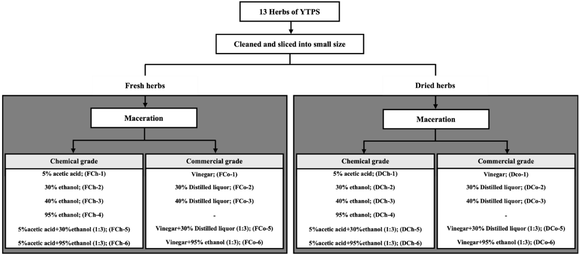
Figure 1 Schematic representation of the YTPS extraction process.
Table 2 Extraction conditions and solvent systems used for YTPS preparation.
Sample |
Chemical-grade solvent |
Code |
Commercial-grade solvent |
Code |
Dried herb |
5% acetic acid |
DCh-1 |
Vinegar |
DCo-1 |
30% ethanol |
DCh-2 |
30% distilled liquor |
DCo-2 |
|
40% ethanol |
DCh-3 |
40% distilled liquor |
DCo-3 |
|
95% ethanol |
DCh-4 |
- |
- |
|
5% acetic acid in 30% ethanol (1:3 v/v) |
DCh-5 |
vinegar diluted in 30% distilled liquor (1:3 v/v) |
DCo-5 |
|
5% acetic acid in 95% ethanol (1:3 v/v) |
DCh-6 |
vinegar diluted in 95% ethanol (1:3 v/v) |
DCo-6 |
|
Fresh herb |
5% acetic acid |
FCh-1 |
Vinegar |
FCo-1 |
30% ethanol |
FCh-2 |
30% distilled liquor |
FCo-2 |
|
40% ethanol |
FCh-3 |
40% distilled liquor |
FCo-3 |
|
95% ethanol |
FCh-4 |
- |
- |
|
5% acetic acid in 30% ethanol (1:3 v/v) |
FCh-5 |
vinegar diluted in 30% distilled liquor (1:3 v/v) |
FCo-5 |
|
5% acetic acid in 95% ethanol (1:3 v/v) |
FCh-6 |
vinegar diluted in 95% ethanol (1:3 v/v) |
FCo-6 |
Note: All extractions were performed by maceration at room temperature for 72 h. Commercial vinegar refers to commercially available vinegar containing approximately 5% acetic acid. Distilled liquor refers to commercially available ethanol-based beverages with specified ethanol concentrations (30% or 40%).
Cell culture
The RAW 264.7 murine macrophage cell lines (ATCC TIB-71, Manassas, VA, USA) was cultured in RPMI 1640 medium supplemented with 10% fetal bovine serum (FBS), and 1% penicillin-streptomycin. Cells were maintained at 37 °C in a humidified atmosphere containing 5% CO₂, and the culture medium was changed every 2 - 3 days. All experimental procedures were conducted at the Center of Excellence in Applied Thai Traditional Medicine Research (CEATMR), Faculty of Medicine, Thammasat University, Thailand.
This study was conducted in accordance with the institutional biosafety regulations of Thammasat University. All procedures were approved by the Institute Biosafety Committee of Thammasat University (Approval No. 076/2018). Experiments were performed under biosafety level 2 (BSL-2) conditions.
Anti-inflammatory activities
Cell viability assay
Cytotoxicity was evaluated using the MTT reduction assay [30]. RAW 264.7 macrophages were seeded into 96-well plates at a density of 1×10⁵ cells/well and incubated for 18 - 24 h at 37 °C in a humidified atmosphere containing 5% CO₂. The medium was then replaced with fresh medium containing various concentrations of the test samples or an equivalent concentration of DMSO (vehicle control). After 24 h of incubation, the supernatant was removed, and 10 µL of MTT solution (5 mg/mL in PBS) was added to each well. The plates were incubated for an additional 2 h under the same conditions. The solution was then removed, and the resulting formazan crystals were dissolved in 100 µL of acidic isopropanol (0.04 M HCl in isopropanol). Absorbance was measured at 570 nm. Cell viability was expressed as a percentage relative to vehicle-treated control. Experiments were performed in 3 independent experiments, each in triplicate. Cytotoxicity results were used to determine the non-cytotoxic concentration range for subsequent anti-inflammatory assays. Only concentrations that maintained cell viability above 70% were selected for further evaluation in nitric oxide (NO), TNF-α, and PGE2 inhibition assays to ensure that the observed effects were not attributable to reduced cell viability.
Nitric oxide assay
NO production in LPS-stimulated RAW 264.7 macrophages was evaluated using a modified method adapted from Tewtrakul and Itharat [30]. Cells were seeded into 96-well plates at a density of 1×10⁵ cells/well and incubated at 37 C in a humidified atmosphere with 5% CO2 for 24 h. The medium was then replaced with 100 µL of fresh medium containing 5 ng/mL LPS and various concentrations of the test samples (0.01 - 100 µg/mL in DMSO). After 24 h of incubation, the supernatant was collected and transferred to a new 96-well plate. Nitrite accumulation, as an indicator of NO production, was determined using Griess reagent (1% sulfanilamide and 0.1% N-(1-naphthyl) ethylenediamine dihydrochloride in 2.5% phosphoric acid). Absorbance was measured at 570 nm. The percentage inhibition of NO production was calculated relative to the LPS-treated control. The half-maximal inhibitory concentration (IC50) values were determined by nonlinear regression analysis using data from 3 independent experiments, each performed in triplicate. Prednisolone was used as a positive control for NO inhibition. Diclofenac was included as a reference non-steroidal anti-inflammatory drug (NSAID) to provide a comparative control.
Determination of TNF-α production
TNF-α production in LPS-stimulated RAW 264.7 macrophages were evaluated using a modified method based on Makchuchit et al. [31], with a Quantikine mouse TNF-α ELISA kit (R&D Systems, MN, USA). Cells were seeded into 96-well plates at a density of 1×10⁵ cells/well and incubated at 37 °C in a humidified atmosphere containing 5% CO₂ for 24 h. The medium was then replaced with fresh medium containing 5 ng/mL LPS and various concentrations of test samples. After 24 h of incubation, 50 µL of supernatant was collected and transferred to an ELISA plate, and TNF-α levels were determined according to the manufacturer’s instructions. Absorbance was measured at 450 nm. The percentage inhibition of TNF-α production was calculated relative to LPS-treated control, and IC₅₀ values were determined by nonlinear regression. All experiments were performed in 2 independent replicates.
Determination of PGE2 production
PGE₂ production in LPS-stimulated RAW 264.7 macrophages were evaluated using a modified method adapted from Tewtrakul and Subhadhirasakul [32], with a PGE₂ ELISA kit (Cayman Chemical, Ann Arbour, MI, USA). Cells were seeded into 96-well plates at a density of 1×10⁵ cells/well and incubated at 37 °C in a humidified atmosphere containing 5% CO₂ for 24 h. The medium was then replaced with fresh medium containing 5 ng/mL LPS and various concentrations of test samples. After 24 h of incubation, 50 µL of supernatant was collected and subjected to competitive ELISA according to the manufacturer’s instructions. Briefly, samples were incubated with PGE₂ tracer and PGE₂ monoclonal antibody at 4 °C for 18 h. After washing, Ellman’s reagent was added, and colour development was allowed to proceed in the dark for 60 - 90 min. Absorbance was measured at 420 nm. The percentage inhibition of PGE₂ production was calculated relative to LPS-treated control, and IC₅₀ values were derived by nonlinear regression. All experiments were performed in 2 independent replicates.
Antioxidant activities
DPPH assay
The DPPH assay was performed according to a modified method described by Yamasaki et al. [33]. In a 96-well plate, 100 µL of extracts at different concentrations (1, 10, 50 and 100 µg/mL) were mixed with 100 L of 0.06 mM DPPH solution. The mixture was incubated in the dark at room temperature for 30 min, and absorbance was measured at 520 nm. Radical scavenging activity was determined based on the reduction in DPPH absorbance. EC50 values were calculated accordingly. Experiments were performed in 3 independent experiments, each in triplicate. Butylated hydroxytoluene (BHT) was used as a reference standard.
ABTS assay
The ABTS assay was performed according to a modified method described by Re et al. [34]. In a 96-well plate, 20 L of extracts at different concentrations (1, 10, 50 and 100 µg/mL) were mixed with 180 µL of ABTS+ solution. The ABTS+ solution was prepared by reacting 7 mM ABTS with 2.45 mM potassium persulfate and allowing the mixture to stand in the dark for 12 - 16 h. Prior to use, the solution was diluted to obtain an absorbance of 0.70 0.05 at 734 nm. After incubation for 6 min in the dark at room temperature, absorbance was measured at 734 nm. Radical scavenging activity was calculated as percentage inhibition, an EC50 values were determined by regression analysis of the dose-response curve. Experiments were performed in 3 independent experiments, each in triplicate. Trolox was used as a reference standard.
Ferric-reducing antioxidant power assay
The FRAP assay was performed according to the method of Benzie and Strain [35] with slight modifications. In a 96-well plate, 20 µL of extract was mixed with 180 µL of freshy prepared FRAP reagent. The FRAP reagent consisted of 300 mM acetate buffer (pH 3.6), 10 mM TPTZ solution in 40 mM HCl, and 20 mM ferric chloride (FeCl3), mixed in a ratio of 10:1:1 (v/v/v), and pre-warmed at 37 C prior to use. After incubation at 37 C for 4 min, absorbance was measured at 593 nm. All samples were analyzed in triplicate in 3 independent experiments. A standard curve was constructed using ferrous sulfate (FeSO4), and results were express as µmol Fe2+ equivalents per gram of extract. Trolox was used as a reference antioxidant, and results were additionally expresses as µmol Trolox equivalent per gram of extract for comparison.
Phytochemical content and ellagic acid quantification
Determination of total phenolic content
The total phenolic content (TPC) of the extracts was determined using a modified Folin-Ciocalteu method [36]. In a 96-well plate, 20 µL of the extract solution was mixed with 100 µL of Folin-Ciocalteu reagent. After 5 min of incubation at room temperature, 80 µL of 7.5% (w/v) sodium carbonate solution was added. The reaction mixture was then incubated in the dark at room temperature for 30 min to allow color development. Absorbance was measured at 765 nm using a microplate reader. All samples were analyzed in triplicate in 3 independent experiments. A calibration curve was constructed using gallic acid standard solutions (2.5 to 100 µg/mL), and results were expressed as mg gallic acid equivalents (GAE) per gram of extract (mg GAE/g extract).
HPLC analysis of Ellagic acid
A stock solution of ellagic acid was prepared at a concentration of 1 mg/mL in methanol (MeOH). A calibration curve was constructed using serial dilutions to obtain standard concentrations of 25, 50, 100, 200, 400, and 800 µg/mL. For sample analysis, crude extracts obtained from different extraction conditions were reconstituted in methanol to a final concentration of 10 mg/mL and filtered prior to injection. Chromatographic separation was performed on a reverse-phase C18 column (Phenomenex Luna-C18, 4.6×250 mm2, 5 microns). The mobile phase consisted of 1% (v/v) formic acid in water (solvent A) and acetonitrile (solvent B). The gradient elution was programmed as follows: 0 - 19 min, 1% B; 20 min, 40% B; 20.1 to 30 min, 80% B; and from 30.1 to 40 min, 1% B for column re-equilibration. The flow rate was maintained at 1.0 mL/min, and total run time was 45 min. An aliquot of 10 µL was injected into the HPLC system. Detection was carried out using a photodiode array detector (DAD) at 254 nm. Quantification of ellagic acid was performed based on the calibration curve.
Statistical analysis
Nitric oxide inhibition and antioxidant properties were performed in 3 independent experiments. TNF-α and PGE2 production were performed in 2 independent experiments. Each concentration was tested in triplicate, and the results are expressed as mean standard error of the mean (SEM). Statistical analysis was performed using one-way analysis of variance (ANOVA). A p-value of less than 0.05 (p < 0.05) was considered statistically significant.
Results and discussion
Extraction yields of YTPS
The extraction yields (% w/w) of fresh and dried YTPS obtained using various solvent systems are summarised in Table 3. As the data were derived from a single extraction replicate, no statistical analysis was performed and the results are presented descriptively.
Overall, extracts prepared from dried plant materials tended to provide higher yields than those from fresh materials, except for extraction with 95% ethanol. This may be attributed to the lower moisture content of dried samples, which increases the relative concentration of extractable constituents. However, the drying process may also result in the degradation or loss of certain thermolabile compounds [37]. Among the tested conditions, the highest yields were observed for DCo-5 (36.83%) and DCh-5 (36.38%), followed by DCo-1 (33.40%), and DCo-6 (32.83%). Chemical-grade solvents generally resulted in slightly higher yields than commercial-grade solvents, although this pattern was not consistent across all conditions. For example, vinegar-based maceration may introduce additional constituents derived from fermentation, which could influence the overall yield [38]. Extraction yield is influenced by multiple factors, including moisture content, solvent polarity and composition, and the chemical characteristics of the phytoconstituents. The relatively higher yields obtained from mixed acid-alcohol solvent systems, followed by acetic acid and 40% ethanol, may be due to improved cell wall disruption and enhanced solubilisation of polar compounds [39-41]. Nevertheless, further replicated experiments are required to confirm these findings.
Anti-inflammatory activities
Osteoarthritis (OA) is a common degenerative joint disorder, particularly among the elderly population worldwide, including in Thailand. The progression of OA is closely associated with chronic inflammation, characterised by increased production of pro-inflammatory mediators such as NO, PGE₂, and TNF-α, which contribute to the generation of reactive oxygen species (ROS) in inflamed articular tissues and chondrocytes [42]. In this study, anti-inflammatory activity of YTPS was evaluated using LPS-stimulated RAW 264.7 macrophages. NO production was assessed across a concentration range, while TNF-α and PGE₂ productions were measured at a single concentration (Figure 2).
Cell viability
The cytotoxicity of YTPS extracts was evaluated using the MTT assay in RAW 264.7 cells. None of the extracts exhibited cytotoxicity at the tested concentrations, indicating that the observed effects were not attributable to reduced cell viability. Based on these results, concentrations that maintained cell viability above 70% were selected for subsequent anti-inflammatory assays. The concentration range of 0.001 - 100 µg/mL was used for nitric oxide (NO) inhibition analysis, while a single non-cytotoxic concentration (100 µg/mL) was selected for TNF-α and PGE2 assays as a preliminary screening. Diclofenac exhibited cytotoxicity at higher concentrations and was therefore evaluated at a lower concentration in subsequent assays.
Inhibition of NO production
Among the tested samples, DCo-6 and DCh-6 exhibited the strongest inhibitory activity against NO production, with IC₅₀ values of 33.80 ± 0.13 and 33.88 ± 1.54 µg/mL, respectively (Figure 2(A)). These were followed by FCh-6 (42.25 ± 2.46 µg/mL), FCo-6 (44.81 ± 2.07 µg/mL), DCh-4 (51.81 ± 0.41 µg/mL), and FCh-4 (53.48 ± 0.97 µg/mL). Dried extracts exhibited significantly greater inhibition of NO production than fresh extracts (p < 0.05). This finding suggests that, under the extraction conditions used, drying may enhance the availability of bioactive constituents associated with NO inhibition [43,44]. In contrast, extracts obtained with 95% ethanol showed similarly activity between dried and fresh materials. Comparison between chemical-grade and commercial-grade solvents revealed no substantial differences in extraction efficiency. Among the extraction conditions evaluated, only 2 solvent systems, mixed acetic acid or vinegar in 95% ethanol (1:3 v/v) and 95% ethanol alone, demonstrated marked inhibition of NO production. Although no previous in vitro studies on YTPS are available, its major component, D. roxburghii, have been reported to inhibit NO production both in vitro and in vivo models [22,45]. Phytochemical studies indicate the presence of alkaloids, flavonoids, phenolic compounds, and terpenoids [46], including ellagic acid [47]. Due to its polar structure, ellagic acid is more readily extracted in polar solvent systems [48], which may partly explain the higher activity observed in these extracts.
Consistent with this, DCh-6 and DCo-6 exhibited significantly greater inhibition of NO production compared with FCh-6, FCo-6, DCh-4, and FCh-4, although all extracts demonstrated inhibitory effects. The greater potency of DCh-6 and DCo-6 is in agreement with a previous clinical study reporting that YTPS extracted with vinegar was more effective than a 40% ethanol extract in reducing pain and improving knee flexion in OA patients [49]. All extracts were less potent than prednisolone with IC50 value of 0.08 ± 0.01 µg/mL (Figure 2(B)). In contrast, diclofenac exhibited minimal inhibitory effect (IC50 > 30 µg/mL) under the same experimental conditions, which is consistent with its primary mechanism as a cyclooxygenase inhibitor rather than a direct modulator of nitric oxide synthesis.
Inhibition of TNF-α production
None of the YTPS extracts inhibited TNF-α production under the tested conditions, suggesting that the anti-inflammatory activity of YTPS may not involve modulation of TNF-α. It should be noted that TNF-α inhibition was evaluated at a single concentration (100 µg/mL) as a preliminary screening, at which all extracts exhibited less than 25% inhibition (Figure 2(C)).
In contrast, ellagic acid has been reported to significantly reduced TNF-α levels in a dose-dependent manner [50]. Prednisolone exhibited strong inhibitory activity with IC50 value of 0.11 0.01 µg/mL (Figure 2(E)). All extracts were significantly lower activity than prednisolone (p < 0.05). Diclofenac also showed minimal activity in this assay, exhibiting only 1.31% inhibition at 30 µg/mL, further supporting that this experimental system is not primary responsive to NSAID-mediated pathways.
Inhibition of PGE2 production
Similarly, none of the YTPS extracts inhibited PGE2 production under the tested conditions, with all samples exhibiting minimal activity (% inhibition < 5% at 100 µg/mL) as presented in Figure 2(D). This indicates that the anti-inflammatory activity of YTPS may not involve cyclooxygenase-mediated pathways.
Ellagic acid has been reported to reduce PGE2 levels in dose-dependent manner [50], but this effect was not observed in YTPS extracts. In contrast, both prednisolone and diclofenac showed strong inhibitory activity, with IC50 values of 0.07 0.01 and 0.004 0.001 µg/mL, respectively (Figure 2(F)). All extracts showed significantly lower activity than the positive controls (p < 0.05).
Taken together, YTPS extracts demonstrated a selective anti-inflammatory profile characterised by inhibition of NO production without significant effects on TNF-α or PGE2. This suggests that the anti-inflammatory activity of YTPS may primarily involve modulation of inducible nitric oxide synthase (iNOS)-related pathways rather than broader inhibition of upstream pro-inflammatory cytokines or cyclooxygenase-mediated pathways.
In osteoarthritis, excessive NO contributes to oxidative stress, cartilage degradation, and chondrocyte apoptosis [42]. Therefore, selective reduction of NO may still be therapeutic relevance, even in the absence of TNF-α or PGE2 inhibition.
This activity profile differs from that of corticosteroids such as prednisolone, which broadly suppress inflammatory mediators, and from NSAIDs such as diclofenac, which primarily target cyclooxygenase pathways. The absence of cytotoxicity further supports that the observed NO inhibition reflects a genuine pharmacological effect.
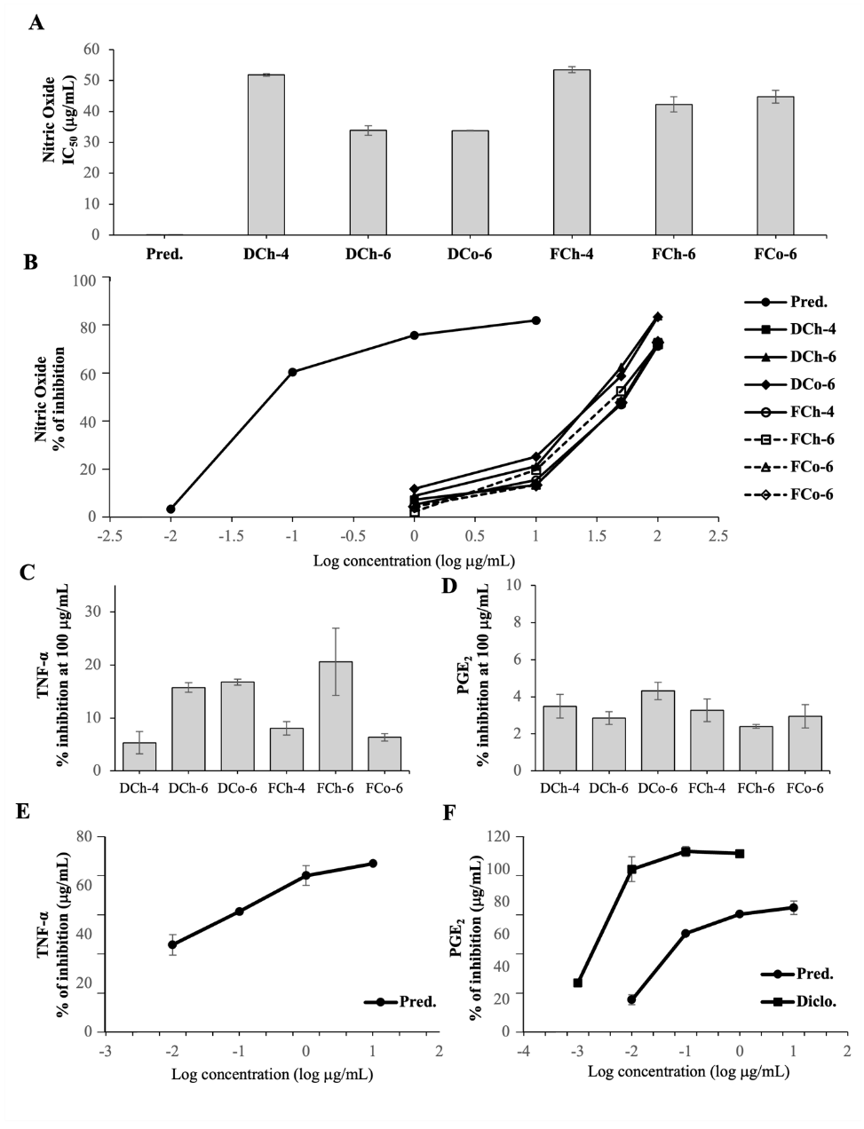
Figure 2 Anti-inflammatory effects of YTPS extracts in LPS-stimulated RAW264.7 macrophages. (A) IC50 values for NO oxide (NO) inhibition, (B) Dose-response curves showing % inhibition of NO production, (C) % inhibition of TNF-α production at 100 µg/mL, (D) % inhibition of PGE2 production at 100 µg/mL, (E) Dose-response curves of prednisolone on TNF-α inhibition, and (F) Dose-response curves of prednisolone and diclofenac on PGE2 inhibition. All data are presented as mean ± SEM. Statistical significance is indicated as follows: (*) p < 0.05 compared to the positive control; different letters indicate significance differences between fresh and dried samples (p < 0.05).
Antioxidant activities
The antioxidant potential of YTPS extracts was evaluated using complementary assays, including DPPH and ABTS radical scavenging assays, and the ferric reducing antioxidant power (FRAP) assay (Table 3).
DPPH free radical scavenging activity assay
All YTPS extracts exhibited DPPH radical scavenging activity, with EC50 values ranging from 7.45 to 21.98 µg/mL (Table 3). Several extracts, particularly FCo‑6, FCh‑6, DCh‑6, DCh‑5, and FCh-1, demonstrated significantly stronger activity than the reference standard butylated hydroxytoluene (BHT) (p < 0.05). In contrast, DCh‑4 and FCo‑3 showed comparatively weaker activity.
Differences between fresh and dried samples were generally modest and dependent on extraction conditions, although some dried extracts showed slightly higher. The effect of solvent grade (chemical vs. commercial) was minimal, indicating that solvent composition rather than grade plays a more important role in DPPH scavenging activity.
ABTS radical cation decolorization assay
In the ABTS assay, YTPS extracts demonstrated EC50 values ranging from 15.63 to 55.51 µg/mL, except for DCh‑4, which showed weak activity (EC50 > 100 µg/mL) (Table 3). Among the tested samples, DCh‑3, DCh‑2, and DCh‑6 exhibited relatively stronger radical scavenging activity.
Similar to the DPPH results, differences between fresh and dried samples were limited, although some dried extracts showed improved activity. Solvent grade did not have a clear impact on antioxidant performance. All extracts showed significantly lower activity than the positive control Trolox (p < 0.05), as expected for crude extracts.
Ferric reducing antioxidant power (FRAP) assay
Consistent with the radical scavenging assays, YTPS extracts demonstrated ferric (Fe3+) reducing capacity. FRAP values ranged from 70.46 to 382.17 mg Fe2+/g extract, while TEAC values ranged from 41.62 to 204.03 mg Trolox/g extract (Table 3). DCh‑3, FCh‑1, and DCh‑2 exhibited the highest reducing capacity, approaching that of BHT, whereas DCh‑4 showed comparatively lower activity.
Dried extracts generally tended to exhibit higher reducing capacity than fresh extracts, although the extent of this difference varied among samples. The influence of solvent grade was inconsistent across samples.
Collectively, the results from the 3 complementary assays confirm that YTPS extracts possess measurable antioxidant activity. Several extracts, particularly DCh‑3, FCh‑6, and DCh‑6, consistently demonstrated relatively high activity across multiple assays.
Extraction with 40% ethanol and the mixed acetic acid in 95% ethanol (1:3 v/v) system was associated with comparatively strong antioxidant activity. This may be due to suitable solvent polarity, which facilitates the extraction of a broad range of amphiphilic phytochemicals. This observation is consistent with previous reports highlighting the importance of solvent polarity in recovering phenolic compounds and antioxidant constituents [39-41].
The observed antioxidant activity may be partly attributed to bioactive polyphenols, including ellagic acid derived from D. roxburghii [47], which has been reported to possess radical scavenging properties [48]. Polyphenolic compounds are well known to contribute to antioxidant activity through electron donation and free radical stabilization mechanism [51]. Although the antioxidant activity of the crude extracts was lower than that of pure reference standards such as Trolox and BHT, measurable activity was still observed, which is consistent with the nature of complex phytochemical mixtures, where synergistic or additive interactions among constituents may contribute to the overall antioxidant activity [52].
Overall, differences between fresh and dried samples were modest, and the effect of solvent grade was limited, suggesting that intrinsic phytochemical composition plays a key role in determining antioxidant activity.
Importantly, the antioxidant activity observed in this study is consistent with the selective anti-inflammatory effects of YTPS, particularly its ability to inhibit NO production. This is relevant in the context of osteoarthritis, where oxidative stress contributes to inflammation and cartilage degradation [42].
Phytochemical content and ellagic acid quantification
Total phenolic content
Phenolic compounds are widely recognised as major phytochemical constituents contributing to antioxidant potential in plant-derived extracts [53]. In this study, total phenolic content (TPC) was determined to characterise the phytochemical profile of YTPS and to examine its association with the observed antioxidant activities.
TPC values ranged from 64.86 to 118.49 mg GAE/g extract, with the exception of FCh-4 and DCh-4, which showed comparatively lower phenolic concentrations (Table 3). Among the tested samples, DCh-3 exhibited the highest phenolic content, followed by DCh-6, DCo-6, and FCo-6. Notably, DCh-3 also demonstrated strong antioxidant activity, consistent with its performance in the ABTS and FRAP assays.
Although TPC reflects the total abundance of phenolic constituents rather than their specific activities, the general agreement between higher TPC values and stronger antioxidant capacity suggests that phenolic compounds contribute, at least in part, to the observed effects.
The influence of solvent grade (chemical versus commercial) on TPC was minimal. In contrast, drying appeared to enhance phenolic recovery in certain extracts, particularly DCh-3 and DCo-3, compared with their fresh extracts (FCh-3 and FCo-3) (p < 0.05). This may be attributed to reduced moisture content and improved extraction efficiency following dehydration. Overall, these findings confirm that YTPS contains appreciable levels of phenolic compounds, which are likely to contribute to its antioxidant properties.
High-performance liquid chromatography (HPLC) analysis
To further characterise the phytochemical basis underlying the observed biological activities, HPLC analysis was performed to identify and quantify the major bioactive constituents. The chromatographic system successfully resolved ellagic acid, with a retention time (RT) of 16.7 min, consistent with that of the reference standard (Figure 3).
Ellagic acid was selected as a phytochemical marker for YTPS extracts, as it is a major constituent of D. roxburghii, the principal component of YTPS formulation [46,47], and has been widely reported to exhibit both antioxidant and anti-inflammatory activities [48,50].
Quantitative analysis revealed that DCh-3 contained the highest ellagic acid content (30.75 ± 6.32 mg/g extract), followed by FCh-6, FCo-6, and DCo-6. The high ellagic acid content in DCh-3 is consistent with its elevated TPC and strong antioxidant performance, particularly in the ABTS and FRAP assays, suggesting that ellagic acid contribute to the observed bioactivity. However, the biological activity of YTPS extracts cannot be attributed to a single compound alone. Given the chemical complexity of herbal formulations, other phytochemical constituents, such as flavonoids and terpenoids, are also likely contribute through additive or synergistic effects.
Comparison across extraction conditions indicated that both drying and solvent composition influenced ellagic acid recovery. Notably, DCh-3 contained significantly higher ellagic acid contents than both its fresh extract (FCh-3) and the corresponding commercial-grade extract (DCo-3) (p < 0.05). This may be explained by enhanced solvent penetration following dehydration, together with the suitable polarity of ethanol-water mixtures, which facilitate the extraction of amphiphilic polyphenols such as ellagic acid.
Collectively, these findings support the use of ellagic acid as a representative bioactive marker for YTPS. However, the overall biological activities are more likely governed by the combined effects of multiple phytochemical constituents rather than a single compound.
To facilitate interpretation across multiple endpoints, a heatmap analysis was performed (Figure 3). The analysis revealed distinct clustering patterns among extracts based on their biological activities and phytochemical profiles. Extracts such as DCh-3 were associated with high antioxidant capacity and phenolic content, whereas DCh-6 was characterised by stronger NO inhibition activity. These finding further support that extraction outcomes are endpoint-dependent, and that no single extraction condition consistently maximized all measured parameters.
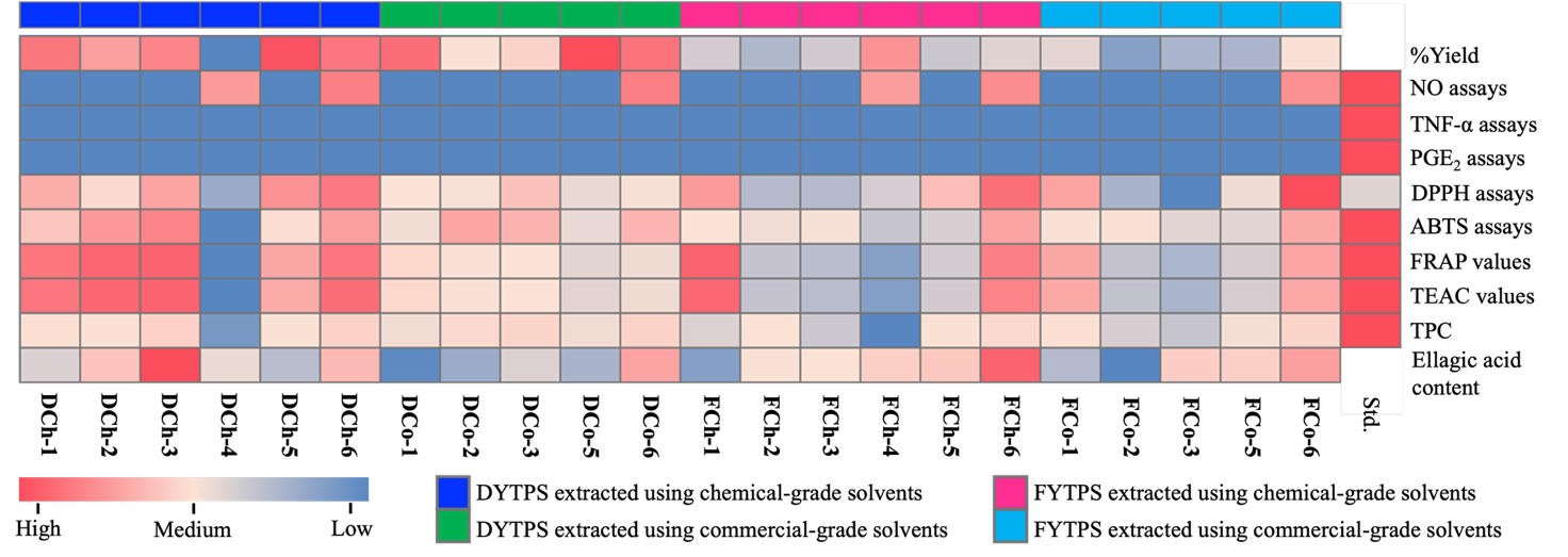
Figure 3 Heat map illustrating the relative bioactivity profiles of YTPS extracts across anti‑inflammatory and antioxidant assays. Data were normalized using min-max scaling within each assay. Red indicates higher activity and blue indicates lower activity after normalization. Anti-inflammatory activity was assessed based on NO, TNF-, and PGE2 inhibition in LPS-stimulated RAW264.7 cells, while antioxidant capacity was evaluated using DPPH, ABTS, FRAP, and TEAC assays Total phenolic content (TPC) and ellagic acid contents are also included for comparison. Detailed quantitative values are presented in Table 3.
Table 3 Percentage yield (% w/w), antioxidant activity, and phytochemical properties of YTPS extracts obtained under different solvent extraction conditions.
Sample |
%Yield (w/w) |
Antioxidant activity |
Phytochemical constituents |
||||
DPPH (EC50, µg/mL) |
ABTS (EC50, µg/mL) |
FRAP value (mg Fe(II)/ g extract) |
TEAC value (mg Trolox/ g extract) |
Total phenolic content (mg GAE/g extract) |
Ellagic acid content (mg/g extract) |
||
DCh-1 |
32.49 |
10.39 ± 0.24b |
27.8 ± 1.61*,b |
373.22 ± 2.80*,b |
199.35 ± 2.00*,b |
98.37 ± 1.79*,a |
13.60 ± 0.56 |
DCh-2 |
28.27 |
11.74 ± 0.30a |
19.56 ± 0.87*,a |
380.81 ± 2.88*,a,b |
203.32 ± 1.55*,a,b |
97.94 ± 0.69* |
17.99 ± 3.83 |
DCh-3 |
31.10 |
10.10 ± 0.48a |
15.63 ± 0.70*,a,b |
382.17 ± 4.27a,b |
204.03 ± 1.48*,a,b |
118.49 ± 2.28*,a |
30.75 ± 6.32a,b |
DCh-4 |
11.62 |
18.05 ± 0.22*,a |
> 100 |
70.46 ± 2.20*,a |
41.62 ± 2.02*,a |
21.62 ± 2.53*,a |
14.07 ± 1.55 |
DCh-5 |
36.38 |
9.57 ± 0.05* |
32.00 ± 3.14*,a |
349.48 ± 2.18*,a,b |
185.87 ± 1.14*,a,b |
93.14 ± 1.96* |
12.67 ± 0.48 |
DCh-6 |
32.54 |
8.79 ± 0.54*,b |
20.48 ± 0.54*,b |
372.93 ± 4.19*,b |
201.33 ± 2.19b |
117.33 ± 0.62* |
19.09 ± 0.91 |
DCo-1 |
33.40 |
12.00 ± 0.07a,b |
37.11 ± 1.85*,b |
326.83 ± 1.36*,a,b |
174.03 ± 0.71*,a,b |
88.64 ± 3.33*,a |
10.20 ± 0.45 |
DCo-2 |
21.00 |
12.29 ± 0.18a |
21.85 ± 0.69*,a |
317.84 ± 2.21*,a,b |
169.34 ± 1.16*,a,b |
106.53 ± 3.10*,a |
11.84 ± 1.23 |
DCo-3 |
22.67 |
11.02 ± 0.04 |
24.32 ± 1.86*,a,b |
321.25 ± 2.14*,a,b |
171.12 ± 1.12*,a,b |
112.88 ± 2.19*,a |
13.65 ± 0.15b |
DCo-5 |
36.83 |
13.07 ± 0.84 |
40.65 ± 0.53* |
283.38 ± 2.63*,a,b |
151.32 ± 1.37*,a,b |
88.84 ± 3.87* |
12.24 ± 0.19 |
DCo-6 |
32.83 |
12.32 ± 0.17a,b |
24.54 ± 0.71*,b |
304.58 ± 3.18*,a,b |
162.41 ± 1.66*,a,b |
115.88 ± 1.73* |
21.47 ± 0.16 |
FCh-1 |
18.62 |
9.83 ± 0.24b |
33.08 ± 2.25* |
381.98 ± 4.44b |
202.87 ± 2.32b |
77.14 ± 1.24*,a,b |
11.24 ± 2.58 |
FCh-2 |
16.55 |
16.31 ± 1.00a |
37.98 ± 3.21*,a |
239.05 ± 1.94*,a |
128.15 ± 1.02*,a |
94.08 ± 2.43*,b |
14.44 ± 2.74 |
FCh-3 |
18.42 |
16.37 ± 0.82a |
35.44 ± 2.07*,a,b |
221.12 ± 3.09*,a,b |
118.78 ± 1.62*,a,b |
68.05 ± 2.11*,a |
14.54 ± 0.86a |
FCh-4 |
29.45 |
14.36 ± 0.44a |
55.51 ± 3.09*,a |
141.63 ± 1.53*,a |
77.23 ± 0.80*,a |
3.69 ± 0.71*,a |
16.60 ± 1.64 |
FCh-5 |
18.05 |
10.88 ± 0.65 |
48.08 ± 2.09*,a |
259.02 ± 3.66*,a |
138.59 ± 1.91*,a |
93.94 ± 2.26* |
17.50 ± 0.15 |
FCh-6 |
19.33 |
8.51 ± 0.65* |
21.39 ± 1.70* |
368.25 ± 4.29*,b |
195.68 ± 2.24*,b |
108.59 ± 4.83* |
28.41 ± 1.55 |
FCo-1 |
19.61 |
10.07 ± 0.10a |
34.19 ± 1.02* |
349.74 ± 3.18*,a,b |
186.01 ± 1.66*,a,b |
98.54 ± 2.73*,a,b |
12.54 ± 0.79 |
FCo-2 |
14.29 |
17.04 ± 0.16a |
34.58 ± 2.00*,a |
235.57 ± 4.38*,a |
126.33 ± 2.29*,a |
74.27 ± 2.73*,a,b |
9.98 ± 1.16 |
FCo-3 |
16.27 |
21.98 ± 2.55* |
44.3 ± 1.54*,a,b |
198.82 ± 2.88*,a,b |
107.12 ± 1.51*,a,b |
64.86 ± 0.76*,a |
16.85 ± 0.39 |
FCo-5 |
16.32 |
12.82 ± 0.75 |
43.90 ± 2.91* |
263.53 ± 3.92*,a |
140.95 ± 2.05*,a |
90.61 ± 1.39* |
16.60 ± 2.39 |
FCo-6 |
20.69 |
7.45 ± 0.14*,a |
22.47 ± 0.85* |
350.17 ± 4.59*,a,b |
186.23 ± 2.40*,a,b |
113.63 ± 0.88* |
21.93 ± 1.76 |
Standard |
- |
13.72 ± 0.39 |
5.34 ± 0.20 |
391.88 ± 1.24 |
209.63 ± 0.65 |
291.70 ± 3.98 |
- |
Note: Antioxidant activities of YTPS extracts were evaluated using DPPH (with BHT as the positive control), ABTS (with Trolox as the positive control), FRAP, and TEAC (with BHT as the reference control). Total phenolic content (TPC) was expressed as mg of GAE/g of extract (with BHT used as a control). Ellagic acid content was expressed as mg/g of extract and quantified by HPLC. All data are presented as the mean ± SEM (n=3). Statistical significance was defined as follows: (*) p < 0.05 compared with the positive control; (a) p < 0.05 between fresh and dried samples; and (b) p < 0.05 between chemical-grade and commercial-grade solvents.
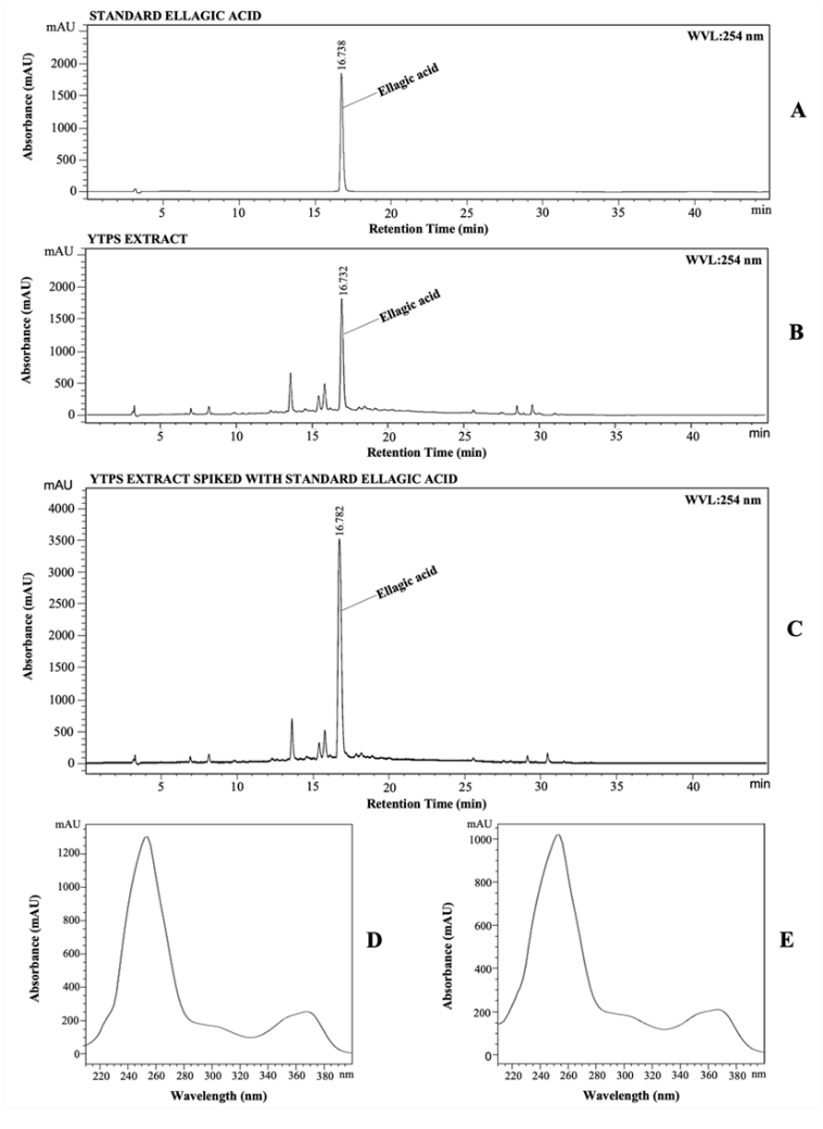
Figure 4 HPLC chromatograms of (A) standard ellagic acid (0.4 mg/mL), (B) DCh-6 extract (10 mg/mL), and (C) DCh-6 extract spiked with ellagic acid standard, showing a consistent peak at a retention time of 16.7 min. Corresponding UV absorption spectra of (D) the standard and (E) ellagic acid in the extract exhibit similar profiles at the onset, apex, and end of the peak.
Conclusions
This study provides experimental evidence supporting the optimisation of extraction conditions for YTPS, with anti-inflammatory activity defined as the primary functional endpoint. Among the evaluated extraction systems, maceration of dried herbs using a chemical-grade solvent consisting of 5% acetic acid in 95% ethanol (1:3 v/v) (DCh-6) exhibited the most pronounced inhibitory effect on nitric oxide (NO) production in LPS-stimulated RAW264.7 cells without inducing cytotoxicity, indicating strong anti-inflammatory potential. However, other evaluated parameters, including total phenolic content, ellagic acid concentration, and antioxidant activities assessed by DPPH, ABTS, and FRAP assays, identified different extracts as having the highest values. These findings indicate that extraction efficiency and bioactivity are endpoint-dependent, rather than governed by a single universally optimal extraction condition. Notably, although DCh-6 did not exhibit the highest total phenolic or ellagic acid content, its antioxidant activities were comparable to those of DCh-3, suggesting that antioxidant efficacy may not be solely dependent on phenolic marker abundance. In contrast, DCh-3 did not demonstrate anti-inflammatory activity in the cellular model.
Collectively, these results highlight the importance of aligning extraction conditions with specific biological targets and support the traditional use of YTPS as a potential candidate for managing osteoarthritis-related inflammation. However, further studies are required to identify additional bioactive constituents and to elucidate the underlying molecular mechanisms.
Acknowledgements
The authors would like to thank the Center of Excellence in Applied Thai Traditional Medicine Research (CEATMR) and the Faculty of Medicine, Thammasat University, Thailand for their support and providing the necessary research facilities.
Declaration of generative AI in scientific writing
The authors declare that generative artificial intelligence tools were used only to refine the language and improve the clarity and readability of this manuscript under direct human oversight. These tools were not applied to the generation of scientific content, data analysis, or the interpretation of experimental results, nor were they assigned authorship or co-authorship. The authors assume full responsibility for the scientific accuracy, integrity, and conclusions of the present study.
CRediT author statement
Pattama Sriumpai: Conceptualization; Methodology; Validation; Formal analysis; Writing - Original draft preparation. Arunporn Itharat: Conceptualization; Methodology; Writing - Reviewing and Editing. Ubonwan Saesiw: Validation; Formal analysis. Srisopa Ruangnoo: Conceptualization; Methodology; Writing - Reviewing and Editing.
References
[1] AM Alshami. Knee osteoarthritis related pain: A narrative review of diagnosis and treatment. International Journal of Health Sciences 2014; 8(1), 85-104.
[2] DJ Hunter, L March and M Chew. Osteoarthritis in 2020 and beyond: A Lancet Commission. The Lancet 2020; 396(10264), 1711-1712.
[3] Thai Bureau of Non-Communicable Diseases, Department of Disease Control and Ministry of Public Health. Non-communicable disease and injury survey report: Annual report. WVO Thailand, Bangkok, 2018.
[4] AM Briggs, MJ Cross, DG Hoy, L Sànchez-Riera, FM Blyth, AD Woolf and L March. Musculoskeletal health conditions represent a global threat to healthy aging: A report for the 2015 World Health Organization world report on ageing and health. Gerontologist 2016; 56(2), 245-255.
[5] R Wittenauer, L Smith and K Aden. Priority medicines for Europe and the World 2013 update. Background Paper 6.12 Osteoarthritis. WHO Essential Medicines and Health Products Information Portal, Switzerland, 2013.
[6] HG Schaible. Mechanisms of chronic pain in osteoarthritis. Current Rheumatology Reports 2012; 14, 549-556.
[7] HG Schaible, F Richter, A Ebersberger, MK Boettger, H Vanegas, G Natura, E Vazquez and GSV Banchet. Joint pain. Experimental Brain Research 2009; 196(1), 153-162.
[8] SL Kolasinski, T Neogi, MC Hochberg, C Oatis, G Guyatt, J Block, L Callahan, C Copenhaver, C Dodge, D Felson and K Gellar. 2019 American College of Rheumatology/Arthritis Foundation guideline for the management of osteoarthritis of the hand, hip, and knee. Arthritis & Rheumatology 2020; 72(2), 220-233.
[9] JP Pelletier, J Martel-Pelletier, F Rannou and C Cooper. Efficacy and safety of oral NSAIDs and analgesics in the management of osteoarthritis: Evidence from real-life setting trials and surveys. Semin Arthritis Rheum 2016; 45(4), 22-27.
[10] M Bardou and AN Barkun. Preventing the gastrointestinal adverse effects of nonsteroidal anti-inflammatory drugs: From risk factor identification to risk factor intervention. Joint Bone Spine 2010; 77(1), 6-12.
[11] Ministry of Public Health. Notification of the Protection and Promotion of Thai Traditional Medicine Committee: The promulgation of national textbooks and national formulas of Thai traditional medicine (No. 1). 2016. Available at: https://www.ratchakitcha.soc.go.th/DATA/PDF/2559/E/007/21.PDF, accessed October 2023.
[12] P Sengupta and J Mukherjee. Terpenoids and related compounds-XI: The structure of roxburgholone, a new triterpenoid constituent of Putranjiva roxburghi. Tetrahedron 1968; 24(20), 6259-6264.
[13] AKA Kumar, SR Chowdhury, TCT Chakrabarti, HK Majumdar, TJT Jha and SMS Mukhopadhyay. A new ellagic acid glycoside and DNA topoisomerase IB inhibitory activity of saponins from Putranjiva roxburghii. Natural Product Communications 2014; 9(5), 675-677.
[14] S Corbett, J Daniel, R Drayton, M Field, R Steinhardt and N Garrett. Evaluation of the anti-inflammatory effects of ellagic acid. Journal of PeriAnesthesia Nursing 2010; 25(4), 214-220.
[15] S Mishra and M Vinayak. Ellagic acid inhibits PKC signaling by improving antioxidant defense system in murine T cell lymphoma. Molecular Biology Reports 2014; 41, 4187-4197.
[16] Department of Thai Traditional and Alternative Medicine, Ministry of Public Health. Kum-Pre-Tat-Phra-Naa-rai. WVO Thailand, Bangkok, Thailand, 2012.
[17] P Kanokkangsadal, M Waipradab, P Toonsakul, K Kanlamyai, R Khun-anake and A Itharat. Clinical efficacy and safety of Ya-Ta-Pra-Sen in treating primary osteoarthritis of knee compared with analgesic cream an open label randomized controlled trial. Thai Journal of Pharmacy Practice 2024; 12(3), 880-890.
[18] S Tewtrakul, S Subhadhirasakul, C Karalai, C Ponglimanont and S Cheenpracha. Anti-inflammatory effects of compounds from Kaempferia parviflora and Boesenbergia Pandurata. Food Chemistry 2009; 115(2), 534-538.
[19] W Pipatrattanaseree, A Itharat, N Mukkasombut and U Saesiw. Potential in vitro anti-allergic, anti-inflammatory and cytotoxic activities of ethanolic extract of Baliospermum montanum root, its major components and a validated HPLC method. BMC Complementary and Alternative Medicine 2019; 19(1), 45.
[20] N Kakatum, N Jaiarree, S Makchuchit and A Itharat. Antioxidant and anti-inflammatory activities of Thai medicinal plants in Sahasthara remedy for muscle pain treatment. Journal of The Medical Association of Thailand 2012; 95, 120-126.
[21] RS Bai and P Sarath. Screening and evaluation of bioactivity of methanolic extract of leaf of Putranjiva roxburghii Wall. (Putranjivaceae). International Journal of Pharmaceutical and Biological Science 2019; 9(1), 1148-1156.
[22] SR Bijekar, M Gayatri and L Rajanna. Evaluation of anti-inflammatory activity of flavonoid fractions from Euphorbiaceae members on raw 264.7 cell lines. Journal of Cytology and Genetics 2015; 16(1), 39-46.
[23] I Sakpakdeejaroen, S Makchuchit and A Itharat. Nitric oxide inhibitory activity of herbal extract formulae for anti-inflammation. Thammasat Medical Journal 2014; 14(1), 7-12.
[24] X Ying, X Chen, S Cheng, Y Shen, L Peng and HZ Xu. Piperine inhibits IL-β induced expression of inflammatory mediators in human osteoarthritis chondrocyte. International Immunopharmacology 2013; 17(2), 293-299.
[25] D Shahwar, MA Raza, A Saeed, M Riasat, FI Chattha, M Javaid and S Ullah. Antioxidant potential of the extracts of Putranjiva roxburghii, Conyza bonariensis, Woodfordia fruiticosa and Senecio chrysanthemoids. African Journal of Biotechnology 2012; 11(18), 4288-4295.
[26] A Bucić-Kojić, M Planinić, S Tomas, L Jakobek and M Šeruga. Influence of solvent and temperature on extraction of phenolic compounds from grape seed, antioxidant activity and colour of extract. International Journal of Food Science & Technology 2009; 44(12), 2394-2401.
[27] S Felhi, A Daoud, H Hajlaoui, K Mnafgui, N Gharsallah and A Kadri. Solvent extraction effects on phytochemical constituents profiles, antioxidant and antimicrobial activities and functional group analysis of Ecballium elaterium seeds and peels fruits. Journal of Food Science and Technology 2017; 37(3), 483-492.
[28] G Ozcan-Sinir, A Ozkan-Karabacak, CE Tamer and OU Copur. The effect of hot air, vacuum and microwave drying on drying characteristic, rehydration capacity, color, total phenolic content and antioxidant capacity of Kumquat (Citrus japonica). Journal of Food Science and Technology 2018; 39, 475-484.
[29] L Zou, F Lu, B Lin, Y Zhou, T Liu and Y Sun. Stability of alkaloids during drying process and their effect on anticoagulating activity of Uncariae Ramulus Cum Uncis. Journal of Analytical Methods in Chemistry 2019; 1, 789515.
[30] S Tewtrakul and A Itharat. Nitric oxide inhibitory substances from the rhizomes of Dioscorea membranacea. Journal of Ethnopharmacology 2007; 109(3), 412-416.
[31] S Makchuchit, R Rattarom and A Itharat. The anti-allergic and anti-inflammatory effects of Benjakul extract (A Thai traditional medicine), its constituent plants and its some pure constituents using in vitro experiments. Biomedicine & Pharmacotherapy 2017; 89, 1018-1026.
[32] S Tewtrakul and S Subhadhirasakul. Effects of compounds from Kaempferia parviflora on nitric oxide, prostaglandin E2 and tumor necrosis factor-alpha productions in RAW264.7 macrophage cell. Journal of Ethnopharmacology 2008; 120(1), 81-84.
[33] K Yamazaki, A Hashimoto, Y Kokusenya, T Miyamoto and T Sato. Electrochemical method for estimating the antioxidative effects of methanol extracts of crude drugs. The Chemical and Pharmaceutical Bulletin 1994; 42(8), 1663-1665.
[34] R Re, N Pellegrini, A Proteggente, A Pannala, M Yang and C Rice-Evans. Antioxidant activity applying an improved ABTS radical cation decolorization assay. Free Radical Biology and Medicine 1999; 26(9-10), 1231-1237.
[35] IF Benzie and JJ Strain. The ferric reducing ability of plasma (FRAP) as a measure of “antioxidant power”: The FRAP assay. Analytical Biochemistry 1996; 239(1), 70-76.
[36] O Folin and V Ciocalteu. On tyrosine and tryptophane determinations in proteins. The Journal of Biological Chemistry 1927; 73, 627-650.
[37] RP Rocha, EC Melo and LL Radünz. Influence of drying process on the quality of medicinal plants: A review. Journal of Medicinal Plants Research 2011; 5(33), 7076-7084.
[38] UF Hutchinson, NP Jolly, BS Chidi, MM Ngongang and SK Ntwampe. Vinegar engineering: A bioprocess perspective. Food Engineering Reviews 2019; 11, 290-305.
[39] L Shi, W Zhao, Z Yang, V Subbiah and HA Suleria. Extraction and characterization of phenolic compounds and their potential antioxidant activities. Environmental Science and Pollution Research 2022; 29(54), 81112-81129.
[40] TL Brown, HE Lemay, BE Bursten, CJ Murphy and PM Woodward. Chemistry: The central science. 12th ed. Pearson Education, Illinois, 2012.
[41] B Zhuang, G Ramanauskaite, ZY Koa and ZG Wang. Like dissolves like: A first-principles theory for predicting liquid miscibility and mixture dielectric constant. Science Advances 2021; 12(7), eabe7275.
[42] MB Goldring and M Otero. Inflammation in osteoarthritis. Current Opinion in Rheumatology 2011; 23(5), 471-478.
[43] N Harbourne, E Marete, JC Jacquier and D O’Riordan. Effect of drying methods on the phenolic constituents of meadowsweet (Filipendula ulmaria) and willow (Salix alba). LWT 2009; 42(9), 1468-1473.
[44] R Lemus-Mondaca, A Vega-Gálvez, P Rojas, K Stucken, C Delporte, G Valenzuela-Barra, RJ Jagus, MV Agüero and A Pasten. Antioxidant, antimicrobial and anti-inflammatory potential of Stevia rebaudiana leaves: Effect of different drying methods. Journal of Applied Research on Medicinal and Aromatic 2018; 11, 37-46.
[45] AL Rajahamsa, KS Deepak, TK Rao, AP Kumar, GS Reddy and MS Potbhare. Muti-model confirmatory evaluation of anti-inflammatory, analgesic and antioxidant activities of Putranjiva roxburghii Wall. International Journal of Advanced Biological and Biomedical Research 2013; 4(12), 921-932.
[46] SR Bijekar, MC Gayatri and L Rajanna. Antimicrobial activity of isolated flavonoid fractions from Drypetes Roxburghii (Wall.) Huresawa and its phytochemical fingerprinting. The International Journal of Innovative Research in Science, Engineering and Technology 2015; 4(7), 6214-6224.
[47] AN Naik, PN Pandya and AU Mankad. A comprehensive review on phytochemicals and medicinal properties of Putranjiva roxburghii Wall. International Association of Biologicals and Computational Digest 2023; 2(1), 234-240.
[48] JE Hayes, P Allen, N Brunton, MN O’Grady and JP Kerry. Phenolic composition and in vitro antioxidant capacity of four commercial phytochemical products: Olive leaf extract (Olea europaea L.), lutein, sesamol and ellagic acid. Food Chemistry 2011; 126(3), 948-955.
[49] K Khuntong, S Fakkham, Y Watcharadul, T Chinakarn and T Imsomboon. A comparison of efficacy of Ya-Tha-Pra-Sen between 40% ethyl alcohol solvent and vinegar solvent for pain reducing in Thai traditional diagnosis of Lom-Jub-Pong-Hang-Kao at Thai traditional and complimentary medicine hospital. Journal of Traditional Thai Medical Research 2022; 8(2), 13-28.
[50] Z Lin, C Lin, C Fu, H Lu, H Jin, Q Chen and J Pan. The protective effect of Ellagic acid (EA) in osteoarthritis: An in vitro and in vivo study. Biomedicine & Pharmacotherapy 2020; 125, 109845.
[51] H Rasouli, MH Farzaei and R Khodarahmi. Polyphenols and their benefits: A review. International Journal of Food Properties 2017; 65, 16-28.
[52] S Wang, KA Meckling, MF Marcone, Y Kakuda and R Tsao. Synergistic, additive, and antagonistic effects of food mixtures on total antioxidant capacities. Journal of Agricultural and Food Chemistry 2011; 59(3), 960-968.
[53] LM Babenko, OE Smirnov, KO Romanenko, OK Trunova and IV Kosakivska. Phenolic compounds in plants: Biogenesis and function. The Ukrainian Biochemical Journal 2019; 91(3), 5-18.