
Trends Sci. 2026; 23(10): 13013
Polyvinyl Chloride and Polyethylene Microplastics Promote Cardiac Oxidative Stress and Histopathological Remodeling in Wistar Rats
Muhammad
Reva Aditya1,
 ,
Athaya Rahmanardi Muhammad1,
,
Athaya Rahmanardi Muhammad1,
Hikmawan
Wahyu Sulistomo2,*,
,
Dian Nugrahenny2,
,
Holipah3,
,
Happy
Kurnia Pematasari4,
,
Cholid Tri Tjahjono5,
,
Veny
Mayangsari5,
and Waqar Ahmed6,
1Master Program in Biomedical Science, Faculty of Medicine, Universitas Brawijaya, Malang, Indonesia
2Department of Pharmacology, Faculty of Medicine, Universitas Brawijaya, Malang, Indonesia
3Department of Public Health, Faculty of Medicine, Universitas Brawijaya, Malang, Indonesia
4Department of Biochemistry and Biomolecular, Faculty of Medicine, Universitas Brawijaya, Malang, Indonesia
5Division of Cardiovascular Prevention and Rehabilitation, Department of Cardiology and Vascular Medicine, Universitas Brawijaya - Dr. Saiful Anwar Teaching Hospital, East Java, Indonesia
6Division of Immunology, Immunity to Infection and Respiratory Medicine, School of Biological Sciences,
Faculty of Biology, Medicine and Health, University of Manchester, Manchester, United Kingdom
(*Corresponding author’s e-mail: [email protected])
Received: 13 December 2025, Revised: 6 January 2026, Accepted: 20 January 2026, Published: 30 April 2026
Abstract
Microplastic pollution is an emerging environmental concern with accumulating evidence of systemic toxicity. Polyethylene (PE) and polyvinyl chloride (PVC) are persistent polymers that may promote oxidative stress and influence redox-sensitive signaling pathways such as nuclear factor kappa-B (NF-κB). This study investigated the cardiac effects of subacute inhalation of PE and PVC microplastics (MPs) in Wistar rats, focusing on oxidative stress, structural alterations, and NF-κB-related responses. Eighteen female Wistar rats were randomly assigned to control, PE, or PVC groups. Exposure groups underwent whole-body inhalation of 15 mg/m³ PE or PVC MPs for 4 h/day, 5 days/week, for 28 days. Cardiac tissue was examined histologically and assessed for malondialdehyde (MDA) levels and p65 NF-κB expression (a marker of NF-κB pathway activity) using immunofluorescence. Microplastic exposure resulted in significant cardiac structural alterations. Mean ventricular wall diameter increased by approximately 48% in the PE group and 75% in the PVC group compared with controls (p < 0.05), indicating more pronounced histopathological remodeling in PVC-exposed animals. Cardiac MDA concentrations increased markedly (control: 0.0189 µmol/g; PVC: 0.272 µmol/g; PE: 0.554 µmol/g), corresponding to roughly 14-fold (PVC) and 29-fold (PE) increases relative to controls (p < 0.001). In contrast, p65 NF-κB expression did not differ significantly among groups (p > 0.05), suggesting that any potential inflammatory pathway involvement remains exploratory and was not statistically demonstrated under subacute exposure conditions. Subacute inhalation of PE and PVC MPs induces oxidative stress and early cardiac remodeling in Wistar rats. Notably, PVC produced greater structural alterations, whereas PE generated higher oxidative stress, indicating distinct cardiotoxic profiles. These findings reflect early cardiac responses, and long-term studies are needed to determine whether sustained exposure leads to persistent inflammation and definitive NF-κB pathway activation.
Keywords: Microplastics, Polyethylene, Polyvinyl chloride, Inhalation exposure, Oxidative stress, NF-kappa B, Cardiotoxicity
Introduction
Plastic pollution has emerged as a pressing global environmental and public health challenge, driven by exponential increases in plastic production, which doubled from 230 million tonnes in 2,000 to 460 million tonnes in 2019 [1,2]. A significant fraction of plastic waste resists recycling, leading to environmental accumulation, fragmentation, and the formation of microplastics (MPs, < 5 mm) and nanoplastics (NPs, < 0.1 µm) through processes such as mechanical weathering and incineration [3-5]. As particle size decreases, the surface-to-volume ratio and chemical reactivity increase, thereby enhancing the potential for biological toxicity [6,7].
Among the diverse polymer types detected in environmental matrices, polyethylene (PE) and polyvinyl chloride (PVC) warrant particular concern due to their large-scale production, environmental persistence, and widespread industrial use [8]. PE, commonly employed in packaging and consumer products, exhibits high hydrophobicity and chemical stability, which may facilitate its bioaccumulation and promote lipid peroxidation in biological membranes [9,10]. PVC, used extensively in construction and manufacturing, contains chlorine and plasticizers such as phthalates, which can leach out and contribute to immunotoxicity, endocrine disruption, and inflammatory signaling [11,12]. Both polymers have been implicated in systemic toxicity, including oxidative stress and activation of redox-sensitive signaling pathways such as nuclear factor kappa-B (NF-κB), a master regulator of inflammation and cellular stress responses [13,14]. NF-κB activation, often assessed via its p65 subunit, is a central mediator in stress-induced tissue injury and has been linked to plastic particle exposure in aquatic and mammalian models [15,16].
Inhalation represents a highly relevant route for human exposure to airborne MPs, with growing evidence indicating pulmonary translocation of inhaled particles into systemic circulation and subsequent distribution to distal organs, including the heart [17]. Previous studies using intravenous or oral MP administration have reported systemic inflammation and oxidative stress [18,19]. However, data on direct cardiotoxic outcomes following respiratory exposure remain sparse, particularly for environmentally
prevalent polymers such as PE and PVC. Moreover, comparative toxicological assessments of these polymers considering their distinct physicochemical properties, additive content, and surface characteristics are lacking, despite their potential to elicit different patterns of cardiac injury [20].
Therefore, this study aimed to investigate and compare the early cardiotoxic effects of subacute inhalation exposure to PE and PVC MPs in a mammalian model. We focused on oxidative damage, assessed via malondialdehyde (MDA) a well-established biomarker of lipid peroxidation and inflammatory signaling through p65 NF-κB expression. We hypothesized that PE, due to its strong lipophilicity, would induce more pronounced oxidative stress, whereas PVC, owing to its structural persistence and additive leaching, would promote greater structural remodeling in cardiac tissue. By integrating histopathological, biochemical, and molecular analyses, this work seeks to clarify the early mechanisms of MP-induced cardiotoxicity and provide experimental evidence supporting airborne MPs as an emerging cardiovascular risk factor. The findings address critical knowledge gaps and may inform future public health strategies and environmental policies aimed at mitigating the cardiovascular risks of airborne plastic pollution.
Materials and methods
Study design and ethical considerations
The present research applies a randomized posttest design and a control group design, which constitutes an authentic experimental design. All research procedures were approved by the Health Research Ethics Commission Medical Faculty of Universitas Brawijaya (authorization No. 79/EC/KEPK-S2/03/2024). The OECD protocol no 412 was followed to determine the number of rats [21]. Eighteen female Wistar rats (Rattus norvegicus) were randomly assigned to 3 groups: PE, PVC, and control. For this study, female rats were selected because previous toxicological evidence indicates that females may display heightened susceptibility to chemical exposures, thereby increasing the sensitivity of the model for detecting treatment-related adverse effects [22]. All animal procedures, including acquisition from the Pharmacology Laboratory, Faculty of Medicine, Universitas Brawijaya, were conducted in strict accordance with the ethical guidelines approved by the institutional Health Research Ethics Commission (Authorization No. 254/EC/KEPK/08/2023).
Exposure chamber model
The PE and PVC MPs exposure was conducted in accordance with our previous study, with minor modifications [23,24]. The Laboratory of Pharmacology, Faculty of Medicine, Universitas Brawijaya, Malang, designed and possessed a chamber (60×60×60 cm3) (Figure 1) that was employed to conduct the MPs exposure of PE and PVC. The animal was able to breathe in an ambient atmosphere that contained PE and PVC MPs as a result of this apparatus. In order to replicate the ambient airstream, the airstream of the device was altered to 1.5 - 2 L/min. In an effort to prevent hypoxia and discomfort, we also supplied oxygen to the chamber. The control group in the laboratory was provided with exclusively filtered air. The equipment's bottom hole was filled with weighed PE and PVC MPs to facilitate coal dust aerosolization. The MPs subsequently circulates and re-enters the chamber through the higher hole. This PE and PVC MPs will be inhaled by rats in a chamber.
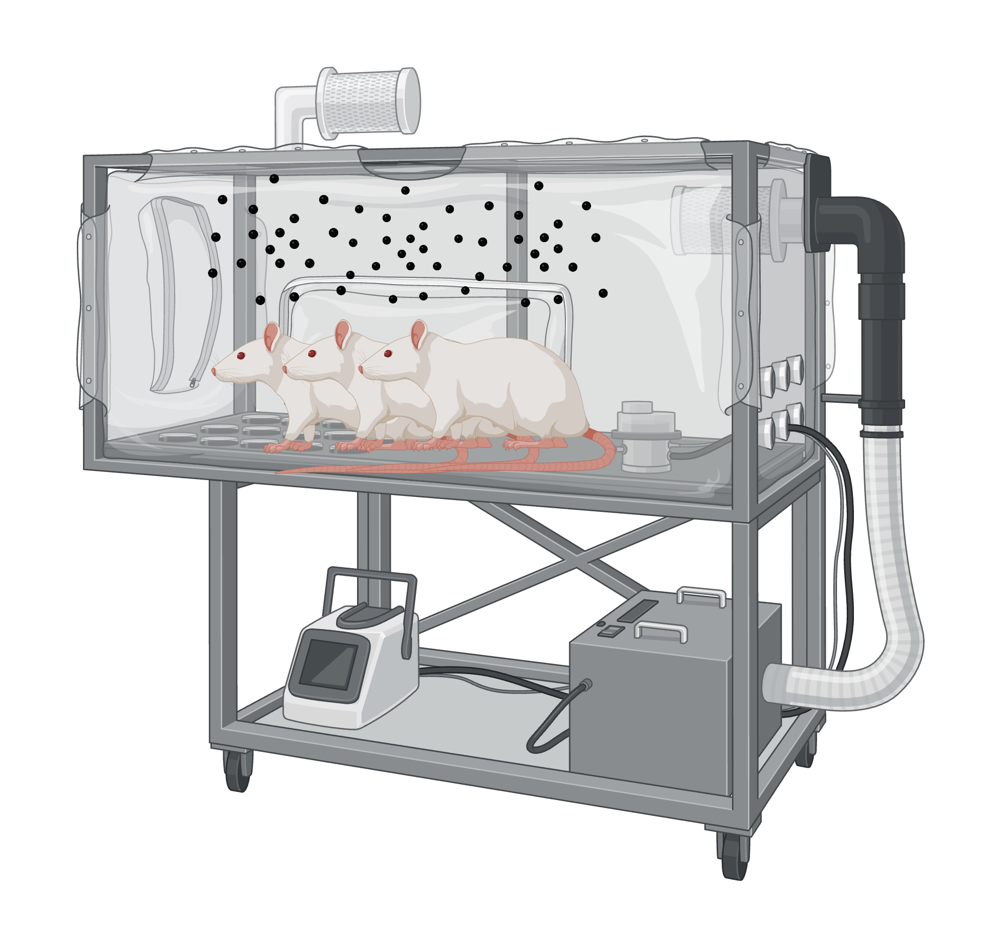
Figure 1 Experimental setup of rats exposed to MPs using an inhalation chamber.
Microplastic characterization
Prior to inhalation exposure, the PE and PVC MPs (production code 2110052, CV Subur Kimia Jaya, Indonesia) were characterized for particle size and morphology via optical microscopy and image analysis at the ECOTON Test Lab using ImageJ software [25,26]. Analysis of 179 PE particles revealed a mean size of 109.87 ± 223.62 µm, with fragments as the dominant morphology (70.39%, n = 126), followed by filaments (27.37%, n = 49), granules (1.68%, n = 3), and fibers (0.56%, n = 1). In parallel, analysis of 459 PVC particles showed a broader size distribution, with a mean size of 1,065.75 ± 9,090.99 µm, and a predominant fragment morphology (81.9%, n = 376), along with pellets (10.5%, n = 48), filaments (3.9%, n = 18), and granules (3.7%, n = 17). These profiles confirm the use of inhalable, irregularly shaped MPs, with PE consisting primarily of smaller fragments and PVC encompassing a wider size range including larger particles.
Microplastic exposure procedure
Rats in the PVC and PE groups were exposed to these characterized MPs via whole-body inhalation using a validated aerosol generation system established in the Pharmacology Laboratory, Faculty of Medicine, Universitas Brawijaya, Malang [27]. Exposure was conducted in a 60×60×60 cm3 chamber over 28 days (4 h/day, 5 days/week). The target concentration of 15 mg/m³ was achieved and maintained through daily gravimetric sampling from the breathing zone, with airflow and environmental conditions strictly controlled to ensure exposure stability. The protocol followed OECD Test Guideline 412 principles [21,25], with concentration referenced against the Occupational Safety and Health Administration (OSHA) permissible exposure limit for particulates not otherwise regulated (PNOR), and was adapted from established inhalation toxicology methods [28,29]. Following exposure, cardiac tissues were harvested for histopathological and morphological analysis after euthanasia.
Histopathological analysis procedure
Heart samples were promptly collected and fixed in 4% paraformaldehyde, followed by serial dehydration, paraffin embedding, and sectioning at 5 μm using a microtome (Figure 2). Sections were deparaffinized, rehydrated, and stained with hematoxylin and eosin (H&E) for morphological assessment. Histopathological analysis was performed via light microscopy. For quantitative morphometry, whole-slide bright-field images were acquired at 20× magnification using an Aperio ScanScope system (Leica Biosystems, Germany). Ventricular wall thickness was directly quantified from these digital slides using the Aperio ImageScope software's linear measurement tool. Measurements were performed in millimeters (mm) on representative cross-sectional areas of the ventricular wall. For each heart, wall thickness was measured at 5 standardized, equidistant points, and the average value was calculated per sample. All measurements were conducted by 2 independent investigators blinded to the experimental groups.
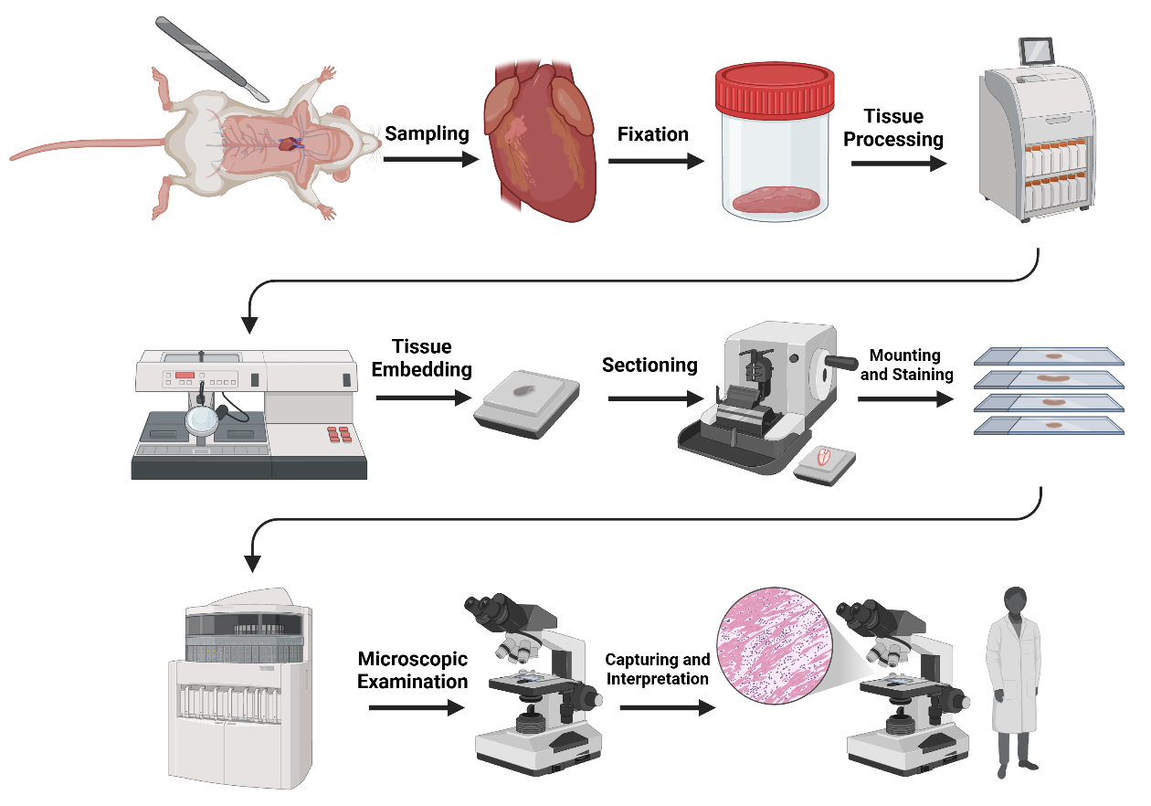
Figure 2 Schematic overview of the histological tissue processing workflow.
MDA concentration analysis procedure
The BIOXYTECH MDA-586® manufacturer’s protocol was followed to determine the MDA concentration in this study (Figure 3). In summary, the alveoli were previously perfused with ice-cold PBS in the absence of blood. Subsequently, the hearts were homogenized in a KCl buffer (7.6%). In order to precipitate the protein, 2.5 L of 10% (w/v) trichloroacetic acid were combined with the homogenate. The supernatant was elevated to a boiling temperature and subsequently mixed with 0.67% TBA for a quarter of an hour after the precipitate was centrifuged. The absorbance of the colored product at 586 nm was determined using a spectrophotometer after it had chilled.
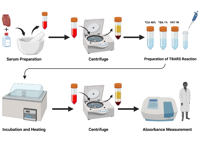
Figure 3 Schematic workflow of the TBARS assay for serum MDA measurement.
Immunofluorescence
Immunofluorescence analysis of cardiac p65 NF‑κB expression was performed on paraffin-embedded tissue sections (5 µm). Sections were deparaffinized and rehydrated by heating at 60 °C for 60 min, followed by sequential immersion in xylene (2×10 min), absolute ethanol (2×10 min), graded ethanol (90%, 80%, 70%; 5 min each), and sterile distilled water (3×5 min). Antigen retrieval was performed by heating slides in 0.01 M sodium citrate buffer (pH 6.0) at 95 - 100 °C for 20 min, followed by cooling at room temperature for 20 min. After washing with PBS (3×5 min), sections were permeabilized with 0.1% PBS‑Triton X‑100 (5×1 min) and blocked with 1% bovine serum albumin (BSA) for 30 min at room temperature. Sections were then incubated overnight at 4 °C with mouse monoclonal anti‑p65 NF‑κB primary antibody (sc‑8008, Santa Cruz Biotechnology, USA) at a dilution of 1:100 in blocking buffer. After washing with PBS (3×5 min), slides were incubated with FITC‑conjugated goat anti‑mouse IgG secondary antibody (Invitrogen, USA) for 30 min at room temperature, followed by nuclear counterstaining with 4′,6‑diamidino‑2‑phenylindole (DAPI, Santa Cruz Biotechnology) at a 1:1,000 dilution for 5 min. After final washes, slides were mounted with antifade mounting medium and coverslipped. Imaging was performed using a fluorescence microscope (Olympus BX51) (Figure 4). For each cardiac sample, 3 random fields were captured at 400× magnification under strictly identical exposure settings. Fluorescence intensity was quantified using ImageJ software by measuring the mean gray value per field. The average intensity per experimental group was calculated and expressed as mean ± standard deviation.
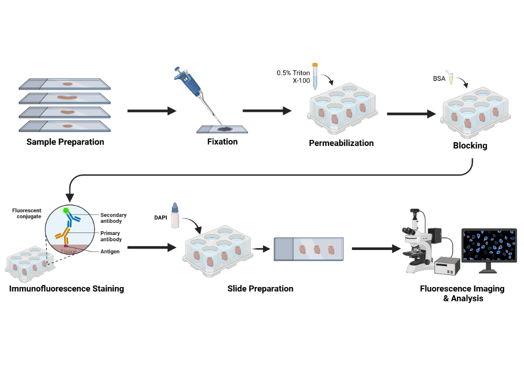
Figure 4 Schematic workflow of the immunofluorescence staining and imaging procedure.
Statistical analysis
All statistical analyses were performed using IBM SPSS Statistics version 25 for Windows. Continuous data (ventricular wall thickness, MDA concentration, and NF-κB fluorescence intensity) are presented as mean ± standard deviation (SD). The normality of data distribution within each experimental group was assessed using the Shapiro-Wilk test, with a p-value > 0.05 indicating no significant deviation from normality. Homogeneity of variances across the control, PE, and PVC groups was evaluated using Levene’s test, where p > 0.05 was considered to satisfy the assumption of equal variance. For datasets meeting both normality and homogeneity assumptions, inter-group comparisons were conducted using 1-way analysis of variance (ANOVA). In cases where the ANOVA yielded a significant result (p < 0.05), Tukey’s honest significant difference (HSD) post hoc test was applied for pairwise comparisons to identify specific group differences. For datasets that violated the assumptions of normality or homogeneity, the non-parametric Kruskal-Wallis test was employed, followed by Dunn’s post hoc test with Bonferroni correction for multiple comparisons. A 2-tailed p-value of less than 0.05 was defined as the threshold for statistical significance for all comparative analyses.
Results and discussion
Cardiac histopathological analysis
Histopathological analysis of ventricular myocardium revealed prominent structural alterations across the experimental groups (Figure 5). Quantitative morphometry demonstrated a significant, polymer-dependent increase in ventricular wall thickness following microplastic exposure. The mean wall thickness was highest in the PVC group (2.40 ± 0.30 mm), intermediate in the PE group (2.03 ± 0.28 mm), and lowest in the Control group (1.37 ± 0.18 mm). Statistical analysis by 1-way ANOVA confirmed a significant overall difference among the groups (p < 0.001). Post hoc comparisons revealed that both the PVC and PE groups exhibited significantly greater wall thickness than the Control group (p < 0.05). Furthermore, the wall thickness in the PVC group was significantly greater than that in the PE group (p < 0.05). This clear progression in effect severity, where PVC induced the greatest change followed by PE and then the control, underscores a distinct and polymer-specific impact on cardiac remodeling.
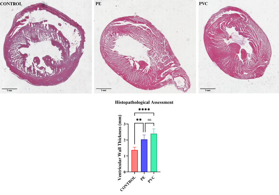
Figure 5 Histopathological analysis and quantification of ventricular wall thickness.
Cardiac p65 NF-κB expression analysis
Immunofluorescence analysis of cardiac p65 NF‑κB expression revealed no statistically significant differences among the control, PE, and PVC groups (Figure 6). The mean fluorescence intensities were comparable across groups (control: 58,684,525 ± 19,666,788; PE: 69,018,490 ± 19,189,405; PVC: 62,836,301 ± 18,498,629). Levene’s test confirmed homogeneity of variances (p = 0.989), and 1‑way ANOVA showed no significant inter‑group variation (p = 0.650). Post‑hoc Tukey’s test confirmed that pairwise comparisons between control, PE, and PVC groups were all non‑significant (p > 0.05). Thus, under the present subacute exposure conditions, cardiac NF‑κB activation was not demonstrably altered by PE or PVC MPs inhalation.
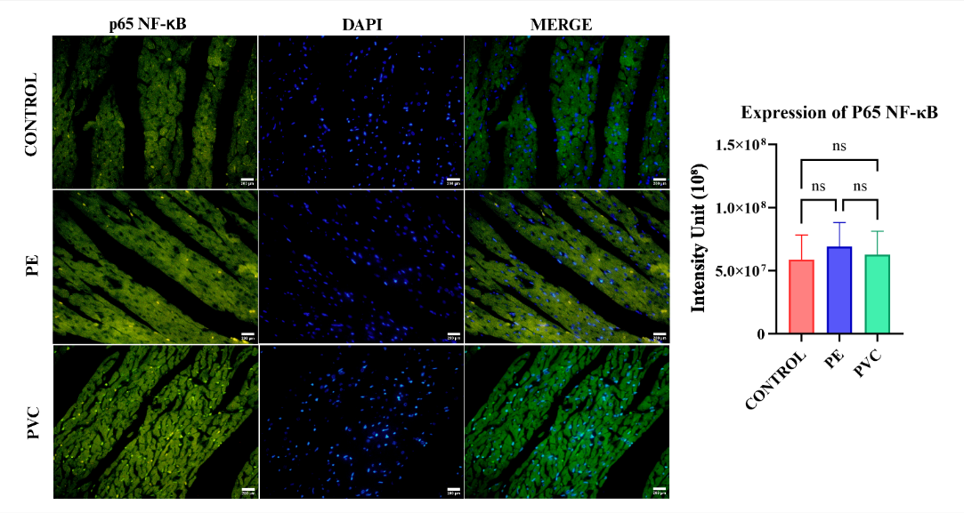
Figure 6 Immunofluorescence results of p65 NF-κB expression in cardiac tissue.
Cardiac MDA concentration
The analysis of cardiac MDA concentrations demonstrated clear differences among the control, PE, and PVC exposure groups (Figure 7). The mean MDA level in cardiac tissue was highest in the PE group (0.554 ± 0.156 ng/mL), followed by the PVC group (0.272 ± 0.149 ng/mL), and the lowest in the control group (0.0189 ± 0.020 ng/mL). One-way ANOVA results revealed a statistically significant difference among the 3 groups (p < 0.001), indicating that MPs exposure significantly influenced oxidative stress levels in cardiac tissue. Post hoc analysis using Tukey’s test showed significant differences between the control and both exposure groups (p < 0.05), as well as between the PE and PVC groups. These findings suggest that both PE and PVC MPs exposure increased lipid peroxidation in cardiac tissue, as reflected by elevated MDA concentrations, with the PE group exhibiting a more pronounced oxidative response than the PVC group.
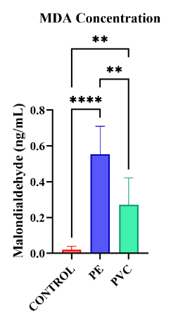
Figure 7 Oxidative stress in cardiac tissue following PE and PVC exposure.
Discussion
The results of this study indicate that inhaled PE and PVC MPs are associated with cardiotoxic changes primarily characterized by oxidative stress, with possible early involvement of inflammatory signaling. Exposure to MPs and NPs via inhalation enables their passage into the systemic circulation through pulmonary capillary translocation, allowing particles to reach distant organs such as the heart [28]. Previous work has demonstrated that inhaled MPs and NPs can migrate from the lungs to cardiac tissue. For example, mice exposed to polystyrene (PS) NPs for 1, 4, and 12 weeks exhibited detectable cardiac accumulation using advanced imaging techniques [30].
Once deposited in cardiac tissue, MPs may induce oxidative stress through disruption of mitochondrial function and dysregulation of cellular redox balance. Impaired redox homeostasis promotes excessive production of reactive oxygen species and depletion of endogenous antioxidants. Elevated oxidative stress can activate pattern-recognition receptors via damage-associated molecular pathways and may influence canonical nuclear factor kappa-B (NF-κB) signaling; however, such activation was not statistically evident in the present study. NF-κB regulates genes associated with immune responses, proliferation, and differentiation, and persistent stimulation of these pathways has the potential to contribute to tissue remodeling over time [13,14,31,32].
In this study, subacute inhalation exposure to PE and PVC MPs at 15 mg/m³ for 28 days resulted in significant cardiac alterations. These were reflected by increased MDA levels and histopathological changes characterized by thickening of the ventricular wall in exposed animals compared with controls. These findings are consistent with previous experimental models. Mice exposed to PS NPs at 16 - 100 µg/day for 1 - 12 weeks demonstrated marked oxidative stress across exposure durations, reductions in antioxidant activity at higher doses, mitochondrial disruption, and structural disorganization of myocardial fibers. Fibrosis became evident with longer exposure periods, accompanied by functional impairment including reduced ejection fraction [30]. Similarly, intratracheal exposure to PS MPs for 4 weeks resulted in increases in ventricular wall thickness and elevated expression of inflammatory markers such as TNF-α [33]. Although NF-κB-related activation has been documented under some exposure scenarios, our findings showed no significant difference in cardiac p65 NF-κB expression across groups. This suggests that oxidative and structural alterations may precede measurable inflammatory activation during subacute exposure. Importantly, the magnitude of increases in MDA and ventricular wall thickness observed here indicates biologically relevant changes, supporting the potential for functional consequences even in the absence of overt inflammation.
A notable observation is the differential toxicity between the 2 polymers. PVC exposure produced more pronounced structural remodeling, whereas PE exposure generated higher oxidative stress. Several mechanistic explanations are plausible. PVC frequently contains chlorine and plasticizers such as phthalates, which can alter membrane integrity and modulate inflammatory signaling [12]. In contrast, PE is chemically simpler and highly hydrophobic, promoting stronger interactions with lipid membranes and enhancing lipid peroxidation, consistent with elevated MDA levels [34]. Surface reactivity may also differ: PE particles tend to adsorb lipophilic molecules and propagate oxidative chain reactions, whereas PVC particles may persist in tissue longer, contributing to gradual structural remodeling. Furthermore, PE has been linked to mitochondrial interference and increased reactive oxygen species, whereas PVC particles may exert greater mechanical or structural stress on ventricular wall due to differences in rigidity and surface chemistry. Collectively, these mechanisms provide a plausible explanation for why PE produced stronger oxidative effects while PVC resulted in more marked histopathological alterations, underscoring that MP-induced cardiotoxicity is polymer-specific rather than uniform across particle types [12,34].
The structural alterations observed in this study warrant further investigation to determine their functional significance. Future research should evaluate cardiac physiology more comprehensively, including heart rate and ejection fraction as indicators of chronotropic and inotropic performance, respectively, as demonstrated in previous MP exposure models [35]. Because the present exposure period was subacute, it is plausible that longer exposure durations are required to elicit more pronounced inflammatory responses or fibrosis, as previously documented following 3-month PS exposure [30]. In addition to NF-κB, alternative pathways such as MAPK, TGF-β, and Nrf2 likely contribute to oxidative-stress-driven remodeling and should be examined in parallel to clarify signaling interactions. Comparing PE and PVC under sub-chronic and chronic inhalation conditions will be particularly important for defining long-term cardiovascular risk profiles.
Study strength and limitation
This study provides comparative evidence on 2 environmentally prevalent MPs using a whole-body inhalation model that closely reflects real-world exposure conditions and potential pulmonary-cardiac translocation. However, several limitations must be considered. The absence of real-time aerosol monitoring during exposure sessions limits the verification of consistent particle concentration and size distribution throughout each inhalation period. Particle characteristics, including size distribution, morphology, surface chemistry, and potential additives or adsorbed pollutants, were not fully characterized despite their known influence on toxicity. Only 1 exposure concentration was utilized, limiting assessment of dose–response relationships and the contribution of micro- versus nanoscale fractions. The MPs employed represented pristine laboratory material, whereas environmental MPs are often aged and chemically altered. Finally, p65 NF-κB alone may not fully capture the breadth of inflammatory signaling responses. Future research incorporating detailed particle profiling, multiple exposure concentrations, mixed polymer systems, and extended exposure durations will be essential for accurately characterizing cardiovascular toxicity associated with airborne MPs.
Conclusions
In conclusion, subacute inhalation of PE and PVC MPs at 15 mg/m³ for 28 days produced significant cardiac alterations in female Wistar rats. Both exposures were associated with myocardial hypertrophy and marked oxidative injury, as reflected by increases in MDA of approximately 14-fold in PVC-exposed rats and 29-fold in PE-exposed rats, together with measurable thickening of the ventricular wall. In contrast, cardiac p65 NF-κB expression did not differ significantly among groups, indicating that clear inflammatory pathway activation was not observed under these subacute exposure conditions.
These findings suggest that PE and PVC induce early oxidative stress–driven cardiac remodeling, with PE exhibiting a stronger oxidative profile whereas PVC produced more pronounced structural changes. A plausible explanation is that the greater hydrophobicity and lipid-interacting properties of PE may enhance membrane lipid peroxidation, while PVC may persist longer in tissue and contribute to structural remodeling through different physicochemical interactions.
Because the present study reflects only short-term exposure, future research is required to verify whether longer durations of inhalation lead to sustained inflammatory activation, progressive fibrosis, and functional impairment, and to clarify the involvement of additional signaling pathways beyond NF-κB. From a broader perspective, these results contribute to growing evidence that airborne MPs may represent an emerging cardiovascular risk factor, underscoring the need for continued environmental monitoring and consideration of regulatory strategies aimed at reducing chronic human exposure.
Acknowledgements
The authors would like to express their outmost gratitude to Brawijaya University, for providing access to their laboratory facilities and for supporting this research. We are deeply thankful of the financial support and the resources provided by Faculty of Medicine of Brawijaya University which were essential to the successful completion of this study.
Declaration of Generative AI in scientific writing
The authors acknowledge the use of generative AI tools (e.g., QuillBot and ChatGPT by OpenAI) in the preparation of this manuscript, specifically for language editing and grammar correction. No content generation or data interpretation was performed by AI. The authors take full responsibility for the content and conclusions of this work.
CRediT author statement
Muhammad Reva Aditya: Conceptualization; Methodology; Investigations, Formal Analysis, Data Curation, Writing - Original Draft, Writing - Review and Editing, Visualization. Athaya Rahmanardi Muhammad: Conceptualization; Methodology; Investigations, Formal Analysis, Data Curation, Writing - Original Draft, Writing - Review and Editing, Project Admnisitration. Hikmawan Wahyu Sulistomo: Conceptualization; Methodology; Writing - Review and Editing, Funding Acquisition, Supervision. Dian Nugrahenny: Supervision. Holipah: Supervision. Happy Kurnia Pematasari: Supervision. Cholid Tri Tjahjono: Supervision. Veny Mayangsari: Supervision. Waqar Ahmed: Supervision.
References
[1] R Kumar, A Verma, A Shome, R Sinha, S Sinha, PK Jha, R Kumar, P Kumar, Shubham, S Das and P Sharma. Impacts of plastic pollution on ecosystem services, sustainable development goals, and need to focus on circular economy and policy interventions. Sustainability 2021; 13(17), 9963.
[2] J Lee, S Kim, SB Park, M Shin, S Kim, MS Kim, G Shin, T Kang, HJ Kim, DX Oh and J Park. Mimicking real-field degradation of biodegradable plastics in soil and marine environments: From product utility to end-of-life analysis. Polymer Testing 2024; 131, 108338.
[3] VG Le, MK Nguyen, HL Nguyen, C Lin, M Hadi, MNT Hung, HG Hoang, KN Nguyen, HT Tran, D Hou, T Zhang and T Zhang. A comprehensive review of micro-and nano-plastics in the atmosphere: Occurrence, fate, toxicity, and strategies for risk reduction. Science of the Total Environment 2023; 904, 166649
[4] Y Yu, N Craig and L Su. A hidden pathway for human exposure to micro-and nanoplastics - the mechanical fragmentation of plastic products during daily use. Toxics 2023; 11(9), 774.
[5] E Dube and GE Okuthe. Plastics and micro/nano-plastics (MNPs) in the environment: Occurrence, impact, and toxicity. International Journal of Environmental Research and Public Health 2023; 20(17), 6667.
[6] S Islam. Toxicity and transport of nanoparticles in agriculture: Effects of size, coating, and aging. Frontiers in Nanotechnology 2025; 7, 1622228.
[7] V Stock, L Böhmert, G Coban, G Tyra, ML Vollbrecht, L Voss, MB Paul, A Braeuning and H Sieg. Microplastics and nanoplastics: Size, surface and dispersant-what causes the effect? Toxicology In Vitro 2022; 80, 105314.
[8] D Pal, R Prabhakar, VB Barua, I Zekker, J Burlakovs, A Krauklis, W Hogland and Z Vincevica-Gaile. Microplastics in aquatic systems: A comprehensive review of its distribution, environmental interactions, and health risks. Environmental Science and Pollution Research 2025; 32(1), 56-88.
[9] DM El-Sherif, MG Eloffy, A Elmesery, M Abouzid, M Gad, HR El-Seedi, M Brinkmann, K Wang and Y Al-Naggar. Environmental risk, toxicity, and biodegradation of polyethylene: A review. Environmental Science and Pollution Research 2022; 29(54), 81166-81182.
[10] P Kowalczyk, K Kadac-Czapska and M Grembecka. Polyethylene Packaging as a Source of Microplastics: Current Knowledge and Future Directions on Food Contamination. Foods 2025; 14(14), 2408.
[11] M Manjunatha, D Seth, S Roy and RB Tangadagi. Utilization of industrial-based PVC waste powder in self-compacting concrete: A sustainable building material. Journal of Cleaner Production 2023; 428, 139428.
[12] MH Kudzin, D Piwowarska, N Festinger and JJ Chruściel. Risks associated with the presence of polyvinyl chloride in the environment and methods for its disposal and utilization. Materials 2023; 17(1), 173.
[13] T Zhang, C Ma, Z Zhang, H Zhang and H Hu. NF‐κB signaling in inflammation and cancer. MedComm 2021; 2(4), 618-653.
[14] J Cornice, D Verzella, P Arboretto, D Vecchiotti, D Capece, F Zazzeroni and G Franzoso. NF-κB: Governing macrophages in cancer. Genes 2024; 15(2), 197.
[15] T Xu, J Cui, R Xu, J Cao and MY Guo. Microplastics induced inflammation and apoptosis via ferroptosis and the NF-κB pathway in carp. Aquatic Toxicology 2023; 262, 106659.
[16] JH Woo, HJ Seo, JY Lee, I Lee, K Jeon, B Kim, K Lee and K Lee. Polypropylene nanoplastic exposure leads to lung inflammation through p38-mediated NF-κB pathway due to mitochondrial damage. Particle and Fibre Toxicology 2023; 20(1), 2.
[17] M Borgatta and F Breider. Inhalation of microplastics - a toxicological complexity. Toxics 2024; 12(5), 358.
[18] R Jia, J Han, X Liu, K Li, W Lai, L Bian, J Yan and Z Xi. Exposure to polypropylene microplastics via oral ingestion induces colonic apoptosis and intestinal barrier damage through oxidative stress and inflammation in mice. Toxics 2023; 11(2), 127.
[19] YL Wang, CC Huang, CM Zheng, WC Liu, YH Lee, HW Chiu and HW Chiu. Polystyrene microplastic-induced extracellular vesicles cause kidney-related effects in the crosstalk between tubular cells and fibroblasts. Ecotoxicology and Environmental Safety 2024; 273, 116098.
[20] L Bridgeman, A Cimbalo, D López-Rodríguez, D Pamies and M Frangiamone. Exploring toxicological pathways of microplastics and nanoplastics: Insights from animal and cellular models. Journal of Hazardous Materials 2025; 490, 137795.
[21] OECD. OECD guidelines on the testing of chemicals: Test no. 412 - 28-day (subacute) inhalation toxicity study. Organisation for Economic Co-operation and Development (OECD), Paris, France, 2018.
[22] R Pohjanvirta, H Miettinen, S Sankari, N Hegde and J Lindén. Unexpected gender difference in sensitivity to the acute toxicity of dioxin in mice. Toxicology and Applied Pharmacology 2012; 262(2), 167-176.
[23] N Kania, E Mayangsari, B Setiawan, D Nugrahenny, F Tony, ES Wahyuni and MA Widodo. The effects of Eucheuma cottonii on signaling pathway inducing mucin synthesis in rat lungs chronically exposed to particulate matter 10 (PM10) coal dust. Journal of Toxicology 2013; 2013, 528146.
[24] RK Saputri, B Setiawan, D Nugrahenny, N Kania, ES Wahyuni and MA Widodo. The effects of Eucheuma cottonii on alveolar macrophages and malondialdehyde levels in bronchoalveolar lavage fluid in chronically particulate matter 10 coal dust-exposed rats. Iranian Journal of Basic Medical Sciences 2014; 17(7), 541-545.
[25] AR Muhammad, MR Aditya, B Lestari and HW Sulistomo. Sub-acute polyethylene microplastic inhalation exposure induced pulmonary toxicity in wistar rats through inflammation and oxidative stress. Toxicology Reports 2025; 14, 102067.
[26] MR Aditya, AR Muhammad, T Astuti, HW Sulistomo and D Nugrahenny. Pulmonary toxicity induced by subacute inhalation of polyvinyl chloride microplastics: Oxidative stress and NF-κB pathway activation in rat models. Iranian Journal of Toxicology 2025; 19(4), 227-234.
[27] B Setiawan, N Kania, D Nugrahenny, N Nurdiana and MA Widodo. Subchronic inhalation of particulate matter 10 coal dust induces atherosclerosis in the aorta of diabetic and nondiabetic rats. Biomarkers and Genomic Medicine 2014; 6(2), 67-73.
[28] S Kauts, S Shabir, S Yousuf, Y Mishra, R Bhardwaj, AA Milibari, SK Singh and MP Singh. The evidence of in-vivo and in-vitro studies on microplastic and nano plastic toxicity in mammals: A possible threat for an upcoming generation? Physics and Chemistry of the Earth, Parts A/B/C 2023; 132, 103511.
[29] CM Cary, TN Seymore, D Singh, KN Vayas, MJ Goedken, S Adams, M Polunas, VR Sunil, DL Laskin, P Demokritou and PA Stapleton. Single inhalation exposure to polyamide micro and nanoplastic particles impairs vascular dilation without generating pulmonary inflammation in virgin female Sprague Dawley rats. Particle and Fibre Toxicology 2023; 20(1), 16.
[30] T Zhang, S Yang, Y Ge, X Wan, Y Zhu, F Yang, J Li, S Gong, Y Cheng, C Hu and Z Chen. Multi-dimensional evaluation of cardiotoxicity in mice following respiratory exposure to polystyrene nanoplastics. Particle and Fibre Toxicology 2023; 20(1), 46.
[31] AJ de Almeida, JC de Oliveira, LV da Silva Pontes, JF de Souza Júnior, TA Gonçalves, SH Dantas, MS de Almeida Feitosa, AO Silva and IA de Medeiros. ROS: Basic concepts, sources, cellular signaling, and its implications in aging pathways. Oxidative Medicine and Cellular Longevity 2022; 2022, 1225578.
[32] X Meng, J Zhang, W Wang, G Gonzalez-Gil, JS Vrouwenvelder and Z Li. Effects of nano-and microplastics on kidney: Physicochemical properties, bioaccumulation, oxidative stress and immunoreaction. Chemosphere 2022; 288, 132631.
[33] Y Zhou, Q Wu, Y Li, Y Feng, Y Wang and W Cheng. Low-dose of polystyrene microplastics induce cardiotoxicity in mice and human-originated cardiac organoids. Environment International 2023; 179, 108171.
[34] M Leela, V Pushpalatha, S Swetha, S Anusuya, S Madhavan, S Eswari, H Thilagam, R Thiagarajan and R Krishnamurthy. Ingestion of weathered high density polyethylene microplastics-induced oxidative stress and modulation of antioxidant responses in post larval stages of Litopenaeus vannamei. Frontiers in Marine Science 2025; 12, 1617070.
[35] A Dimitriadi, C Papaefthimiou, E Genizegkini, I Sampsonidis, S Kalogiannis, K Feidantsis, DC Bobori, G Kastrinaki, G Koumoundouros, DA Lambropoulou and GZ Kyzas. Adverse effects polystyrene microplastics exert on zebrafish heart-molecular to individual level. Journal of Hazardous Materials 2021; 416, 125969.