
Trends Sci. 2026; 23(10): 13244
Phytochemical Profile and In Vitro Antioxidant, Anti-Inflammatory, Antidiabetic, and Antibacterial Activities of Nauclea orientalis Stem Bark Extracts
Do Van Mai1, Chong Kim Thien Duc2, Nguyen Van Ky3, Thai Viet Tang4,
Nguyen Duy Tuan4, Nguyen Huynh Bich Lieu4 and Tran Chi Linh4,*
1Faculty of Pharmacy, College of Health Sciences, Nam Can Tho University, Can Tho City 94000, Viet Nam
2Faculty of Health Sciences, College of Natural Sciences, Can Tho University, Can Tho City 94000, Viet Nam
3Department of Chemistry, Faculty of Science, Chulalongkorn University, Bangkok 10330, Thailand
4Faculty of Medicine, College of Health Sciences, Nam Can Tho University, Can Tho City 94000, Viet Nam
(*Corresponding author’s e-mail: [email protected])
Received: 1 January 2026, Revised: 27 January 2026, Accepted: 10 February 2026, Published: 15 April 2026
Abstract
Nauclea orientalis, locally known as Gao Vang in Vietnam, is widely distributed in Southeast Asia and has been traditionally used in ethnomedicine to treat fever, inflammation, and skin disorders. Although phytochemical investigations of Nauclea species and the Rubiaceae family have been reported, studies systematically linking polyphenol-enriched fractions with multiple bioactivities remain limited. This study aimed to determine the total polyphenol and flavonoid contents and to evaluate the antioxidant, anti-inflammatory, antidiabetic, and antibacterial activities of different extracts of N. orientalis, including ethanol extract, hexane, ethyl acetate, and dichloromethane fractions. Among the tested extracts, the ethyl acetate fraction exhibited the highest total polyphenol and flavonoid contents. Consistently, this fraction showed strong antioxidant activity, with IC50 values ranging from 31.63 ± 0.40 to 54.61 ± 0.72 µg/mL across representative radical scavenging assays, comparable to trolox. In addition, notable anti-inflammatory activity was observed through inhibition of nitric oxide scavenging capacity, with IC50 values of 84.96 ± 1.48 µg/mL, along with superior protective effects in red blood cell membrane stabilization and protein denaturation models. This fraction also demonstrated significant antidiabetic potential through effective inhibition of α-amylase and α-glucosidase, as well as pronounced antibacterial activity. High-performance liquid chromatography analysis qualitatively identified gallic acid, quercetin, and rutin as representative constituents. Overall, these findings highlight the ethyl acetate fraction of N. orientalis as a source with multifunctional bioactivities, providing new pharmacological insight beyond previously reported phytochemical studies of this genus.
Keywords: Nauclea orientalis, Antibacterial, Antidiabetic, Anti-inflammatory, Antioxidant
Introduction
Oxidative stress, characterized by a redox imbalance where the generation of reactive oxygen (ROS) or nitrogen species (RNS) overwhelms endogenous antioxidant defenses, is a fundamental driver of cellular damage. This failure in redox homeostasis compromises phospholipid membranes, proteins, and nucleic acids, contributing to the pathogenesis of various chronic diseases [1]. While the
body employs both enzymatic (e.g., superoxide dismutase, catalase) and non-enzymatic defenses (e.g., glutathione), these systems are often insufficient under pathological conditions [2]. Consequently, reinforcing these defenses with exogenous antioxidants from plant sources is critical to mitigate protein structural alterations and restore biological function.
Similarly, inflammation serves as an essential defense mechanism mediated by cytokines (TNF-α, IL-6) and effectors like nitric oxide (NO) and prostaglandin E2 (PGE2) [3,4]. However, its dysregulation forms a pathological triad with oxidative stress and diabetes mellitus, particularly type 2 diabetes [5]. In this vicious cycle, hyperglycemia-induced ROS exacerbates insulin resistance, while chronic inflammation further amplifies metabolic dysfunction and impairs immune responses [6]. Thus, breaking this feedback loop between oxidative stress and inflammation is a key therapeutic strategy.
Despite epidemiological projections indicating a rise in global diabetes prevalence from 8.8% in 2015 to 10.4% by 2040 [7], current management strategies remain limited. Type 1 and type 2 diabetes are distinct in etiology [8], yet pharmacological interventions for both, as well as NSAIDs used for inflammation, are frequently associated with adverse long-term effects, including gastrointestinal and cardiovascular risks [9,10]. Furthermore, the molecular crosstalk where ROS activate pro-inflammatory transcription factors like NF-κB [11] necessitates the development of multi-target agents capable of simultaneously modulating both oxidative and inflammatory pathways.
Natural bioactive compounds, particularly polyphenols and flavonoids, have emerged as promising candidates to address this complex etiology [5]. Nauclea orientalis (Gao vang), a Rubiaceae species widely distributed in Southeast Asia, has been traditionally utilized for fever and inflammatory conditions. While recent phytochemical investigations have characterized the alkaloid and phenolic profiles of its barks and leaves [12,13], the stem bark remains largely unexplored. We hypothesize that the stem bark may harbor a distinct phytochemical profile with potent bioactivity capable of disrupting the oxidative-inflammatory axis in diabetes. Therefore, this study aims to bridge this knowledge gap by evaluating the antioxidant, anti-inflammatory, antidiabetic, and antibacterial potential of N. orientalis stem bark, contributing to the development of novel plant-based therapeutics.
Materials and methods
Plant materials
The stem bark of N. orientalis (Figure 1) was collected from Binh Thuy District, Can Tho City, Viet Nam. Botanical identification was performed by Dr. Nguyen Thi Ai Lan, Deputy Director of the Institute of Biotechnology, Tra Vinh University, based on morphological characteristics. A voucher specimen (code: CT_Nor202304010019) was deposited at the Biology Laboratory, Faculty of Medicine, College of Health Sciences, Nam Can Tho University.
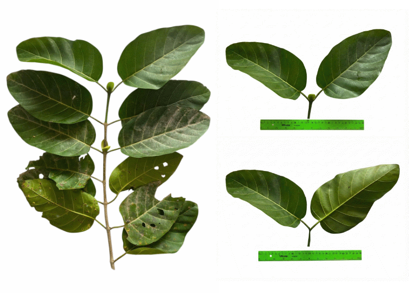
Figure 1 N. orientalis.
Processing of plant materials and preparation of crude extracts
After collection, 10 kg of fresh N. orientalis stem bark (NOSB) was dried at 55 °C and then finely ground to obtain 4.4 kg of dry powder. The powder was standardized using a 60-mesh sieve. Moisture content was determined using a YOKE DSH-10A moisture analyzer (YOKE, China), yielding a value of 7.27 ± 0.17%. The dried powder was subsequently stored at 4 °C until further use. For extraction, 1 kg of the stem bark powder was macerated with ethanol at a solid-to-solvent ratio of 1:10 (w/v), and the process was repeated three times. The combined extracts were concentrated to obtain 120 g of crude ethanol extract. From this extract, 100 g was subjected to successive liquid-liquid partitioning with solvents of increasing polarity, namely n-hexane, dichloromethane, and ethyl acetate, yielding 10.4 g, 12.2 g, and 20.4 g of the respective fractions. The moisture contents of the ethanol extract and the n-hexane, dichloromethane, and ethyl acetate fractions were measured using the same moisture analyzer, with values of 6.73 ± 0.15%, 7.00 ± 0.20%, 6.87 ± 0.15%, and 6.60 ± 0.10%, respectively.
Quantitative determination of total polyphenol content and total flavonoid content
The total polyphenol content (TPC) and total flavonoid content (TFC) of NOSB extracts were quantitatively determined following the standardized spectrophotometric procedures described by Duc et al. [1], with modifications [1]. The concentrations of TPC and TFC were calculated using their respective calibration curves, with gallic acid employed as the reference standard for TPC (expressed as mg gallic acid equivalents per gram of extract, mg GAE/g extract; y = 0.0091x + 0.0179, R2 = 0.9994) and quercetin used as the reference standard for TFC (expressed as mg quercetin equivalents per gram of extract, mg QE/g extract; y = 0.0178x + 0.0029, R2 = 0.9998).
In vitro evaluation of antioxidant activity
The in vitro antioxidant activity of NOSB extracts was evaluated using modified procedures adapted from a previous study [14]. Antioxidant capacity was assessed through multiple complementary assays, including total antioxidant capacity (TAC), reducing power (RP), ferric reducing antioxidant power (FRAP), DPPH radical scavenging, and ABTS●+ radical scavenging. Ascorbic acid was employed as the positive control in all assays. The half-maximal inhibitory concentration (IC50, µg/mL) and antioxidant effectiveness were calculated from absorbance at the corresponding wavelengths.
In vitro evaluation of anti-inflammatory activity
The in vitro anti-inflammatory activity of NOSB extracts was evaluated using complementary assays, including red blood cell (RBC) membrane stabilization, inhibition of bovine serum albumin (BSA) denaturation, and suppression of nitric oxide (NO●) scavenging activity in a cell-free chemical system, following the methods described by Tran et al. [15], with modifications [15]. Diclofenac was used as the reference standard (positive control) for the RBC and BSA assays, and Trolox was employed as the positive control for NO● scavenging, while 10% dimethyl sulfoxide (DMSO) served as the negative control. The inhibitory effects on RBC membrane stabilization, BSA denaturation, and NO● radical generation were expressed as inhibition percentage (%) and half-maximal inhibitory concentration (IC50, μg/mL), calculated from absorbance measurements at the corresponding wavelengths.
In vitro evaluation of antidiabetic activity
The in vitro antidiabetic activity of NOSB extracts was evaluated through α-amylase and α-glucosidase inhibition assays, following the methods described by Linh et al. [16], with modifications [16]. The absorbance of the reaction mixtures was measured using a Thermo Scientific Multiskan GO UV/Vis microplate spectrophotometer (USA) at 660 nm for the α-amylase assay and 405 nm for the α-glucosidase assay. To eliminate potential colorimetric interference from phenolic compounds, a sample blank containing the extract and buffer (without enzyme) was prepared for each concentration. The background absorbance of this blank was subtracted from the absorbance of the test sample before calculating the percentage of inhibition. The inhibitory effects on both enzymes were expressed as inhibition percentage (%) and half-maximal inhibitory concentration (IC50, μg/mL). Acarbose was used as the positive control.
In vitro evaluation of antibacterial activity
The in vitro antibacterial activity of NOSB extracts was evaluated using agar well diffusion, broth microdilution, and droplet counting methods, following the procedures described by Ngan et al. [17], with modifications [17]. The antibacterial assays were conducted against the following bacterial strains: Listeria innocua ATCC 33090, Staphylococcus aureus ATCC 6538, Pseudomonas aeruginosa ATCC 27853, and Escherichia coli ATCC 25922TM. Ciprofloxacin was used as the positive control, while 10% DMSO served as the negative control. Antibacterial efficacy was assessed based on inhibition zone diameter, minimum inhibitory concentration (MIC), and minimum bactericidal concentration (MBC).
Qualitative profiling by HPLC–PDA
Qualitative HPLC–PDA analysis was conducted on a Waters Alliance e2695 system with a 2998 PDA detector using a C18 column (150×4.6 mm, 5 µm) at 35 °C. Samples (10 µL) were eluted at 1.0 mL/min with a gradient of 0.1% H3PO4 (A) and acetonitrile (D) over 20 min. PDA detection was set at 190 - 600 nm; chromatograms were recorded at 270 nm and UV-Vis spectra (200 - 400 nm) were used for qualitative identification. Gallic acid, rutin, and quercetin (Sigma-Aldrich) were used as reference standards based on retention times and UV-Vis spectra.
Statistical analysis
Data are presented as mean ± standard error. Statistical analysis was performed using one-way ANOVA followed by Tukey’s post hoc test in Minitab 16. All assays were conducted in triplicate in three independent experiments, with statistical significance set at p < 0.05.
Results and discussion
Quantitative analysis of total polyphenol and total flavonoid content in NOSB
The present study quantified total polyphenol content (TPC) and total flavonoid content (TFC) in NOSB extracts obtained using solvents of different polarities, including ethanol extract (EE), ethyl acetate fraction (EF), n-hexane fraction (HF), and dichloromethane fraction (DF). As summarized in Table 1, extraction efficiency showed a clear dependence on solvent polarity for both TPC and TFC. Among the tested solvents, the EF exhibited the highest levels of TPC (179.36 ± 6.62 mg GAE/g extract) and TFC (146.28 ± 3.09 mg QE/g extract), followed by EE and DF, whereas the HF contained the lowest amounts.
This trend can be attributed to differences in solvent polarity, which strongly influence the solubility and recovery of polyphenol and flavonoid compounds. Moderately polar/polar solvents such as ethyl acetate and ethanol are more compatible with the chemical nature of these compounds [18,19], thereby enhancing extraction efficiency, whereas non-polar solvents as n-hexane are less effective [20]. Given the substantially higher TPC and TFC observed in the EF, it can be reasonably anticipated that this fraction may exhibit stronger biological activities than the other extracts. This expectation is based on the well-established role of polyphenols and flavonoids as key bioactive constituents in plant-derived extract [21]. Accordingly, the outstanding TPC and TFC of the EF support its potential to exhibit stronger biological activities than the other fractions.
Table 1 TPC and TFC values of NOSB extracts.
Samples |
TPC (mg GAE/g extract) |
TFC (mg QE/g extract) |
Ethanol extract |
129.54b ± 5.22 |
115.48b ± 2.68 |
n-Hexane fraction |
50.24d ± 3.12 |
|
Dichloromethane fraction |
90.53c ± 5.50 |
62.20c ± 2.18 |
Ethyl acetate faction |
179.36a ± 6.62 |
146.28a ± 3.09 |
Note: Values in the same row with different letters indicate statistically significant differences (p < 0.05)
Antioxidant activity of NOSB extracts
As shown in Table 2A, all extracts of N. orientalis exhibited antioxidant activity in the ABTS●+, DPPH, FRAP, and TAC assays. However, pronounced differences in potency were observed among them. Overall, the EF demonstrated the strongest antioxidant activity across all assays, whereas the HF consistently showed the weakest performance. Particularly, in the ABTS●+ assay, the EF exhibited an IC50 value of 42.28 ± 0.52 µg/mL, which was approximately 2.3-fold lower than that of HF (96.77 ± 5.17 µg/mL) and 1.7-fold lower than that of DF (72.86 ± 1.74 µg/mL), indicating a markedly stronger radical-scavenging capacity. Similarly, in the DPPH assay, the EF showed nearly two-fold greater activity than HF and approximately 1.6-fold higher activity than DF, while also outperforming the EE. A comparable trend was observed in the FRAP assay, where the antioxidant potential of the EF (IC50 = 44.94 ± 1.50 µg/mL) was approximately 2.2 times stronger than that of HF and 1.7 times stronger than that of DF, reflecting a superior electron-donating ability. Notably, the EF exhibited potent reducing power with an IC50 value comparable to the PSC (49.43 ± 0.59 µg/mL), a promising finding given the multi-component nature of the fraction compared to the pure standard. Finally, in the TAC assay, the EF again displayed the lowest IC50 value (31.63 ± 0.40 µg/mL), corresponding to more than a two-fold increase in total antioxidant capacity compared with HF and approximately 1.8-fold higher activity than DF.
Across all antioxidant assays, the overall order of activity was consistently EF > EE > DF > HF. The outstanding antioxidant performance of the EF closely corresponds to its significantly higher TPC and TFC, which were approximately 3 - 5 times greater than those of the HF. This quantitative relationship suggests that elevated TPC and TFC levels likely contribute to the observed in vitro antioxidant capacity [22]. Consequently, the EF can be considered the most potent antioxidant fraction among the tested samples, reflecting both its enriched chemical composition and strong in vitro antioxidant capacity. These findings are consistent with a recent study, in which five extracts and fractions obtained from Ixora duffii leaves, including the ethanol extract, n-hexane fraction, dichloromethane fraction, ethyl acetate fraction, and water fraction, were evaluated. In that study, the ethyl acetate fraction, which exhibited the highest total polyphenol, total flavonoid, total tannin, and total alkaloid contents, also demonstrated the strongest antioxidant activity across five assays (ABTS●⁺, DPPH, RP, FRAP, and TAC), with IC50 values ranging from 8.91 ± 0.16 to 16.11 ± 0.20 µg/mL [5]. The close agreement between these results and those of the present study further supports the reliability and consistency of the current findings.
Table 2 In vitro antioxidant, anti-inflammatory, and antidiabetic activities of N. orientalis extracts.
Methods |
The IC50 (μg/mL) values |
||||
EE |
HF |
DF |
EF |
PSC |
|
A: Antioxidant activity |
|||||
ABTS●+ |
50.90c ± 0.99 |
96.77a ± 5.17 |
72.86b ± 1.74 |
42.28d ± 0.52 |
8.73e ± 0.25 |
DPPH |
72.37c ± 1.59 |
100.53a ± 1.50 |
86.10b ± 0.70 |
54.61d ± 0.72 |
6.49e ± 0.29 |
FRAP |
61.69c ± 1.29 |
98.96a ± 1.97 |
76.14b ± 0.80 |
44.94d ± 1.50 |
49.43d ± 0.59 |
TAC |
36.25c ± 0.60 |
69.73a ± 1.60 |
56.53b ± 0.34 |
31.63d ± 0.40 |
9.97e ± 0.05 |
B: Anti-inflammatory activity |
|||||
NO● |
109.34c ± 1.40 |
147.75a ± 1.33 |
126.74b ± 0.65 |
84.96d ± 1.48 |
61.33e ± 4.10 |
RBCs |
32.61c ± 0.29 |
62.87a ± 2.52 |
49.13b ± 1.20 |
28.62d ± 0.40 |
50.19b ± 1.20 |
BSA |
44.90c ± 0.89 |
60.99a ± 1.29 |
58.31a ± 1.85 |
36.46d ± 0.38 |
51.09b ± 0.69 |
C: Antidiabetic activity |
|||||
α-Amylase |
61.67c ± 1.41 |
96.60a ± 2.95 |
77.15b ± 1.22 |
53.18d ± 0.49 |
7.23e ± 0.40 |
α-Glucosidase |
21.07c ± 0.51 |
31.99a ± 0.34 |
28.28b ± 0.98 |
16.5d ± 0.35 |
4.14e ± 0.01 |
Anti-inflammatory activity of NOSB extracts
The anti-inflammatory effects of the extracts were evaluated using NO● inhibition, RBCs membrane stabilization, and BSA denaturation assays (Table 2B). Among the tested samples, the EF consistently exhibited the strongest anti-inflammatory activity, with IC50 values of 84.96 ± 1.48 µg/mL (NO●), 28.62 ± 0.40 µg/mL (RBCs), and 36.46 ± 0.38 µg/mL (BSA). These values were approximately 1.1 - .3-fold lower than those of the EE and 1.5 - 1.7-fold lower than those of the DF, indicating a pronounced enhancement of anti-inflammatory potential. Besides, the HF demonstrated the lowest anti-inflammatory activity among the tested extracts. Notably, the EF exhibited substantial protective effects comparable to the PSC in the RBCs membrane stabilization and BSA denaturation assays, a promising result considering the complex nature of the fraction compared to the pure standard, while its NO● inhibitory activity remained approximately 1.4-fold weaker. Overall, the observed activity trend (EF > EE > DF > HF) parallels the distribution of TPC and TFC, supporting the hypothesis that these polyphenolic compounds are key contributors to the fractionʼs bioactivity.
Numerous studies have established that polyphenols and flavonoids exert anti-inflammatory effects through both direct and indirect mechanisms [23,24]. Beyond their capacity to scavenge ROS and alleviate oxidative stress, these compounds can modulate key inflammation-related pathways by suppressing pro-inflammatory mediators, stabilizing cellular membranes, and preventing protein denaturation [25]. Given the close interplay between oxidative stress and inflammatory signaling, the strong antioxidant activity of polyphenol- and flavonoid-rich fractions likely contributes to their enhanced anti-inflammatory potential. Accordingly, the potent anti-inflammatory effects of N. orientalis fractions with high TPC and TFC may be attributed to the antioxidative properties of these compounds, which are known to play a protective role in mitigating inflammatory processes [26,27].
Antidiabetic activity of NOSB extracts
The antidiabetic potential of the extracts was evaluated through α-amylase and α-glucosidase inhibition assays (Table 2C). Solvent polarity markedly influenced enzyme inhibitory efficacy. Among all extracts, the EF exhibited the strongest inhibitory activity, with IC50 values of 53.18 ± 0.49 µg/mL for α-amylase and 16.5 ± 0.35 µg/mL for α-glucosidase. Based on IC50 ratios, EF was approximately 1.8-fold (α-amylase) and 1.9-fold (α-glucosidase) more potent than the HF, 1.4-fold and 1.7-fold more potent than the DF, and ~1.15-fold and 1.3-fold more potent than the EE, respectively (Table 2C). Although EF was less active than the positive control “acarbose” (IC50 α-amylase = 7.23 ± 0.40 µg/mL and IC50 α-glucosidase = 4.14 ± 0.01 µg/mL), it demonstrated substantial inhibitory potential for other extracts. The observed increase in enzyme inhibition from non-polar to moderately polar solvents correlated with higher TPC and TFC, suggesting that these compounds play an important role in modulating carbohydrate-hydrolyzing enzymes and contributing to the antidiabetic activity.
Polyphenol compounds are known to inhibit carbohydrate-hydrolyzing enzymes through non-covalent interactions, such as hydrogen bonding and hydrophobic interactions, which may alter enzyme conformation and reduce catalytic efficiency [28]. Flavonoids, in particular, have been reported to preferentially inhibit α-glucosidase, thereby delaying intestinal glucose release and absorption [29]. In addition to direct enzyme inhibition, polyphenols exert antidiabetic effects by alleviating oxidative stress and inflammation associated with insulin resistance [30]. A 2024 study optimized a polyphenol-rich extract from Curcuma zedoaria rhizomes and evaluated its activity using α-amylase and α-glucosidase inhibition assays. The optimized extract showed markedly stronger antidiabetic activity than the non-optimized extract, with IC50 values ranging from 5.89 ± 0.23 to 9.62 ± 0.11 µg/mL, whereas the non-optimized extract exhibited weaker inhibition (IC50 = 65.09 ± 0.99 µg/mL for α-amylase and 56.77 ± 0.09 µg/mL for α-glucosidase). These results highlight the pivotal role of polyphenols in antidiabetic activity and support the consistency of the present findings [31].
Antibacterial activity of NOSB extracts
The antibacterial activity of the extracts/fractions was assessed using the agar diffusion method as a preliminary screen, followed by MIC/MBC assays (Tables 3 and 4). Among all samples, the EF exhibited the strongest antibacterial activity against all tested strains. At 1024 µg/mL, it produced the largest inhibition zones against Staphylococcus aureus (30.83 ± 0.49 mm) and Listeria innocua (27.27 ± 0.68 mm), while smaller zones were observed for Escherichia coli (18.80 ± 0.46 mm) and Pseudomonas aeruginosa (19.47 ± 0.50 mm). To ensure accurate assessment regarding the diffusion limitations of non-polar extracts, broth microdilution was employed. Consistently, the EF showed the lowest MIC values among all extracts (64 µg/mL for L. innocua and S. aureus, 128 µg/mL for E. coli, and 256 µg/mL for P. aeruginosa). Based on the classification criteria reported by Duc et al. [5], MIC values were interpreted as follows: very good activity (40 - 80 µg/mL), good activity (80 - 160 µg/mL), and moderate activity (160 - 320 µg/mL). In contrast, the MIC results confirmed that HF and DF exhibited weak or no antibacterial activity. The higher efficacy of the EF may be attributed to its enrichment in polyphenol and flavonoid compounds, which are known to exert antibacterial effects through membrane disruption and inhibition of essential cellular processes.
A clear difference in susceptibility between Gram-positive and Gram-negative bacteria was observed. Gram-positive strains (L. innocua and S. aureus) were more sensitive to the active extracts, as indicated by larger inhibition zones and lower MIC/MBC values, whereas Gram-negative bacteria (E. coli and P. aeruginosa) showed reduced susceptibility. This difference can be attributed to structural variations in the bacterial cell envelope, particularly the presence of an outer lipopolysaccharide-rich membrane in Gram-negative bacteria, which acts as an additional permeability barrier limiting the diffusion of antibacterial compounds [32]. The prominent antibacterial activity of the EF is likely linked to its higher TPC and TFC, suggesting that these solvent-selective metabolites play a role in the observed bacterial inhibition. Polyphenol and flavonoid compounds are known to exert antibacterial effects through membrane disruption [33,34], enzyme inhibition, interference with nucleic acid synthesis, and induction of oxidative stress in pathogenic microorganisms, thereby contributing to the enhanced antibacterial potential of this fraction.
Table 3 Diameter of inhibition zones (mm) of extract/fractions and ciprofloxacin at at different concentrations (µg/mL).
L. innocua |
Diameter of inhibition zones (mm) of extract/fractions at different concentrations (µg/mL) |
||||
64 |
128 |
256 |
512 |
1024 |
|
EE |
- |
12.17d ± 0.59 |
14.23c ± 0.91 |
16.97b ± 0.72 |
19.27a ± 0.32 |
HF |
- |
- |
- |
- |
14.60a ± 0.10 |
DF |
- |
- |
- |
- |
16.83a ± 0.68 |
EF |
12.63e ± 0.25 |
15.10d ± 1.04 |
17.90c ± 0.61 |
22.07b ± 0.51 |
27.27a ± 0.68 |
Ciprofloxacin |
Diameter of inhibition zones (mm) of ciprofloxacin at different concentrations (µg/mL) |
||||
1 |
2 |
4 |
8 |
16 |
|
- |
- |
- |
22.17b ± 0.58 |
27.23a ± 0.57 |
|
S. aereus |
Diameter of inhibition zones (mm) of extract/fractions at different concentrations (µg/mL) |
||||
64 |
128 |
256 |
512 |
1024 |
|
EE |
- |
14.40d ± 0.17 |
17.07c ± 0.42 |
20.43b ± 0.06 |
23.17a ± 0.68 |
HF |
- |
- |
- |
12.10b ± 0.53 |
15.10a ± 0.78 |
DF |
- |
- |
- |
13.43b ± 0.90 |
18.53a ± 1.00 |
EF |
16.80e ± 0.53 |
19.73d ± 0.59 |
22.77c ± 0.64 |
25.93b ± 1.16 |
30.83a ± 0.49 |
Ciprofloxacin |
Diameter of inhibition zones (mm) of ciprofloxacin at different concentrations (µg/mL) |
||||
1 |
2 |
4 |
8 |
16 |
|
- |
- |
20.40c ± 1.15 |
26.30b ± 0.66 |
31.50a ± 0.95 |
|
E. coli |
Diameter of inhibition zones (mm) of extract/fractions at different concentrations (µg/mL) |
||||
64 |
128 |
256 |
512 |
1024 |
|
EE |
- |
- |
12.90c ± 0.66 |
16.77b ± 0.50 |
19.43a ± 0.51 |
HF |
- |
- |
- |
- |
- |
DF |
- |
- |
- |
- |
- |
EF |
- |
12.07d ± 0.61 |
14.20c ± 0.17 |
16.90b ± 0.60 |
18.80a ± 0.46 |
Ciprofloxacin |
Diameter of inhibition zones (mm) of ciprofloxacin at different concentrations (µg/mL) |
||||
1 |
2 |
4 |
8 |
16 |
|
- |
11.13c ± 1.01 |
15.73b ± 0.59 |
17.60b ± 1.13 |
21.77a ± 0.55 |
|
P. aeruginosa |
Diameter of inhibition zones (mm) of extract/fractions at different concentrations (µg/mL) |
||||
64 |
128 |
256 |
512 |
1024 |
|
EE |
- |
- |
- |
13.57b ± 0.55 |
17.70a ± 0.36 |
HF |
- |
- |
- |
- |
- |
DF |
- |
- |
- |
- |
- |
EF |
- |
- |
13.33c ± 0.96 |
16.90b ± 0.72 |
19.47a ± 0.50 |
Ciprofloxacin |
Diameter of inhibition zones (mm) of ciprofloxacin at different concentrations (µg/mL) |
||||
1 |
2 |
4 |
8 |
16 |
|
- |
- |
- |
16.80b ± 0.53 |
20.47a ± 0.15 |
|
Note: Within the same row, values marked with different superscript letters represent statistically significant differences (p > 0.05). The symbol “–” denotes the absence of inhibitory activity
Table 4 MIC and MBC values of extract/fractions and ciprofloxacin.
Samples |
P. aeruginosa |
E. coli |
L. innocua |
S. aureus |
||||
MIC |
MBC |
MIC |
MBC |
MIC |
MBC |
MIC |
MBC |
|
EE |
512 |
1024 |
256 |
512 |
128 |
256 |
128 |
256 |
HF |
- |
- |
- |
- |
1024 |
2048 |
512 |
1024 |
DF |
- |
- |
- |
- |
1024 |
2048 |
512 |
1024 |
EF |
256 |
512 |
128 |
256 |
64 |
128 |
64 |
128 |
Ciprofloxacin |
8 |
16 |
2 |
4 |
8 |
16 |
4 |
8 |
Note: “–” represents data that have not been determined
Qualitative HPLC-PDA profiling of compounds in the EF of NOSB
Based on the biological activity results presented above, the EF demonstrated markedly superior performance compared to the EE, HF, and DF. To further elucidate the potential phytochemical basis underlying this enhanced bioactivity, a qualitative analysis of the EF was conducted using HPLC-PDA. The HPLC chromatogram of the EF exhibited several distinct peaks, among which three major peaks showed retention times comparable to those of the reference standards: gallic acid, rutin (quercetin-3-O-rutinoside), and quercetin (retention times of standards: 4.27 min for gallic acid, 9.42 min for rutin, and 12.51 min for quercetin (Figure 2(a)); corresponding peaks in the EF observed at 4.41 min, 9.66 min, and 12.59 min (Figure 2(b)), respectively). The tentative identification of these compounds was therefore based on the similarity of their retention times under identical chromatographic conditions.
From a structural standpoint, gallic acid is a simple phenolic acid characterized by a trihydroxybenzoic acid core, containing three hydroxyl groups directly attached to an aromatic ring. This structural configuration is known to confer strong hydrogen-donating capacity and electron delocalization, properties that have been widely associated with antioxidant and free radical scavenging activities [35]. In addition, phenolic acids such as gallic acid have been frequently reported to exhibit anti-inflammatory, antibacterial, and antidiabetic potentials, which may contribute to antidiabetic-related effects through modulation of oxidative stress and inflammatory pathways [36].
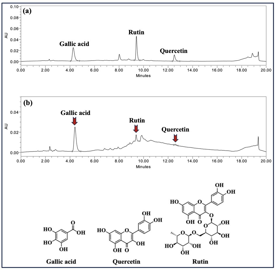
Figure 2 HPLC chromatograms illustrating (a) reference standards of gallic acid, rutin, and quercetin, and (b) their qualitative identification in the ethyl acetate fraction (EF).
Besides, quercetin and rutin belong to the flavonoid class of polyphenolic compounds, possessing a characteristic C6–C3–C6 flavonol backbone with multiple hydroxyl substitutions [14]. Quercetin, as the aglycone form, exhibits a planar and highly conjugated structure that is commonly linked to antioxidant, anti-inflammatory, and antimicrobial activities, as well as inhibitory effects against carbohydrate-hydrolyzing enzymes [37]. Rutin, a glycosylated derivative of quercetin containing a rutinose moiety, retains the fundamental flavonoid scaffold while displaying modified physicochemical properties, such as enhanced solubility and stability, which have been reported to influence its biological behavior [38]. Both compounds have been widely documented for their potential roles in antioxidant defense, inflammation regulation, and metabolic disorder-related bioactivities.
In summary, the qualitative HPLC results indicate that gallic acid, quercetin, and rutin may contribute to the enhanced biological activities of the EF, providing mechanistic support for its superior performance.
Limitations and propose future directions
Despite the comprehensive evaluation of N. orientalis extracts, several limitations should be acknowledged. First, the biological assessments were primarily restricted to in vitro models. Specifically, the anti-inflammatory activity was based on cell-free assays, and the antidiabetic evaluation focused on inhibition capacity without performing kinetic characterization to determine the mode of enzyme inhibition. Consequently, future research should incorporate macrophage models and detailed kinetic analyses to elucidate cellular mechanisms [39,40], followed by in vivo studies to confirm efficacy and safety [41]. Second, while representative polyphenols were identified qualitatively, their quantitative contributions and synergistic interactions remain to be explored through compound isolation and HPLC-based profiling. Finally, to facilitate practical application, future work should establish an optimized extraction process [1] integrated with life cycle assessment and economic analysis [42] to evaluate feasibility for large-scale production.
Conclusions
The present study validates N. orientalis stem bark as a significant source of bioactive phytochemicals. Fractionation with ethyl acetate proved effective in enriching polyphenols and flavonoids, resulting in this fraction exhibiting the most prominent antioxidant, anti-inflammatory, antidiabetic, and antibacterial activities among the investigated extracts. These biological effects were strongly correlated with the total polyphenol and flavonoid contents, suggesting that compounds such as gallic acid, quercetin, and rutin, identified via qualitative analysis, likely contribute to the observed properties. Collectively, these in vitro findings provide scientific evidence supporting the traditional use of N. orientalis and underscore the potential of the ethyl acetate fraction as a candidate for developing plant-based therapeutic agents.
Acknowledgements
The authors gratefully acknowledge Dr. Nguyen Thi Ai Lan for her valuable support in the taxonomic identification of the plant material.
CRediT author statement
Tran Chi Linh: Conceptualization; Methodology; Investigation; Data curation; Writing - Original Draft Preparation; Writing - Review & Editing; Supervision. Do Van Mai: Methodology; Investigation; Data curation, Writing - Original Draft Preparation. Chong Kim Thien Duc: Conceptualization; Investigation; Visualization; Formal analysis. Nguyen Van Ky: Investigation; Validation. Thai Viet Tang: Resources; Validation. Nguyen Duy Tuan: Data curation; Visualization. Nguyen Huynh Bich Lieu: Visualization; Validation.
References
[1] CKT Duc, NT Tuan, TC Linh, HVT Luong and DT Pham. Optimization of ultrasound-assisted extraction of Ehretia asperula leaves with antioxidant and anti-diabetic activities and assessment of the process environmental impacts. Sustainable Chemistry and Pharmacy 2025; 45, 102039.
[2] N Chandimali, SG Bak, EH Park, HJ Lim, YS Won, EK Kim, SI Park and SJ Lee. Free radicals and their impact on health and antioxidant defenses: A review. Cell Death Discovery 2025; 11, 19.
[3] ME Kim and JS Lee. Advances in the regulation of inflammatory mediators in nitric oxide synthase: Implications for disease modulation and therapeutic approaches. International Journal of Molecular Sciences 2025; 26(3), 1204.
[4] NK Bhol, MM Bhanjadeo, AK Singh, UC Dash, RR Ojha, S Majhi, AK Duttaroy and AB Jena. The interplay between cytokines, inflammation, and antioxidants: Mechanistic insights and therapeutic potentials of various antioxidants and anti-cytokine compounds. Biomedicine & Pharmacotherapy 2024; 178, 117177.
[5] CKT Duc, DT Pham, HBL Nguyen and CL Chan. Evaluation of phytochemical screening, ‘Antibacterial, Antioxidant, Anti-Inflammatory, and Antidiabetic Activities’ of sequentially extracted ixora duffii leaves extract. Trends in Sciences 2025; 22(7), 10028.
[6] A Caturano, M Rocco, G Tagliaferri, A Piacevole, D Nilo, GD Lorenzo, I Iadicicco, M Donnarumma, R Galiero, C Acierno, C Sardu, V Russo, E Vetrano, C Conte, R Marfella, L Rinaldi and FC Sasso. Oxidative stress and cardiovascular complications in Type 2 Diabetes: From pathophysiology to lifestyle modifications. Antioxidants (Basel) 2025; 14(1), 72.
[7] K Ogurtsova, JDR Fernandes, Y Huang, U Linnenkamp, L Guariguata, NH Cho, D Cavan, JE Shaw and LE Makaroff. IDF Diabetes Atlas: Global estimates for the prevalence of diabetes for 2015 and 2040. Diabetes Research and Clinical Practice 2017; 128, 40-50.
[8] SA Antar, NA Ashour, M Sharaky, M Khattab, NA Ashour, RT Zaid, EJ Roh, A Elkamhawy and AA Al-Karmalawy. Diabetes mellitus: Classification, mediators, and complications; A gate to identify potential targets for the development of new effective treatments. Biomedicine & Pharmacotherapy 2023; 168, 115734.
[9] B Gieroba, A Kryska and A Sroka-Bartnicka. Type 2 diabetes mellitus - conventional therapies and future perspectives in innovative treatment. Biochemistry and Biophysics Reports 2025; 42, 102037.
[10] H Ribeiro, I Rodrigues, L Napoleão, L Lira, D Marques, M Veríssimo, JP Andrade and M Dourado. Non-steroidal anti-inflammatory drugs (NSAIDs), pain and aging: Adjusting prescription to patient features. Biomedicine & Pharmacotherapy 2022; 150, 112958.
[11] S Liu, J Liu, Y Wang, F Deng and Z Deng. Oxidative stress: Signaling pathways, biological functions, and disease. MedComm 2025; 6(7), 70268.
[12] Z Zhang, HN ElSohly, MR Jacob, DS Pasco, LA Walker and AM Clark. New indole alkaloids from the bark of Nauclea orientalis. Journal of Natural Products 2001; 64(8), 1001-1005.
[13] T Kanchanapoom, P Sahakitpichan, N Chimnoi, C Petchthong, W Thamniyom, P Nangkoed and S Ruchirawat. Monoterpene alkaloid glycosides from the leaves of Nauclea orientalis. Phytochemistry Letters 2021; 41, 83-87.
[14] CKT Duc, TC Linh, NQC Thanh, PQ Nhien, DTX Trang, LT Danh and NT Tuan. Isolation and evaluation of the antioxidant capacity of compounds from Ehretia asperula Zoll. & Moritzi. Indonesian Journal of Chemistry 2024; 24(4), 1206-1217.
[15] LC Tran, CKT Duc, TT Nguyen, DT Pham, DT Luu, TTP Truong and TTX Dai. Anti-inflammatory and antioxidant properties of endophytic bacteria isolated from Paramignya trimera: Detection and identification of bioactive compounds. Journal of Biologically Active Products from Nature 2025; 15(4), 432-454.
[16] TC Linh, DT Pham, DV Mai, NHB Lieu, PK Dinh and CKT Duc. Investigating the in vitro anti-obesity activity of the ultrasonic-assisted extraction process of alkaloid- rich extract from Mansoa alliacea flowers using response surface methodology. Natural Resources for Human Health 2025; 5(3), 265-278.
[17] LTM Ngan, JK Moon, JH Kim, T Shibamoto and YJ Anh. Growth-inhibiting effects of Paeonia lactiflora root steam distillate constituents and structurally related compounds on human intestinal bacteria. World Journal of Microbiology and Biotechnology 2012; 28, 1575-1583.
[18] JE Lee, JTM Jayakody, JI Kim, JW Jeong, KM Choi, TS Kim, C Seo, I Azimi, J Hyun and B Ryu. The influence of solvent choice on the extraction of bioactive compounds from asteraceae: A comparative review. Foods 2024; 13(19), 3151.
[19] E Gil-Martín, T Forbes-Hernández, A Romero, D Cianciosi, F Giampieri and M Battino. Influence of the extraction method on the recovery of bioactive phenolic compounds from food industry by-products. Food Chemistry 2022; 378, 131918.
[20] D Palaiogiannis, T Chatzimitakos, V Athanasiadis, E Bozinou, DP Makris and SI Lalas. Successive solvent extraction of polyphenols and flavonoids from Cistus creticus L. Leaves. Oxygen 2023; 3, 274-286.
[21] D Ciupei, A Colişar, L Leopold, A Stănilă and ZM Diaconeasa. Polyphenols: From classification to therapeutic potential and bioavailability. Foods 2024; 13(24), 4131.
[22] M Rudrapal, SJ Khairnar, J Khan, AB Dukhyil, MA Ansari, MN Alomary, FM Alshabrmi, S Palai, PK Deb and R Devi. Dietary polyphenols and their role in oxidative stress-induced human diseases: Insights into protective effects, antioxidant potentials and mechanism(s) of action. Frontiers in Pharmacology 2022; 13, 806470.
[23] JM Al-Khayri, GR Sahana, P Nagella, BV Joseph, FM Alessa and MQ Al-Mssallem. Flavonoids as potential anti-inflammatory molecules: A review. Molecules 2022; 27(9), 2901.
[24] N Yahfoufi, N Alsadi, M Jambi and C Matar. The Immunomodulatory and anti-inflammatory role of polyphenols. Nutrients 2018; 10(11), 1618.
[25] A Kulkarni, CC Kulkarni, SR Pradeep, J Poyya, AK Kudva, V Radhakrishnan and AS Khandagale. Role of anti-inflammatory and antioxidant properties of natural products in curing cardiovascular diseases. Current Issues in Molecular Biology 2025; 47(11), 955.
[26] K Jomova, SY Alomar, R Valko, J Liska, E Nepovimova, K Kuca and M Valko. Flavonoids and their role in oxidative stress, inflammation, and human diseases. Chemico-Biological Interactions 2025; 413, 111489.
[27] M Krawczyk, I Burzynska-Pedziwiatr, LA Wozniak and M Bukowiecka-Matusiak. Impact of polyphenols on inflammatory and oxidative stress factors in diabetes mellitus: Nutritional antioxidants and their application in improving antidiabetic therapy. Biomolecules 2023; 13(9), 1402.
[28] D de Paulo Farias, FF de Araújo, IA Neri-Numa and GM Pastore. Antidiabetic potential of dietary polyphenols: A mechanistic review. Food Research International 2021; 145, 110383.
[29] E Barber, MJ Houghton and G Williamson. Flavonoids as human intestinal α-Glucosidase inhibitors. Foods 2021; 10(8), 1939.
[30] F Shahidi and R Danielski. Review on the role of polyphenols in preventing and treating Type 2 Diabetes: Evidence from in vitro and in vivo studies. Nutrients 2024; 16(18), 3159.
[31] CL Tran, VM Do, VT Huynh, XN Hong and KTD Chong. Optimization of polyphenol extraction conditions from rhizomes of Curcuma zedoaria with antioxidant, anti-inflammatory and anti-diabetic activities in vitro. Trends in Sciences 2024; 21(9), 8544.
[32] R Suručić, M Travar, TK Vasović, JR Selgrad, L Suručić, M Momčilović, MP Stojiljković and R Škrbić. In vitro and in silico analysis of differential antibacterial activity of pomegranate polyphenols against gram-positive and gram-negative bacteria. Antibiotics 2025; 14(9), 912.
[33] Y Liu, J Zhu, Z Liu, Y Zhi, C Mei and H Wang. Flavonoids as promising natural compounds for combating bacterial infections. International Journal of Molecular Sciences 2025; 26(6), 2455.
[34] LD Rossi, G Rocchetti, L Lucini and A Rebecchi. Antimicrobial potential of polyphenols: Mechanisms of action and microbial responses—a narrative review. Antioxidants 2025; 14(2), 200.
[35] B Badhani, N Sharma and R Kakkar. Gallic acid: A versatile antioxidant with promising therapeutic and industrial applications. RSC Advances 2015; 5(32), 27540-27557.
[36] M Hadidi, R Liñán-Atero, M Tarahi, MC Christodoulou and F Aghababaei. The potential health benefits of gallic acid: Therapeutic and food applications. Antioxidants 2024; 13(8), 1001.
[37] EJ Carrillo-Martinez, FY Flores-Hernández, AM Salazar-Montes, HF Nario-Chaidez and LD Hernández-Ortega. Quercetin, a Flavonoid with great pharmacological capacity. Molecules 2024; 29(5), 1000.
[38] A Ganeshpurkar and AK Saluja. The pharmacological potential of rutin. Saudi Pharmaceutical Journal 2017; 25(2), 149-164.
[39] Y Tian, S Zhou, R Takeda, K Okazaki, M Sekita and K Sakamoto. Anti-inflammatory activities of amber extract in lipopolysaccharide-induced RAW 264.7 macrophages. Biomedicine & Pharmacotherapy 2021, 141, 111854.
[40] YF Zeng, J Lee, YX Si, L Yan, TR Kim, GY Qian, ZR Lü, ZM Ye and SJ Yin. Inhibitory effect of Zn2+ on α-glucosidase: Inhibition kinetics and molecular dynamics simulation. Process Biochemistry 2012 47(12), 2510-2517.
[41] TC Linh, CKT Duc, NT Tuan and DTX Trang. Study on the toxicity and in vitro and in vivo antidiabetic activities of Miliusa velutina stem bark ethanolic extract. Phytomedicine Plus 2025; 5(4), 100879.
[42] CKT Duc, NT Tuan, TC Linh, HVT Luong and DT Pham. Sustainable recovery of cellulose from lotus seedpods: Integrating process optimization and environmental-preliminary economic analysis. Bioresource Technology 2026, 441, 133603.