
Trends Sci. 2026; 23(10): 13201
Design of A Stable Microemulsion with Phenolic Compounds from Acanthus ebracteatus for Hair Tonic Applications
Nakuntwalai Wisidsri, Khemjira Jarmkom, Wongnapa Nakyai,
Monsicha Khuanekkaphan and Suradwadee Thungmungmee*
Faculty of Integrative Medicine, Rajamangala University of Technology Thanyaburi, Pathum Thani 12130, Thailand
(*Corresponding author’s e-mail: [email protected])
Received: 29 December 2025, Revised: 13 February 2026, Accepted: 20 February 2026, Published: 15 April 2026
Abstract
Botanical ingredients have been increasingly incorporated into cosmeceutical hair tonic formulations due to their potential to promote hair growth. Acanthus ebracteatus Vahl. has attracted interest as a functional natural ingredient with potential hair growth-enhancing effects. This study aims to evaluate the capacity of A. ebracteatus extract (AEE) to promote the proliferation of human hair follicle dermal papilla cells (HDPCs) and to develop a hair tonic AEE microemulsion (AEE-ME). Total phenolic content (TPC) of AEE was determined using the Folin-Ciocalteu assay. The cytotoxicity and proliferation of HDPCs treated with AEE were assessed using a resazurin reduction assay. Microemulsions were constructed using pseudo-ternary phase diagrams composed of isononyl isononanoate (oil), deionized water, and various mass ratios of laureth-9 (surfactant) and ethanol (cosurfactant) (Smix). AEE-ME was characterized by droplet size, polydisperse index (PDI), zeta potential, pH, conductivity, and viscosity, as well as TPC. The stability of AEE-ME was evaluated over 12 weeks. The results showed that AEE contained TPC at 220.71 ± 9.28 mg of gallic acid equivalents (GAE)/g. AEE was safe for HDPCs at concentrations ranging from 3.125 to 200 μg/mL. HDPCs proliferation increased significantly after 48 h of incubation compared to the control. Microemulsion regions expanded as the mass ratio of Smix increased. AEE was incorporated into a suitable microemulsion base (MB1) composed of 70% Smix (2:1), 20% water, and 10% oil. The droplet size, PDI, zeta potential, pH, electrical conductivity, and viscosity were 97.37 ± 2.46 nm, 0.38 ± 0.01, −2.16 ± 0.06 mV, 6.20 ± 0.02, 24.07 ± 0.55 S/cm, and 22.1 ± 1.5 MPa, respectively. TPC value of AEE-ME was 294.32 ± 6.44 mg GAE/g. All parameters remained stable after storage at 4 °C, ambient temperature, and 45 °C. Therefore, hair tonic AEE-ME may be used for hair loss protection with high stability.
Keywords: Acanthus ebracteatus, Human hair follicle dermal papilla cells, Cell viability, Hair proliferation, Pseudo-ternary phase diagram, Hair tonic microemulsion, Stability
Introduction
Hair loss, or alopecia, is a multifactorial condition with significant psychosocial and clinical implications. The hair growth cycle, composed of the anagen (growth), catagen (regression), and telogen (resting) phases, is tightly regulated by molecular and cellular mechanisms. Disruptions in this cycle can result in excessive hair shedding, thinning, or baldness [1]. Among the primary causes of hair loss, genetic predisposition is particularly important. Androgenetic alopecia, the most common form, results from increased
follicular sensitivity to dihydrotestosterone (DHT), leading to miniaturization of hair follicles and progressive thinning [2]. Hormonal fluctuations, such as those associated with thyroid disorders, pregnancy, and menopause, also contribute to alterations in hair growth dynamics [3]. Nutritional deficiencies and oxidative stress have been increasingly recognized as contributing factors. Insufficient intake of essential micronutrients, including iron, zinc, and biotin, can impair keratin synthesis and follicle metabolism [4,5]. Moreover, reactive oxygen species (ROS), generated by ultraviolet (UV) radiation, environmental toxins, or metabolic processes, may damage follicular cells and accelerate hair follicle aging [6,7]. Stress and chronic inflammation further exacerbate these effects, promoting premature entry of hair follicles into the telogen phase [8,9]. In addition, iatrogenic factors such as chemotherapy remain well-documented triggers of temporary or permanent alopecia [10]. Given the interplay of genetic, hormonal, nutritional, environmental, and psychological factors, hair loss represents a complex condition requiring multidisciplinary approaches. Recent studies focusing on molecular signaling pathways, including oxidative stress responses, growth factors, and inflammatory mediators, have opened new possibilities for targeted therapies and preventive strategies [6,11]. At present, several topical or synthetic agents are available for the treatment of hair loss, such as minoxidil and finasteride. However, previous reports have indicated that these agents may be associated with various adverse effects, including skin irritation, erythema, scalp irritation, facial hypertrichosis, allergic contact dermatitis, pruritus, elevated liver enzyme levels, nocturnal enuresis, testicular pain, and headaches [12]. Consequently, natural-derived substances have gained increasing attention as alternative therapeutic options for hair loss management, as they are generally considered to offer improved safety profiles with fewer undesirable side effects.
Acanthus ebracteatus Vahl. is a plant that belongs to the Acanthaceae family. It has been used in traditional medicine as an astringent, expectorant, stimulant, and to heal skin diseases by using the whole plant, as a cough remedy, for paralysis, and for wound healing by the root, as a snakebite cure by the tender shoots and leaves, and as a treatment for preventing alopecia and treating rheumatism with the leaves [13]. A. ebracteatus leaves have been reported to contain phytochemical constituents, including phenolics and phenolic glycosides such as caffeic acid, dihydroferulic acid 4-O-glucuronide, chlorogenic acid, kelapayoside A, verbasoside, and flavonoids and flavonoid glycosides like luteolin, apigenin, saponarin, baicalin, and tetramethylquercetin 3-rutinoside [14]. Total phenolic and flavonoid contents of A. ebracteatus leavesʼ extract have been investigated for their relation to antioxidant activities [14,15]. Moreover, its compounds have shown anti-bacterial, anti-cancer, and anti-neurodegenerative properties [14,16]. In terms of hair treatment, A. ebracteatus has been found to promote dermal papilla cell proliferation, prevent cell apoptosis, and display anti-inflammatory activity related to hair loss [17]. Topical application of the ethanolic extract of A. ebracteatus on wounded skin in rats has been shown to promote wound healing and accelerate the recovery process. Additionally, the extract demonstrated anti-inflammatory effects at the wound site. Notably, it was found to enhance the expression of a key growth factor, vascular endothelial growth factor (VEGF), which is known to play an important role in angiogenesis. This upregulation of VEGF expression may also potentially support and promote hair follicle development and hair growth. Although several studies have reported and supported the biological activities of the extract, its application in a stable nano-particulate formulation has not yet been documented.
Microemulsions are promising drug delivery systems characterized by optical isotropy, low viscosity, and thermodynamic stability. They typically consist of droplets with sizes ranging from 10 to 200 nm, resulting in a transparent appearance [18]. Microemulsions can be used for several applications, such as food, textile, cosmetics, and pharmaceutical industries, due to their advantages, including improved stability, increased solubility of hydrophilic and lipophilic drugs, enhanced bioavailability of poorly soluble drugs, controlled drug release, and ease in preparation [19,20]. Typically, microemulsions are colloidal dispersions composed of oil, aqueous, surfactant, and cosurfactant components in suitable ratios. Synthetic and natural bioactive compounds that promote hair growth and decrease hair loss have been formulated into hair tonic microemulsions [21,22]. In general, physicochemical parameters of microemulsions, such as particle size, polydisperse index (PDI), zeta potential, pH, and viscosity, affect the formulation’s properties. For example, the small size of microemulsion, low PDI value, and electrostatic repulsion, which represent the zeta potential, provide advantages such as thermal stability, translucency, and long-term stability [21,23]. Moreover, a smaller size enhances skin penetration and promotes biological performance. In addition, the formulation pH was within a suitable range, thereby minimizing the risk of scalp irritation and itching [24]. Therefore, the development of microemulsion formulations with favorable physicochemical properties is essential for predicting and ensuring the stability, efficacy, and safety of the developed formulation, thereby serving as an indicator of overall product quality.
Therefore, this study aimed to investigate hair dermal papilla proliferation caused by high phenolic compounds from A. ebracteatus extract. Since none of these reports or commercial products included the use of A. ebracteatus loaded in hair tonic microemulsion, a stable A. ebracteatus extract-loaded hair tonic microemulsion was studied. This developed formulation represents an initial step toward the development of effective and safe topical products containing natural hair growth-promoting agents, with the potential to be further advanced and translated into commercially viable applications.
Materials and methods
Chemicals and reagents
Commercial-grade 95% ethanol, analytical-grade 99% ethanol, dimethyl sulfoxide (DMSO), and methanol were purchased from RCI Labscan, Thailand. Resazurin and gallic acid were obtained from Sigma-Aldrich, USA. Human hair follicle dermal papilla cells (HDPCs) and growth medium, including fetal calf serum, bovine pituitary extract, basic fibroblast growth factor, and insulin, were purchased from Promocell, Germany. The cosmetic-grade ingredients, including isononyl isononanoate and laureth-9, were purchased from MySkinRecipes, Thailand.
Plant extraction
A. ebracteatus was collected from Chatuchak Market, Bangkok, Thailand. This plant was identified by Assist. Prof. Dr. Pranee Nangngam, an expert in plant botany, Department of Biology, Faculty of Science, Naresuan University. A voucher specimen (PNU 6081) was deposited at the Faculty of Science, Naresuan University, Thailand. The fresh leaves were dried in a hot air oven at 50 °C for 24 h. The dried plant was ground into powder and then macerated with 95% ethanol for 7 days. The extract was filtered using filter paper connected to a vacuum pump. Then, the filtrate was evaporated using a rotary evaporator under reduced pressure at 45 °C. The percentage of yield of A. ebracteatus crude extract (AEE) was calculated. AEE was stored at −20 °C until use.
%yield = [weight of extract (g) / weight of matter (g)] × 100 (1)
Total Phenolic Content (TPC) determination
The TPC of AEE was determined using the Folin-Ciocalteu assay [25]. Twenty µl of the extract was well-mixed with 100 μl of 10% Folin-Ciocalteu’s reagent. After incubating the mixture at room temperature for 7 min, 100 μl of 7.5% sodium carbonate was added. The mixture was continuously incubated at room temperature for 45 min. The absorbance of the mixture was then measured at 765 nm using a Microplate reader CALIOstar (BMG LABTECH, Germany). TPC value was determined using a gallic acid calibration curve and expressed as milligrams of gallic acid equivalents per gram of dry extract (mg GAE/g).
Cytotoxicity
Human hair follicle dermal papilla cells (HDPCs) were cultured in follicle dermal papilla cell growth medium at 37 °C, 5% CO2. The cytotoxicity of HDPCs was determined by resazurin reduction assay [25]. HDPCs were seeded in a 96-well plate at a concentration of 4×10⁵ cells/mL and incubated at 37 °C with 5% CO2 for 24 h. Then, AEE at concentrations of 3.125, 12.5, 25, 50, 100, and 200 µg/mL was applied. After 24 h of incubation, 50 µg/mL resazurin solution, dissolved in DMDM, was added and incubated for an additional 4 h. The fluorescent intensity of the reaction mixture was determined at 560 nm against 600 nm. The percentage of cell viability was calculated using the formula.
%Cell viability=[(FIcontrol-FIsample)/FIcontrol]×100 (2)
Cell proliferation measurement
The cell proliferation assay was slightly modified from the method described by Hughes et al. [26]. Non-cytotoxic concentrations of AEE were used throughout the experiment to evaluate their effects on cell proliferation. HDPCs at 4×10⁵ cells/mL were seeded into a 96-well plate and incubated at 37 °C with 5% CO2 for 24 h. After 24 and 48 h, cell viability was assessed using the resazurin reduction assay. The percentage of cell viability is expressed by the formula, which can be interpreted as an indication of cell proliferation.
%Cell viability=[(FIsample-FIcontrol)/FIsample]×100 (3)
Preparation of microemulsions
Construction of pseudo-ternary phase diagram
A pseudo-ternary phase diagram was developed to identify the microemulsion formation region [27]. Microemulsions consisting of isononyl isononanoate (oil), laureth-9 (surfactant), ethanol (cosurfactant), and deionized water (water) were prepared using a water titration method. Laureth-9 and ethanol were mixed to form Smix and prepared in different mass ratios: 1:1, 2:1, 3:1, 1:2, and 1:3. Smix was weighed and then mixed with oil under a magnetic bar at 200 rpm. The water phase was added dropwise into the mixtures until they formed transparent and homogeneous microemulsions. This process continued until the transparent liquid turned translucent and turbid. The mixtures were classified as microemulsions when they appeared as clear, homogeneous liquids, and low viscosity by visualization. The transparent regions represented by the microemulsion were finally determined through the pseudo-ternary phase diagram after being retained for one night. Then ratios of the components exhibiting microemulsion characteristics were plotted on a pseudo-ternary phase diagram using CHEMIX School software v12.5 (Arne Standnes, Bergen, Norway).
Formulation and characterization of microemulsion bases (MB) and A. ebracteatus extract microemulsion (AEE-ME)
The appropriate microemulsion bases (MB), prepared using Smix, oil, and water in the weight ratio based on the center of the microemulsion region and with Smix limited to 70% to prevent skin irritation, were formulated (Table 1). The formulation with the smallest droplet size and a PDI less than 0.3 was chosen to incorporate with AEE. Then, an accurately weighed amount equivalent to 0.50% (w/w) of AEE was added to the mixture of Smix and isononyl isononanoate. Next, the mixture was stirred until homogeneous. Water was added and stirred for 15 min at 200 rpm. Finally, the AEE-ME was obtained.
Characterization of MB and AEE-ME
Firstly, six MB form pseudo-ternary phase diagrams were evaluated. The isotropic property was assessed using cross-polarized light microscopy (Olympus U-HSEXP, Tokyo, Japan). Then, droplet size, zeta potential, and polydispersity index (PDI) were measured without dilution using a dynamic light scattering Nanoparticle Analyzer (NanoParticle SZ-100V2 Series, HORIBA, Japan) at 25 °C with a scattering angle of 173° [28]. The AEE-ME was additionally evaluated for viscosity, pH, and electrical conductivity using a Brookfield® rheometer (Model: R/S-CPS plate & plate), pH meter, and conductivity meter, respectively.
Total phenolic content (TPC) of AEE-ME
The TPC of AEE-ME was determined using the Folin–Ciocalteu assay as described above. The 500 mg of AEE-ME was weighed and mixed with 1 mL of 95% ethanol using a vortex. Then, the mixture was centrifuged at 10,000 rpm for 10 min. The supernatant was used to determine TPC using a gallic acid calibration curve. The TPC of AEE-ME was expressed as milligrams of gallic acid equivalents per gram of microemulsion (mg GAE/g).
Stability of AEE-ME
The stability of the AEE-ME was assessed under different storage conditions, including 4 °C, ambient temperature, and 45 °C, over a period of 12 weeks [28]. The evaluation focused on physicochemical stability parameters, such as color, odor, phase separation, droplet size, zeta potential, PDI, pH, and electrical conductivity. These parameters were measured at specific time points: 0, 4, 8, and 12 weeks.
Statistical analysis
All experiments were performed in triplicate. The results are shown as means and standard deviations (SD). One-way analysis of variance (ANOVA), followed by Tukey’s post-hoc test (SPSS version 22.0), was used to compare groups. The p-values less than 0.05 indicate significant differences.
Results and discussion
Plant extraction
The solvent for plant extraction is important for extracting bioactive compounds. According to the study by Wisuitiprot et al. [37], extraction with 95% ethanol yielded a higher level of the biomarker of A. ebracteatus, namely verbascoside, compared with extraction using 50% ethanol, hexane, or water [17]. Verbascoside markedly inhibited the release of pro-inflammatory cytokines in LPS-stimulated murine macrophage RAW 264.7 cells, as well as IL‑1α and IL-6 production in UVB-irradiated dermal papilla cells. Moreover, it showed a protective effect against apoptosis in dermal papilla cells, suggesting its potential application in anti-hair loss therapies [17]. Therefore, 95% ethanol was used to extract A. ebracteatus leaves. A viscous A. ebracteatus leaf crude extract (AEE) was brown-green in color. A high percentage yield of AEE was found at 15.80%. The high extraction yield indicates that the extract has strong potential for practical applications and demonstrates economic feasibility for commercial utilization. Over the past decades, botanical or natural raw materials have been considered to address batch-to-batch quality variability and ensure the efficacy and safety of finished products [29]. Many factors, such as cultivation position, climate, harvest time, and extraction process, influence this quality. Therefore, a chromatographic fingerprint can be used to evaluate batch-to-batch quality variability of the extract. Consequently, chromatographic fingerprinting techniques, such as high-performance liquid chromatography (HPLC) or mass spectrometry, may be considered in subsequent studies to support quality control of the extract and improve batch-to-batch consistency of its bioactivity.
Total Phenolic Content (TPC) of AEE
Previous studies have reported a significant association between phenolic content and biological activity. Accordingly, plants rich in phenolics and flavonoids are widely recognized for their strong antioxidant, anti-inflammatory, antimicrobial, and anticancer activities [30,31]. Moreover, several studies have demonstrated the beneficial effects of plant extracts with high phenolic content on the proliferation of human dermal papilla cells and the promotion of hair growth [32,33]. In the results, the TPC value of AEE was quantified using the calibration equation of y = 3.1401x + 0.0148 (R2 = 0.9970) and was found to be 220.71 ± 9.28 mg GAE/g. Although the TPC obtained in this study differed from that reported in previous studies, for example, TPC in 70% ethanolic extract of A. ebracteatus leaf was found to be 138.20 ± 0.10 mg GAE/g dry extract [14], while the 80% methanolic extract showed TPC of 344.67 ± 2.06 mg GAE/g DW [15], ethanol is considered a safer solvent for use than methanol, owing to its lower toxicity and greater suitability for applications involving human exposure. Therefore, AEE still exhibited a level of TPC that is anticipated to exert biological activity associated with the promotion of hair growth.
Cytotoxicity
The cytotoxic effect of AEE on dermal papilla cells was assessed using a resazurin reduction assay. This assay is based on the intracellular reduction of the nonfluorescent dye resazurin to the fluorescent product resorufin by metabolically active, viable cells, reflecting their intrinsic reducing power. This method offers a simple, sensitive, non-destructive, and cost-effective approach for cell viability assessment and is well-suited for quantitative and high-throughput applications [34]. In HDPCs exposed to the extract at concentrations ranging from 3.125 to 200 µg/mL for 24 h, AEE did not induce cell death, as indicated by a cell viability percentage that was not significantly different from that of the control group (0.2% DMSO), suggesting its non-cytotoxic nature (Figure 1). This result aligns with a previous study reporting that A. ebracteatus extract, obtained with 95% ethanol, showed no cytotoxic effects at concentrations from 0.98 to 500 µg/mL by MTT assay [17].
Cell proliferation
In this study, AEE was considered suitable for anti-hair loss applications that encouraged the growth of dermal papilla cells. The growth of HDPCs was assessed at 24 and 48 h using non-cytotoxic concentrations of AEE identified in initial testing, specifically 3.125 - 200 µg/mL. The results showed that, after incubation with the extract for 24 and 48 h, cell viability was maintained across all tested concentrations compared to the untreated control group (Figure 1). Additionally, AEE showed a trend toward increased hair follicle cell proliferation at concentrations from 12.5 to 200 µg/mL, with increases of 109% to 122%. These results may be attributed to the presence of phenolic compounds in the extract. Previous phytochemical investigations have indicated that A. ebracteatus leaves contain a range of bioactive phenolic compounds. Among these, caffeic acid has been reported to promote keratinocyte proliferation and stimulate hair growth in murine models through induction of the anagen phase and enhancement of hair shaft elongation [35]. In addition, an amide derivative of caffeic acid, N-[3,5-bis(trifluoromethyl)phenyl], has been identified as a non-steroidal inhibitor of steroid 5α-reductase type 1 (SRD5A1). Overexpression of 5α-reductase leads to excessive production of dihydrotestosterone (DHT), a key factor implicated in the pathogenesis of androgen-dependent conditions, including androgenic alopecia [36]. In this study, AEE did not evaluate cell proliferation in each cell cycle. However, the study by Wisuitiprot et al. [37] revealed that A. ebracteatus leaf extract at a concentration of 125 - 500 µg/mL promotes dermal papilla cell proliferation and increases the synthesis (S) and mitosis (G2/M) phases in the cell cycle stages [37]. In addition, verbascoside has been identified as a biomarker in A. ebracteatus leaf extracts. Verbascoside, a phenylethanoid glycoside, functions as a natural transient receptor potential vanilloid-3 (TRPV3) inhibitor with protective effects on hair follicles. It has been shown to prevent testosterone-induced apoptosis in dermal papilla cells, thereby supporting follicle survival, and is regarded as a potential active ingredient for anti-hair loss applications [38]. Chlorogenic acid has also been identified as an active constituent of A. ebracteatus leaves [14]. Previous studies have demonstrated that chlorogenic acid, isolated from Morus alba root extract, promotes induction of the anagen phase in hair follicle models by upregulating the production of growth factors, including VEGF, FGF7, HGF, and FGF2. These factors facilitate the proliferation of HFDPCs and support the telogen-to-anagen transition through enhanced secretion of angiogenic paracrine mediators [39]. Collectively, these findings suggest that A. ebracteatus leaves possess potential application as a source of active ingredients in hair tonic formulations to promote hair follicle growth.
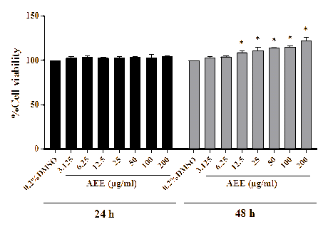
Figure 1 Human hair follicle dermal papilla cells (HDPCs) viability cells after 24-hour and 48-hour incubation with A. ebracteatus extract (AEE). Data show the mean ± SD (n=3). *p < 0.05 indicates a significant difference compared with control (0.2% DMSO).
Pseudo-ternary phase diagram
Microemulsion compositions were selected with a safety profile in commercial cosmetic products. Non-ionic surfactants are commonly utilized in microemulsions and other formulations in cosmetics and pharmaceutical products due to their non-toxic, low-cost, and biocompatible properties [40]. Laureth-9 is a polyethylene glycol ether of lauryl alcohol (called polyoxyethylene lauryl ether) that contains 9 ethylene oxide units in its molecule and is used as a non-ionic surfactant or emulsifier in cosmetics and personal care products. Its structure consists of a hydrophobic lauryl (C12) tail and a hydrophilic polyoxyethylene chain, which reduces interfacial tension between oil and water and stabilizes oil in water (o/w) microemulsion. Previous studies revealed that non-ionic polyoxyethylene lauryl ether structures, such as Brij-35 and Brij-30, were used to prepare microemulsions [41,42]. Ethanol, a short-chain alcohol, is widely used in herbal hair tonic formulations because of its properties, such as a phytochemical solubilizer, a permeation enhancer, and leaving a dry feel after use [43,44]. In microemulsions, ethanol is often used as a cosurfactant because it helps expand the microemulsion region and prevents the formation of gels, precipitates, and liquid crystals [45-47]. In this study, laureth-9 and ethanol were used as the surfactant and cosurfactant, respectively. Different mass ratios of laureth-9 and ethanol (Smix) 1:1, 2:1, 3:1, 1:2, and 1:3 were examined. The pseudo-ternary phase diagrams, constructed using isononyl isononanoate (oil), deionized water, and various mass ratios of Smix, are shown in Figure 2. The result showed that when increasing the laureth-9 ratio from 1:1 to 3:1, microemulsion regions rose from 11.46% to 22.81%. These results are consistent with Thungmungmee et al. (2025) study, which demonstrated that increasing the ratio of Tween 80 (surfactant) to ethanol (cosurfactant) leads to an expansion of the microemulsion region [27]. This finding indicates that an elevated surfactant concentration reduces the interfacial tension between the oil and aqueous phases, thereby facilitating the formation of microemulsions [48]. In contrast, increasing the proportion of cosurfactant was found to decrease the microemulsion region, which shrank from 11.46% to 7.60% and 6.43% at Smix mass ratios of 1:1, 1:2, and 1:3, respectively. This change may be due to the disruption of the interfacial film between the oil and aqueous phases, ultimately leading to the demulsification of the microemulsion system. Therefore, the relative amounts of surfactant and cosurfactant are key factors in determining the system’s phase behavior.
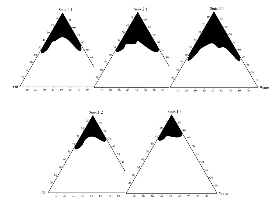
Figure 2 Pseudo-ternary phase diagrams constructed by isononyl isononanoate (oil), deionized water (water), and different mass ratios of laureth-9 and ethanol (Smix) 1:1, 2:1, 3:1, 1:2, and 1:3. The black areas represent microemulsion regions.
Formulation and characterization of MB and AEE-ME
To identify a suitable microemulsion base for the development of a hair tonic containing AEE, six formulations (MB1-MB6) were selected from the pseudo-ternary phase diagram at Smix ratios of 2:1 and 3:1, as presented in Table 1. These formulations were chosen based on their broad microemulsion regions, which facilitate the incorporation of AEE or other additives while preserving the microemulsion structure. Firstly, all formulations were determined to have isotropic properties by cross-polarized light microscopy. No birefringence or liquid crystal feature was observed under polarized light microscopy, indicating that they were indeed microemulsions [49].
Table 1 Formulation composition (by weight) with corresponding particle size, zeta potential, and PDI.
Formula |
Ratio of Smix |
%Smix (Laureth-9: Ethanol) |
%Water |
%Oil (Isononyl isononanoate) |
Particle size (nm) |
Zeta potential (mV) |
Polydisperse index (PDI) |
MB1 |
2:1 |
70 |
20 |
10 |
5.00 ± 0.18 |
‒1.68 ± 0.31 |
0.21 ± 0.01 |
MB2 |
2:1 |
70 |
15 |
15 |
10.62 ± 0.32 |
‒2.08 ± 0.32 |
0.23 ± 0.03 |
MB3 |
2:1 |
70 |
10 |
20 |
39.16 ± 1.09 |
‒1.22 ± 0.03 |
0.24 ± 0.02 |
MB4 |
3:1 |
70 |
20 |
10 |
101.70 ± 0.50 |
‒0.84 ± 0.10 |
0.20 ± 0.02 |
MB5 |
3:1 |
70 |
15 |
15 |
105.62 ± 0.54 |
‒1.08 ± 0.11 |
0.23 ± 0.01 |
MB6 |
3:1 |
70 |
10 |
20 |
163.60 ± 0.40 |
‒2.67 ± 0.11 |
0.21 ± 0.03 |
Hair follicle-targeted drug delivery systems enable controlled drug release and improved therapeutic efficacy with minimal adverse effects, representing a promising approach for treating hair follicle-associated disorders, such as hair loss and androgenetic alopecia [50]. Carrier particle size is a critical determinant of penetration efficiency, with keratinocyte-mediated mechanical pumping—driven by hair movement—enhancing follicular deposition. Nanoparticles with a mean diameter of 400 - 700 nm have been identified as optimal for deep follicular penetration, whereas smaller particles (~20 nm) accumulate more rapidly but tend to migrate into deeper skin layers over time [50]. These findings highlight the importance of tailoring particle size to achieve preferential retention within the hair follicle. Salimi et al. [51] revealed that valproic acid microemulsion with particle sizes ranging from 10.25 to 24.7 nm penetrates easily into the hairy guinea pig skin [51]. According to Cai et al. [23], a rosemary oil-based microemulsion with a droplet size of approximately 24 nm was found to improve the penetration of active compounds such as quercetin and adenosine through the skin, as well as their retention within the dermal layer, particularly within hair follicles [23]. In the animal model, this formulation significantly improves hair regeneration on days 7 and 14. The microemulsion containing 0.3% (w/w) cayenne pepper ethanolic extract exhibited a mean droplet size of 93.6 nm and effectively promoted hair growth in a rat model, resulting in a statistically significant increase in hair length of 1.09 cm after 30 days of treatment [52]. Subongkot et al. [53] demonstrated, using confocal laser scanning microscopy, that finasteride-loaded microemulsions predominantly penetrated the skin via the intercluster pathway, with additional contributions from the intercellular and transcellular routes [53]. These penetration mechanisms account for the enhanced skin permeation observed for microemulsions with small droplet sizes, approximately 80 nm. In the present study, the mean droplet size of MB formulations (MB1-MB6) was from 5.00 to 163.60 nm (Table 1). Therefore, all MB formulations might penetrate the skin through the intercluster and transfollicular pathways into hair follicles. Additionally, based on our observations, a higher oil content tends to increase particle size, as seen with MB1, MB2, and MB3, which contain 10%, 15%, and 20%, respectively. Their particle sizes are 5.00 ± 0.18 nm, 10.62 ± 0.32 nm, and 39.16 ± 1.09 nm, due to insufficient surfactant concentration to reduce the size [54]. In this study, at a given water content, the droplet size increased as Smix increased. For example, at a 20% water concentration with Smix 2:1, the size was 5.00 ± 0.18 nm, whereas at a 20% water concentration with Smix 3:1, the size was 101.70 ± 0.50 nm. This could be due to an improper ratio of surfactant to cosurfactant at the interfacial film [48]. Moreover, the smaller size tends to remain uniformly dispersed for a long time, thereby reducing the likelihood of coalescence and enhancing the long-term stability of the system [55]. The PDI value is crucial for evaluating the uniformity of microemulsions, with a range from 0 to 1. A value near zero indicates a narrow size distribution, which reduces droplet-droplet interactions and coalescence, resulting in high stability. The results showed that the PDI values of MBs were around 0.2, which is below 0.5 and indicates homogeneity [56]. Zeta potential values near zero indicate minimal electrostatic repulsion, which increases the likelihood of particles aggregating or flocculating, potentially reducing colloidal stability. However, the neutral values, ranging from −2.67 to −0.84 mV, may be due to the presence of nonionic surfactant (laureth-9) in the formulations, which provides steric stabilization. Thus, nonionic surfactants impart steric stabilization, preventing adjacent particles from agglomerating and thereby enhancing particle stability [57]. Based on the results of all MB formulations, smaller droplet sizes tend to be more stable than larger ones. Therefore, MB1, which had the lowest average droplet size of 5.00 ± 0.18 nm and showed a monodisperse distribution with a PDI of 0.21 ± 0.01, was chosen to incorporate AEE and evaluate the microemulsion properties.
The AEE-ME was confirmed to be isotropic. The droplet size of AEE-ME (94.63 ± 5.21 nm) increased more than tenfold compared to MB1 (6.83 ± 0.30 nm) (Table 2). This phenomenon may be due to the high extract content, which can dissolve within the internal phase, external phase, and their interfaces. Leanpolchareanchai et al. [58] also found that the incorporation of Thai mangos seed kernel extract into microemulsion formulations affects the droplet size by increasing from 8 - 29 nm to 9 - 70 nm [58]. However, the microemulsion exhibited greater penetration through porcine skin, up to 60-fold, into both the epidermal and dermal layers, compared with the solution form. Consequently, the droplet size of AEE-ME was still suitable for hair follicle use. After incorporating AEE into MB, the PDI slightly increased, showing a direct correlation with particle size. The zeta potential values of MB and AEE-ME were comparable, remaining close to neutrality. The AEE-ME was slightly more acidic than the MB formulation—possibly due to AEE’s phytochemicals such as (R)-(+)-2-pyrrolidone-5-carboxylic acid, caffeic acid, and chlorogenic acid [1]. However, the pH value of 6.20 ± 0.02 for the AEE-ME was within the compatible pH range of 5 - 7 for human scalp and hair shaft proteins [58]. Therefore, this hair tonic is within the ideal pH range for maintaining healthy hair. Conductivity measurements serve as an indicator of the type of microemulsions, distinguishing between oil-in-water (o/w) and water-in-oil (w/o) systems. Results showed conductivity values of 20.13 ± 0.90 S/cm and 24.07 ± 0.55 S/cm for MB and AEE-ME, respectively, which are lower than 100 μS/cm, confirming their classification as water-in-oil (w/o) microemulsions [54]. The incorporation of the extract resulted in a slight increase in conductivity, likely due to the presence of ionized moieties of phytochemicals, such as phenolic compounds, in the AEE extract [60]. However, this value does not change the type of microemulsions. The viscosity, which affects product spreadability on the skin and flow behavior during dispensing from the packaging, stayed constant. Incorporation of AEE in MB slightly increased viscosity, but it was not significantly different.
Total Phenolic Content (TPC) of AEE-ME
TPC in the formulation exhibited the strongest association with antioxidant activity [61]. TPC in AEE-ME was measured to assess the potential of its bioactive compounds in the formulation. The TPC value of AEE-ME was 294.32 ± 6.44 mg GAE/g. This suggests that AEE-ME contributes to antioxidant activity and associated effects.
Stability of AEE-ME
Regarding microemulsions, particle size, PDI, and zeta potential are key parameters that significantly affect the stability [18]. Based on the stability study of the AEE-ME stored at different temperatures—namely 4 °C, ambient temperature, and 45 °C—with evaluations conducted at 0, 4, 8, and 12 weeks, it was observed that the color and odor remained unchanged across all storage conditions. The formulation showed no signs of phase separation or turbidity throughout the 12-week stability testing period. As shown in Tables 2 and 3, droplet size, zeta potential, PDI, pH, conductivity, and viscosity exhibited no statistically significant changes, indicating the favorable properties and stability of the microemulsion. Based on the observed stability results, the developed microemulsion remained stable even under elevated temperature conditions (45 C). In comparison, previous studies have reported that a microemulsion composed of trilaureth-4 phosphate, propylene glycol, water, isononyl isononanoate, and Dendrobium formosum extract showed changes in droplet size along with a decrease in viscosity when stored at 45 °C after a 2-month stability study [28]. The absence of significant changes in droplet size and viscosity in the present formulation suggests a favorable stability profile under high-temperature storage conditions. The viscosity stability of the formulation can be attributed to the hydration of the surfactant’s hydrophilic chains surrounding the droplets and to the interaction between the extract and the components [62]. An appropriate water content in AEE-ME facilitates hydrogen bonding among these hydrated chains, strengthening inter-droplet interactions and yielding optimal viscosity. In addition, phenolic and flavonoid glycosides in the extract may interact with the interfacial film through hydrogen bonding with polar residues and hydrophobic interactions with nonpolar regions, thereby reinforcing the interfacial network, enhancing its mechanical strength, and contributing to overall formulation stability. Therefore, the favorable and stable physicochemical properties of AEE-loaded microemulsion suggest its suitability for topical application, particularly in hair tonic formulations. However, prolonged exposure to elevated temperatures may compromise the chemical stability of the active compounds. Therefore, storage at room temperature or at lower temperatures is recommended, as these conditions may further enhance the overall stability of the product.
Table 2 Droplet size, polydisperse index (PDI), and zeta potential of microemulsion base (MB) and AEE microemulsion (AEE-ME) at time points of stability test; starting time (0), 4 week (4), 8 week (8), and 12 week (12), at 4 °C, ambient temperature (AT), and 45 °C.
Sample |
Particle size (nm) (n=3) |
Polydisperse index (PDI) (n=3) |
Zeta potential (mV) (n=3) |
||||||
4 C |
AT |
45 C |
4 C |
AT |
45 C |
4 C |
AT |
45 C |
|
MB0 |
6.83 ± 0.30 |
6.83 ± 0.30 |
6.83 ± 0.30 |
0.25 ± 0.04 |
0.25 ± 0.04 |
0.25 ± 0.04 |
‒1.68 ± 0.05 |
‒1.69 ± 0.03 |
‒1.69 ± 0.03 |
MB4 |
7.02 ± 0.63 |
6.89 ± 0.65 |
6.43 ± 0.55 |
0.25 ± 0.02 |
0.21 ± 0.10 |
0.25 ± 0.12 |
‒1.71 ± 0.10 |
‒1.72 ± 0.08 |
‒1.70 ± 0.06 |
MB8 |
7.21 ± 0.13 |
7.18 ± 0.22 |
7.32 ± 0.42 |
0.26 ± 0.05 |
0.22 ± 0.09 |
0.28 ± 0.11 |
‒1.63 ± 0.07 |
‒1.70 ± 0.03 |
‒1.72 ± 0.11 |
MB12 |
7.11 ± 0.44 |
7.07 ± 0.38 |
7.41 ± 0.54 |
0.24 ± 0.07 |
0.23 ± 0.06 |
0.30 ± 0.12 |
‒1.68 ± 0.09 |
‒1.68 ± 0.04 |
‒1.73 ± 0.13 |
AEE-ME0 |
97.37 ± 2.46 |
97.37 ± 2.46 |
97.37 ± 2.46 |
0.38 ± 0.01 |
0.38 ± 0.01 |
0.38 ± 0.01 |
‒2.16 ± 0.06 |
‒2.16 ± 0.06 |
‒2.16 ± 0.06 |
AEE-ME4 |
96.28 ± 3.78 |
99.16 ± 2.88 |
100.09 ± 1.87 |
0.39 ± 0.09 |
0.38 ± 0.05 |
0.40 ± 0.11 |
‒2.15 ± 0.13 |
‒2.21 ± 0.11 |
‒2.23 ± 0.19 |
AEE-ME8 |
95.97 ± 2.44 |
96.00 ± 4.59 |
98.56 ± 3.66 |
0.36 ± 0.10 |
0.39 ± 0.13 |
0.42 ± 0.13 |
‒2.14 ± 0.08 |
‒2.23 ± 0.09 |
‒2.25 ± 0.05 |
AEE-ME12 |
96.33 ± 4.75 |
95.17 ± 5.05 |
99.93 ± 4.38 |
0.39 ± 0.11 |
0.39 ± 0.08 |
0.41 ± 0.10 |
‒2.16 ± 0.07 |
‒2.22 ± 0.10 |
‒2.26 ± 0.10 |
Table 3 The pH, conductivity, and viscosity of microemulsion base (ME) and AEE microemulsion (AEE-ME) at time points of stability test; starting time (0), 4 week (4), 8 week (8), and 12 week (12), at 4 °C, ambient temperature (AT), and 45 °C.
Sample |
pH (n=3) |
Conductivity (S/cm) (n=3) |
Viscosity (MPa) (n=3) |
||||||
4 C |
AT |
45 C |
4 C |
AT |
45 C |
4 C |
AT |
45 C |
|
MB0 |
6.20 ± 0.02 |
6.20 ± 0.02 |
20.13 ± 0.90 |
20.13 ± 0.90 |
20.13 ± 0.90 |
19.6 ± 2.3 |
19.6 ± 2.3 |
19.6 ± 2.3 |
|
MB4 |
6.22 ± 0.09 |
6.20 ± 0.05 |
6.15 ± 0.12 |
20.43 ± 1.10 |
22.27 ± 0.50 |
20.67 ± 0.91 |
21.6 ± 1.2 |
20.1 ± 1.0 |
19.6 ± 1.2 |
MB8 |
6.11 ± 0.04 |
6.16 ± 0.08 |
6.20 ± 0.05 |
21.20 ± 2.01 |
20.30 ± 1.20 |
22.40 ± 1.25 |
20.5 ± 1.3 |
21.0 ± 1.3 |
18.2 ± 1.6 |
MB12 |
6.13 ± 0.04 |
6.14 ± 0.09 |
6.22 ± 0.07 |
19.87 ± 1.26 |
21.73 ± 2.02 |
20.63 ± 0.85 |
20.1 ± 0.9 |
21.0 ± 0.6 |
18.9 ± 2.1 |
AEE-ME0 |
5.95 ± 0.02 |
5.95 ± 0.02 |
5.95 ± 0.02 |
24.07 ± 0.55 |
24.07 ± 0.55 |
24.07 ± 0.55 |
22.1 ± 1.5 |
22.1 ± 1.5 |
22.1 ± 1.5 |
AEE-ME4 |
5.93 ± 0.08 |
5.96 ± 0.03 |
6.03 ± 0.05 |
27.83 ± 1.89 |
29.40 ± 1.14 |
28.67 ± 0.67 |
22.5 ± 2.0 |
21.8 ± 0.8 |
20.3 ± 1.8 |
AEE-ME8 |
5.95 ± 0.03 |
5.98 ± 0.03 |
6.02 ± 0.06 |
27.97 ± 1.89 |
29.60 ± 1.31 |
29.13 ± 0.47 |
21.6 ± 1.8 |
21.9 ± 1.9 |
19.6 ± 2.1 |
AEE-ME12 |
5.99 ± 0.06 |
5.99 ± 0.02 |
6.00 ± 0.02 |
27.97 ± 1.50 |
29.03 ± 1.07 |
28.57 ± 2.59 |
22.9 ± 1.1 |
22.8 ± 2.3 |
20.5 ± 2.0 |
Conclusions
Based on the experimental results, A. ebracteatus significantly enhanced hair follicle cell proliferation, showing an increase of 109% - 122% at concentrations ranging from 12.5 to 200 μg/mL after 48 h of incubation, without inducing cytotoxic effects compared with the untreated control. These findings indicate its potential application as an active ingredient in hair tonic formulations to promote hair follicle growth. To improve skin permeation and therapeutic efficacy, microemulsion technology was employed in the formulation development. Microemulsions composed of isononyl isononanoate, deionized water, and varying mass ratios of laureth-9 and ethanol were developed, with the microemulsion region expanding from 11.46% to 22.81% as the Smix ratio increased from 1:1 to 3:1. The developed AEE-ME exhibited favorable physicochemical properties, including appropriate particle size, low PDI, suitable zeta potential, pH, and viscosity, and showed good stability throughout the 12-week storage period. In addition, the formulation contained phenolic compounds, which are recognized as biologically active constituents. Therefore, the developed microemulsion-based hair tonic formulation has promising potential for commercial application in cosmeceutical and pharmaceutical products. Nevertheless, skin permeation and clinical studies were not conducted in this study. Consequently, further in vivo studies in animal models or human volunteers are required to confirm the efficacy and safety of the hair tonic application.
Acknowledgements
The authors gratefully acknowledge the financial support provided by Rajamangala University of Technology Thanyaburi (Grant No. 256300022125), Thailand. Additionally, the authors thank the Faculty of Integrative Medicine, Rajamangala University of Technology Thanyaburi, Thailand for providing research facilities and technical support for this study.
Declaration of Generative AI in Scientific Writing
During manuscript preparation, the authors used ChatGPT to improve clarity and language. All assisted content was reviewed and revised by the authors, who take full responsibility for the accuracy and integrity of the published work.
CRediT Author Statement
Nakuntwalai Wisidsri: Conceptualization; Methodology; Validation; Writing - Reviewing and Editing. Khemjira Jarmkom: Investigation; Data curation. Wongnapa Nakyai: Investigation; Data curation. Monsicha Khuanekkaphan: Resources; Data curation. Suradwadee Thungmungmee: Conceptualization; Methodology; Investigation; Software; Writing - Original draft and Editing.
References
[1] N Natarelli, N Gahoonia and RK Sivamani. Integrative and mechanistic approach to the hair growth cycle and hair loss. Journal of Clinical Medicine 2023; 12(3), 893.
[2] A Kidangazhiathmana and P Santhosh. Pathogenesis of androgenetic alopecia. Clinical Dermatology Review 2022; 6(2), 69-74.
[3] AK Gupta, V Economopoulos, A Mann, T Wang and P Mirmirani. Menopause and hair loss in women: Exploring the hormonal transition. Maturitas 2025; 198, 108378.
[4] EL Guo and R Katta. Diet and hair loss: Effects of nutrient deficiency and supplement use. Dermatology Practical & Conceptual 2017; 7(1), 1-10.
[5] HM Almohanna, AA Ahmed, JP Tsatalis and A Tosti. The role of vitamins and minerals in hair loss: A review. Dermatology and Therapy 2019; 9(1), 51-70.
[6] A Liang, Y Fang, L Ye, J Meng, X Wang, J Chen and X Xu. Signaling pathways in hair aging. Frontiers in Cell and Developmental Biology 2023; 11, 1278278.
[7] T Samra, RR Lin and AD Maderal. The effects of environmental pollutants and exposures on hair follicle pathophysiology. Skin Appendage Disorders 2024; 10(4), 262-272.
[8] W Liang, Y Zhao, B Cai, Y Huang, X Chen, N Ni, Y Wang, Z Lin, C Lin and K Huang. Psychological stress induces hair regenerative disorders through corticotropin-releasing hormone-mediated autophagy inhibition. Biochemical and Biophysical Research Communications 2024; 699, 149564.
[9] N Peyravian, S Deo, S Daunert and JJ Jimenez. The inflammatory aspect of male and female pattern hair loss. Journal of Inflammation Research 2020; 13, 879-881.
[10] A Freites-Martinez, J Shapiro, CVD Hurk, S Goldfarb, JJ Jimenez, AM Rossi, R Paus and ME Lacouture. Hair disorders in cancer survivors. Journal of the American Academy of Dermatology 2019; 80(5), 1199-1213.
[11] F Du, J Li, S Zhang, X Zeng, J Nie and Z Li. Oxidative stress in hair follicle development and hair growth: Signalling pathways, intervening mechanisms and potential of natural antioxidants. Journal of Cellular and Molecular Medicine 2024; 28(12), 18486.
[12] MS Nestor, G Ablon, A Gade, H Han and DL Fischer. Treatment options for androgenetic alopecia: Efficacy, side effects, compliance, financial considerations, and ethics. Journal of Cosmetic Dermatology 2021; 20(12), 3759-3781.
[13] P Matos, MT Batista and A Figueirinha. A review of the ethnomedicinal uses, chemistry, and pharmacological properties of the genus Acanthus (Acanthaceae). Journal of Ethnopharmacology 2022; 293, 115271.
[14] OJ Olatunji, OO Olatunde, TJ Jayeoye, S Singh, S Nalinbenjapun, S Sripetthong, W Chunglok and C Ovatlarnporn. New insights on Acanthus ebracteatus Vahl: UPLC-ESI-QTOF-MS profile, antioxidant, antimicrobial and anticancer activities. Molecules 2022; 27(6), 1981.
[15] S Khabuanchalad and P Saleethong. Comparative analysis of phenolic compounds, carotenoid, and antioxidant activity of leaf and stem of Acanthus ebracteatus Vahl (Sea Holly). Journal of Health Science and Alternative Medicine 2024; 6(02), 96-106.
[16] V Rakkhitawatthana, C Sillapachaiyaporn, S Nilkhet, J Brimson and T Tencomnao. Effect of Thai medicinal plants Acanthus ebracteatus Vahl. Carthamus tinctorius L. and Streblus asper Lour. on neurite outgrowth activity in Neuro-2A cells. Journal of Associated Medical Sciences 2022; 56(1), 71-84.
[17] V Wisuitiprot, K Ingkaninan, P Chakkavittumrong, W Wisuitiprot, N Neungchamnong, R Chantakul and N Waranuch. Effects of Acanthus ebracteatus Vahl. extract and verbascoside on human dermal papilla and murine macrophage. Scientific Reports 2022; 12, 1491.
[18] N Suhail, A Alzahrani, WJ Basha, NA Kizilbash, AH Zaidi, J Ambreen and MK Hassan. Microemulsions: Unique properties, pharmacological applications, and targeted drug delivery. Frontiers in Nanotechnology 2021; 3, 754889.
[19] GK Sahu, H Sharma, A Gupta and CD Kaur. Advancements in microemulsion based drug delivery systems for better therapeutic effects. International Journal of Pharmaceutical Sciences and Developmental Research 2015; 1(1), 8-15.
[20] W Poomanee, W Chaiyana, RR Wickett and P Leelapornpisid. Stability and solubility improvement of Sompoi (Acacia concinna Linn.) pod extract by topical microemulsion. Asian Journal of Pharmaceutical Sciences 2017; 12(4), 386-393.
[21] A Muangsanguan, W Ruksiriwanich, P Linsaenkart, P Jantrawut, P Rachtanapun, K Jantanasakulwong, SR Sommano, K Sringarm, C Arjin, M Sainakham and JM Castagnini. Synergistic phytochemical and pharmacological actions of hair RiseTM microemulsion: A novel herbal formulation for androgenetic alopecia and hair growth stimulation. Plants 2024; 13(19), 2802.
[22] J Heroweti, DN Wibowo, H Khoeriyah and R Adelia. Hair growth activity of hair tonic preparations containing cinnamon essential oil (Cinnamomum burmanii) and virgin coconut oil. Jurnal Ilmu Kefarmasian Indonesia 2023; 21(2), 186-192.
[23] W Cai, K Cai, Z Li, X Zhang, S Gong, L Liao, J Gao, Q Xu, E Liu, J Liang and Y Huang. Rosemary oil-based microemulsion co-delivering quercetin and adenosine promotes hair regeneration in androgenetic alopecia via multiple actions. Chemical Engineering Journal 2026; 528, 172193.
[24] NA Aliudin, SR David and R Rajabalaya. Formulation and in vitro efficacy evaluation of polyherbal hair tonics for enhancing hair health on various hair types. Biomedical and Pharmacology Journal 2024; 17(4), 2643-2660.
[25] S Thungmungmee and N Wisidsri. Protective effects of avocado peel and seed extracts against UVB-damaged fibroblasts for the development of an anti-photoaging nanoemulgel. Scientific Reports 2025; 15, 28006.
[26] K Hughes, R Ho, S Greff, E Filaire, E Ranouille, C Chazaud, G Herbette, JF Butaud, JY Berthon and P Raharivelomanana. Hair growth activity of three plants of the Polynesian cosmetopoeia and their regulatory effect on dermal papilla cells. Molecules 2020; 25(19), 4360.
[27] S Thungmungmee, B Sritularak, N Wisidsri, N Kanana and N Nimmano. Exploring anti-aging potential of Dendrobium species and novel microemulsion delivery of Dendrobium kentrophyllum extract for anti-aging effect. Nanotechnology, Science and Applications 2025; 18, 157-171.
[28] S Thungmungmee, B Sritularak, N Wisidsri and N Nimmano. Preparation and evaluation of Dendrobium formosum extract-loaded microemulsions for anti-aging purpose. Scientific Reports 2025; 15, 41451.
[29] H Xiong, LX Yu and H Qu. Batch-to-batch quality consistency evaluation of botanical drug products using multivariate statistical analysis of the chromatographic fingerprint. AAPS PharmSciTech 2013; 14(2), 802-810.
[30] J Lu, X Fu, T Liu, Y Zheng, J Chen and F Luo. Phenolic composition, antioxidant, antibacterial and anti-inflammatory activities of leaf and stem extracts from Cryptotaenia japonica Hassk. Industrial Crops and Products 2018; 122, 522-532.
[31] N Maheshwari and MC Sharma. Anticancer properties of some selected plant phenolic compounds: Future leads for therapeutic development. Journal of Herbal Medicine 2023; 42, 100801.
[32] S Park, N Choi, LNH Trang, M Oh, Y Oh, JH Sung and SH Kim. Dermal papilla cell proliferation of phytochemicals isolated from chestnut shells (Castanea crenata). Plants 2023; 12(5), 1018.
[33] WA Boisvert, M Yu, Y Choi, GH Jeong, YL Zhang, S Cho, C Choi, S Lee and BH Lee. Hair growth-promoting effect of Geranium sibiricum extract in human dermal papilla cells and C57BL/6 mice. BMC Complementary Medicine and Therapies 2017; 17, 109.
[34] B Vieira-Da-Silva and M Castanho. Resazurin reduction-based assays revisited: Guidelines for accurate reporting of relative differences on metabolic status. Molecules 2023; 28(5), 2283.
[35] S Miyata, Y Oda, C Matsuo, H Kumura and K Kobayashi. Stimulatory effect of Brazilian propolis on hair growth through proliferation of keratinocytes in mice. Journal of Agricultural and Food Chemistry 2014; 62(49), 11854-11861.
[36] ACK Lin, P Netcharoensirisuk, K Sanachai, W Sukma, C Chansriniyom, C Chaotham, W De-Eknamkul, T Rungrotmongkol and S Chamni. Caffeic acid N-[3,5-bis(trifluoromethyl)phenyl] amide as a non-steroidal inhibitor for steroid 5α-reductase type 1 using a human keratinocyte cell-based assay and molecular dynamics. Scientific Reports 2022; 12, 20858.
[37] V Wisuitiprot, K Ingkaninan, W Wisuitiprot, J Srivilai, P Chakkavittumrong and N Waranuch. Effects of some medicinal plant extracts on dermal papilla cells. Journal of Cosmetic Dermatology 2022; 21(11), 6109-6117.
[38] X Sun, H Qi, H Wu, Y Qu and K Wang. Anti-pruritic and anti-inflammatory effects of natural verbascoside through selective inhibition of temperature-sensitive Ca2+-permeable TRPV3 channel. Journal of Dermatological Science 2020; 97(3), 229-231.
[39] J Hyun, J Im, SW Kim, HY Kim, I Seo and SH Bhang. Morus alba root extract induces the anagen phase in the human hair follicle dermal papilla cells. Pharmaceutics 2021; 13(8), 1155.
[40] R Parveen, N Akhtar, MA Farooq, S Ghayas, R Bushra, DH Khan and M Aquib. Preparation of microemulsion containing Lycopersicon esculentum extract: In vitro characterization and stability studies. Pakistan Journal of Pharmaceutical Sciences 2019; 32(4), 1821-1827.
[41] RC Racovita, MD Ciuca, D Catana, C Comanescu and O Ciocirlan. Microemulsions of nonionic surfactant with water and various homologous esters: Preparation, phase transitions, physical property measurements, and application for extraction of tricyclic antidepressant drugs from aqueous media. Nanomaterials 2023; 13(16), 2311.
[42] S Chouhan, LS Chauhan and H Khambete. Effect of structural parameters of Brij surfactants on self-emulsification of poorly soluble drug. International Journal of Applied Pharmaceutics 2024; 16(4), 218-230.
[43] Leny, K Fitri, YK Lase, I Hafiz and B Iskandar. Formulation of hair tonic from ethanol extract of Sea Hibiscus (Hibiscus tileaceus L.) leaves in promoting hair growth on guinea pig (Cavia porcellus). Journal of Drug Delivery and Therapeutics 2022; 12(2), 1-5.
[44] H Abadi, HS Winata, Parhan, VE Diana, A Chan and R Haryani. Hair tonic formulation of clove leaves (Syzygium aromaticum) ethanol extract and the effectiveness on rabbit hair growth. International Journal of Applied Pharmaceutics 2020; 12(6), 245-248.
[45] MR Patel, RB Patel, JR Parikh, AB Solanki and BG Patel. Investigating effect of microemulsion components: In vitro permeation of ketoconazole. Pharmaceutical Development and Technology 2011; 16(3), 250-258.
[46] P Szumala. Structure of microemulsion formulated with monoacylglycerols in the presence of polyols and ethanol. Journal of Surfactants and Detergents 2015; 18(1), 97-106.
[47] ML Klossek, J Marcus, D Touraud and W Kunz. The extension of microemulsion regions by combining ethanol with other cosurfactants. Colloids and Surfaces A: Physicochemical and Engineering Aspects 2013; 427, 95-100.
[48] S Khajitkhajonwong and W Leesajakul. Optimization and characterization of indomethacin-loaded plai oil microemulsion using a 23 full factorial design. The Thai Journal of Pharmaceutical Sciences 2023; 47(4), 8.
[49] ALM Carvalho, JA da Silva, AAM Lira, TMF Conceição, R de Souza Nunes, RL Cavalcanti de Albuquerque Junior, VHV Sarmento, LB Leal and DP de Santana. Evaluation of microemulsion and lamellar liquid crystalline systems for transdermal zidovudine delivery. Journal of Pharmaceutical Sciences 2016; 105(7), 2188-2193.
[50] Y Gu, Q Bian, Y Zhou, Q Huang and J Gao. Hair follicle-targeting drug delivery strategies for the management of hair follicle-associated disorders. Asian Journal of Pharmaceutical Sciences 2022; 17(3), 333-352.
[51] A Salimi, S Jafarian, A Salimi and SM Soleymani. Formulation and evaluation of valproic acid microemulsions for enhanced transfollicular delivery in guinea pig skin. Journal of Cosmetic Dermatology 2025; 24(2), 16685.
[52] D Anggraini, NH Sandi and M Djohari. Formulation of microemulsion of cayenne pepper (Capsicum frutescens L.) ethanol extract and hair growth activity test. Majalah Obat Tradisional 2024; 29(2), 114-120.
[53] T Subongkot, N Charernsriwilaiwat, R Chanasongkram, K Rittem, T Ngawhirunpat and P Opanasopit. Development and skin penetration pathway evaluation using confocal laser scanning microscopy of microemulsions for dermal delivery enhancement of finasteride. Pharmaceutics 2022; 14(12), 2784.
[54] FR Ali, MH Shoaib, RI Yousuf, SA Ali, MS Imtiaz, L Bashir and S Naz. Design, development, and optimization of dexibuprofen microemulsion based transdermal reservoir patches for controlled drug delivery. BioMed Research International 2017; 2017(1), 4654958.
[55] M Tagavifar, S Herath, UP Weerasooriya, K Sepehrnoori and G Pope. Measurement of microemulsion viscosity and its implications for chemical enhanced oil recovery. SPE Journal 2017; 23(1), 66-83.
[56] F Dehghani, N Farhadian, S Golmohammadzadeh, A Biriaee, M Ebrahimi and M Karimi. Preparation, characterization and in-vivo evaluation of microemulsions containing tamoxifen citrate anti-cancer drug. European Journal of Pharmaceutical Sciences 2017; 96, 479-89.
[57] G Coneac, V Vlaia, I Olariu, AM Muţ, DF Anghel, C Ilie, C Popoiu, D Lupuleasa and L Vlaia. Development and evaluation of new microemulsion-based hydrogel formulations for topical delivery of fluconazole. AAPS PharmSciTech 2015; 16(4), 889-904.
[58] J Leanpolchareanchai, K Padois, F Falson, R Bavovada and P Pithayanukul. Microemulsion system for topical delivery of Thai mango seed kernel extract: Development, physicochemical characterisation and ex vivo skin permeation studies. Molecules 2014; 19(11), 17107-17129.
[59] SS Adav, ARYL Wu and KW Ng. Insights into structural and proteomic alterations related to pH-induced changes and protein deamidation in hair. International Journal of Cosmetic Science 2024; 47(2), 281-296.
[60] AS Yoon and P Sakdiset. Development of microemulsions containing Glochidion wallichianum leaf extract and potential for transdermal and topical skin delivery of gallic acid. Scientia Pharmaceutica 2020; 88(4), 53.
[61] D Chatziharalambous, C Kaloteraki, P Potsaki, O Papagianni, K Giannoutsos, DI Koukoumaki, D Sarris, K Gkatzionis and AE Koutelidakis. Study of the total phenolic content, total antioxidant activity and in vitro digestibility of novel wheat crackers enriched with cereal, legume and agricultural by-product flours. Oxygen 2023; 3(2), 256-273.
[62] J Pan, Y Tang, Z Liang, Y Cao and Y Chen. Improvement in the stability of perilla seed oil microemulsion and its role in fat accumulation reduction in Caenorhabditis elegans. Colloids and Interfaces 2025; 9(5), 56.