
Trends
Sci.
2026;
23(11):
13641
Toward Sustainable Biosensing: Green-Synthesized Gold Nanoparticles from Ocimum Americanum Enhance the Performance of NcCDH-Based Lactose Biosensor
Sudarma Dita Wijayanti1,*, Josephine Laurentia Liem1, Haya Nurhidayah1,
Wirya Sarwana2, Yulizah Hanifah3, Fenty Nurtyastuti Eka Pertiwi1,
Nur
Ida Panca Nugrahini1
 and
Wisnu Arfian Anditya Sudjarwo4
1Department of Food Science and Biotechnology, Universitas Brawijaya, East Java 65145, Indonesia
2Interdiciplinary Research Center for Hydrogen Technologies and Carbon Management (IRC-HTCM),
King Fahd University of Petroleum and Minerals, Dhahran 31261, Saudi Arabia
3Research Center for Catalysis, National Research and Innovation Agency (BRIN), Banten 15314, Indonesia
4Institute of Biophysics, BOKU University, Vienna 1190, Austria
(*Corresponding author’s e-mail: [email protected])
Received: 18 February 2026, Revised: 12 April 2026, Accepted: 19 April 2026, Published: 1 June 2026
Abstract
The increasing demand for sustainable biosensing calls for alternative approaches that integrate green chemistry and nanotechnology. This study reports an eco-friendly and facile method for synthesizing gold nanoparticles (AuNPs) utilizing Ocimum americanum leaves extract as a bioreductant. Furthermore, we demonstrate their application to improve the performance of lactose biosensor employing cellobiose dehydrogenase from Neurospora crassa (NcCDH) as a promising approach for lactose detection in the dairy industry. Ultrasound-assisted extraction of dehydrated Ocimum americanum leaves for 3 min shows highest antioxidant activity (IC50 = 83.53) which was subsequently used as the bioreductant for the green synthesis of AuNPs. Characterization of AuNPs spectra, zeta potential and imaging results confirmed that the chemically synthesized AuNPs were monodisperse and stable while green synthesized AuNPs were smaller but polydisperse. Electrodeposition at varying concentration of chitosan showed highest current response at 0.1% chitosan, while highest immobilization efficiency (40.51 ± 3.56%) was achieved at 0.5% chitosan. Cyclic voltammetry characterization revealed 2 distinct electron transfer pathways of NcCDH via the FAD and heme domains. The incorporation of chemically synthesized AuNPs increased current responses by 327.77% (FAD) and 14.23% (heme), whereas Green-AuNPs yielded 549.74% (FAD) and 14.23% (heme) in comparison to the modified SPCEs without AuNPs. Chemical-AuNPs resulted in higher sensitivity, but Green-AuNPs provided lower detection limit to the biosensor analytical performance. Both chemically and green synthesized AuNPs-modified electrodes showed good stability up to 21 days, exceeding the control electrode which showed 2.5-times higher activity lost within 21 days. Our findings confirmed that AuNPs, particularly Green-AuNPs, significantly enhance the analytical performance of NcCDH-based lactose biosensor by facilitating electron transfer between the enzyme and electrode through both the FAD and heme domain.
Keywords: AuNPs, Biosynthesis, Electrochemical, Biosensor, Neurospora crassa, Cellobiose dehydrogenase, Ocimum americanum
Introduction
A biosensor is a selective analytical tool that involves communication between a bioreceptor and physical transducer to produce detectable signals. In its operation, the specific interaction between a target analyte and its bioreceptor induces physicochemical changes at the transducer surface, which are then converted into measurable signals [1]. Enzyme-based biosensors are particularly interesting among different types of biosensors because of their remarkable specificity at recognizing substrates. The interaction between enzyme and analyte creates detectable signals that are directly related to the concentration of analyte. Enzymatic biosensors commonly employ an amperometric approach, in which redox reactions produce electrical currents measured by an electrode. The combination of high selectivity of the enzymes and remarkable sensitivity of the electrochemical detection method makes this approach the most studied branch in biosensor research [2].
In food industries, enzymatic biosensors have been used to detect monosaccharides, such as glucose oxidase (GOx)-based biosensor, which has been used to detect glucose in fruit juice, nectar, honey, and wine [3]. Recent studies also validate that the biosensor shows results that are highly consistent with the conventional detection method, High Performance Liquid Chromatography (HPLC) [4]. However, disaccharide detection requires a more complex approach. Lactose, a glucose-galactose disaccharide, stands as one of the most critical disaccharides in the food industry and serves as a fundamental carbohydrate in milk and dairy products. Detection of maltose became crucial to maintain dairy product quality, especially in lactose-free milk. According to the Italian Ministry of Health, the threshold of lactose concentration to be labelled “lactose-free milk” is lower than 0.01% and has been adopted in a few other European countries [5]. The development of enzyme-based biosensors for lactose detection mostly involves cascade reactions, which combine hydrolysis by -galactosidase followed by oxidation by glucose oxidase. However, these systems face several problems, such as the instability of the enzyme, cross-reactivity, and complexity in the immobilization process. In order to overcome these limitations, a single-enzyme reaction system employing cellobiose dehydrogenase (CDH) becomes a promising alternative, and cellobiose dehydrogenase from Neurospora crassa (NcCDH) has been reported to be able to detect lactose [6].
Cellobiose dehydrogenase (CDH, EC 1.1.99.18) is a hemoflavoenzyme produced extracellularly by fungi such as Neurospora crassa, which plays a crucial role in lignocellulose degradation. This enzyme has a wide range of substrates it may work with, which include -1,4-glycosidic bond linkages such as lactose, cellobiose, and glucose [7]. CDH consists of 2 functional domains structurally, a flavin-binding dehydrogenase domain (DH) that catalyzes substrate oxidation and a cytochrome domain (cyt) containing heme b that enables direct electron transfer (DET) to electrodes [8-10]. NcCDH present in different phylogenetic classes such as Class II variants (IIA and IIB), have been produced recombinantly in Pichia pastoris exhibiting high specific activity (21.2 U/mg) [6,11]. NcCDH have been proven to have a good sensitivity to lactose (Km: 22 - 91 mM). It can detect lactose through 2 different electron transfer mechanisms, which are Direct Transfer Electron (DET) and Mediated Electron Transfer (MET) [12]. DET can occur via intramolecular electron transfer from FADH2 (DH) to heme b (cyt) and be detected by the electrode. MET can be employed if heme b in cyt domain are inaccessible due to immobilization issues [13] or domain flexibility or movement [14]. Redox mediators such as 1,4-benzoquinone and 2,6-dichlorophenolindophenol (DCIP) are used as electron acceptors from FADH2 to reduced mediators that can be detected by electrode [7]. Due to its unique characteristics and its ability to detect low substrate concentrations without requiring enzyme cascade reaction, NcCDH is an exceptionally suitable biocatalyst for developing efficient lactose biosensors.
One of the main challenges in developing enzymatic biosensors is the immobilization of enzymes on the electrode surface. Chitosan is widely used for enzyme immobilization via entrapment. However, conventional entrapment methods generally result in uneven enzyme distribution and uncontrolled film thickness [15]. Electrodeposition provides an alternative immobilization strategy to address this limitation. Chitosan is sensitive to pH (pKa ≈ 6.3), water-soluble, and present in cationic form at pH below the pKa, which lets it interact with other negatively charged molecules via electrostatic forces. During electrodeposition at a negative charge, chitosan is reduced, resulting in a localized increase in pH near the surface of the electrode. Chitosan becomes insoluble when the pH goes above the pKa, producing a thin and porous film that entraps the enzymes [16]. This strategy allows us to control the thickness and stability of the film, making it more porous because of the formation of H2 gas when H+ is reduced [17].
In this study, we incorporated gold nanoparticles (AuNPs) into the chitosan matrix to solve the limitations that arise from performing entrapment methods to immobilize the enzymes, such as low sensitivity due to the limited substrate diffusion. Adding AuNPs to the system will greatly improve the electrical conductivity and speed up the electron transfer in the detection system. Thus, combining chitosan biocompatibility with AuNPs’ catalytic properties may help to improve the performance of biosensors while mitigating the problems caused by diffusion [18]. Synthesis of AuNPs has been carried out using physical, chemical, and green methods, which allow control over particle size, shape, and surface chemistry of the AuNPs. Chemical reduction is the most common method used for synthesizing AuNPs, which uses gold salts, such as HAuCl4 in aqueous solution, reduced by sodium citrate or ascorbic acid. Variation of reducing agent concentration is commonly used to tune the size of the nanoparticles. The chemical reductant used in this method allows more control of particle size but unfortunately raises environmental and safety concerns. The biological approach, or green method is carried out by utilizing plant extracts, microorganisms, or enzymes. It is considered an alternative, cleaner method in comparison to the chemical method [19]. Plant source extracts, such as those from basil leaf (Ocimum americanum), have potential for green synthesis of AuNPs due to their phytochemical compounds (flavonoids, phenolics, terpenoids, and polyphenols) [20-24]. To date, Ocimum americanum has also been reported effective in synthesizing AgNPs (8 - 55 nm), MnONPs (13.4 nm), and ZnONPs (20.07 ± 0.34 nm) [25-27]. This study aimed to evaluate the potential of incorporating green-synthesized AuNPs in a CDH-based lactose biosensor onto screen-printed carbon (SPCEs) through electrodeposited-chitosan entrapment in comparison with the chemically synthesized AuNPs. This application of an eco-friendly, plant-mediated synthesis of gold nanoparticles (AuNPs) using Ocimum americanum leaf extract as a bioreductant, to the best of our knowledge, has not been previously carried out for electrochemical biosensor application. This study is the first investigation utilizing O. americanum-derived Green-AuNPs as a nanomaterial mediator in an enzyme-based electrochemical biosensor employing cellobiose dehydrogenase (NcCDH) for lactose detection. The findings of this study may offer insights on the development of sustainable biosensing integrating eco-friendly nanomaterials with advanced biocatalytic systems, with potential for food industry application.
Materials and methods
Fresh Ocimum americanum leaves were obtained from a local farmer in Malang Regency, East Java, Indonesia. Cellobiose dehydrogenase from Neurospora crassa (NcCDH) was kindly provided by Laboratory of Food Biotechnology, BOKU University, Austria. Screen Printed Carbon Electrodes (SPCEs, c110) with a geometrical surface area of 0.1256 cm2. The SPCEs consist of working electrode: Carbon, auxiliary electrode: Carbon and reference electrode: silver, were purchased from Metrohm, Switzerland. SPCEs were washed using ultrapure water, followed by sonicating in ethanol for 5 min prior to further modification. Medium molecular weight of chitosan, acetic acid (CH3COOH), HAuCl4, sodium citrate (Na3C6H5O7), K2HPO4, KH2PO4, Potassium chloride (KCl), lactose monohydrate, 2,6-Dichlorophenolindophenol (in 10% ethanol), sodium fluoride (NaF), Coomassie Brilliant Blue G-250, ethanol (95%), phosphoric acid (85%), and nitrogen gas were all purchased from Merck in their highest purity form available, and all solutions were prepared with ultrapure water (Millipore, Germany).
Preparation of Ocimum americanum extract
The sample of fresh basil (Ocimum americanum) leaves was cleaned by washing them with distilled water, and then the moisture was removed by wiping the leaves with paper towel. Three different batches of variations were prepared to assess which preparation treatment was best for sample extraction. For the first batch, fresh leaves were dehydrated at 40 °C for 8 h and subsequently ground into fine powder using a blender. The leaf powder was then ready to be extracted. For the second batch, the fresh leaves were crushed using a chopper prior to extraction. The following extraction steps for each of sample were using the ultrasound-assisted extraction (UAE) method for different time durations (1, 3, 5, 10 min) by adding water at a 1:20 (w:v) ratio to the sample, followed by filtration through Whatman No. 42 filter paper. The extract produced was then centrifuged (3 min, 5,000 rpm) in order to remove impurities, followed by sonicating for 15 min. Finally, the third batch was prepared by adding 15% of maltodextrin to the extract and freeze-drying the mixture for 17 h. All the extracts (liquid and powder) were ultimately kept at 4 °C before further analysis.
Determination of antioxidant activity of Ocimum americanum extract
Determination of antioxidant activity of the sample extract was conducted using the DPPH (2,2-diphenyl-1-picrylhydrazyl) method with minor modification [28,29]. Different extract concentrations of 6.25, 12.5, 25, 50, and 100 ppm were prepared, followed by mixing each of the concentrations with 150 µL of 0.1 mM DPPH using a vortex. All the mixtures were then incubated for 30 min under dark conditions at room temperature. Measurements were performed using 96-well microplate reader, and absorbances were observed at 517 nm. The antioxidant activity of each sample was assessed using the IC50 value, which indicates the concentration needed to inhibit 50% DPPH radical activity. The lower the IC50 value, the higher the scavenging activity of the sample.
Synthesis of chemically and green AuNPs
Gold nanoparticles (AuNPs) were both green and chemically synthesized following the modified Turkevich synthesis method as reported [30]. Briefly, 6.3 mL of MilliQ water (MQW) was mixed with 0.028 mL of 0.5 mM of HAuCl4 followed by heating the mixture to 90 C while stirring at 200 rpm. For the chemical synthesis, 0.17 mL of 1% sodium citrate was added as a reductant under a continuous stirring condition for 2 min, followed by the addition of 0.28 mL of HAuCl4. For the green synthesis, 0.75 mL of Ocimum americanum leaf extract, was used as the reducing agent under the same condition and equivalent volume. In both methods, heating was maintained for 30 min until the color of the colloidal solution changed from yellow to cherry red, which indicates the final formation of nanoparticles with the expected size. The synthesized AuNPs were purified by mixing 200 µL of the colloidal solution with 800 µL MQW, followed by 9000×g for 15 min by centrifugation. The resulting pellet was resuspended in 1 mL MQW, centrifuged at 5000×g for 10 min, and finally diluted in 100 µL MQW, which was stored at 4 °C in aluminum-foil covered vials.
Characterization of chemically and green-AuNPs
The formation of AuNPs was confirmed through observation of their absorbance spectra using UV-Vis spectrophotometry in the 400 - 800 nm range. Spectra data was collected using a UV-Vis spectrophotometer (Shimadzu UV-1280, Kyoto, Japan). The zeta potentials were characterized using a Particle Size Analyzer (HORIBA NanoPartica SZ-100) to obtain surface charges information. The nanoparticle formation and size were characterized using Transmission Electron Spectrophotometry (F200X G2 (S)TEM, ThermoFischer Scientific, Massachusetts, US). For Fourier-transform infrared spectroscopy (FT-IR) analysis, the AuNPs were centrifuged to separate the solid AuNPs from the aqueous solvent, and the excess water was subsequently dried in an oven at 40 °C for 48 h. FT-IR (TENSOR II Spectrometer, Bruker Optics, Billerica, US) spectra analysis was conducted in the range of 4,500 to 500 cm−1 with resolution of 4 cm.
Electrode modification and biosensor fabrication
Screen-printed carbon electrode modification was conducted through chitosan entrapment via electrodeposition. All solutions were sonicated for 30 min, followed by purging with nitrogen gas for 15 min prior to measurements. In order to evaluate the effect of chitosan concentration on the immobilization of NcCDH onto the electrode, varied concentrations of chitosan solution (0% - 0.5%) were applied to modify the electrode. As much as 10 L of chemical-AuNPs, 10 L of different concentrations of chitosan solution (0% - 0.5%) and 10 L NcCDH enzyme solution (10 U/mg) were mixed together. About 30 L of the mixture was carefully drop-casted onto the SPCE surface, followed by electrodeposition, performed by applying a constant potential of −1 V for 50 s to ensure the formation of a stable layer. The modified SPCE was then rinsed with phosphate buffer (pH 6) and nitrogen-dried before storage at 4 C as illustrated in Figure 1. The best concentration of chitosan was further used for fabrication of biosensors incorporating green-AuNPs.
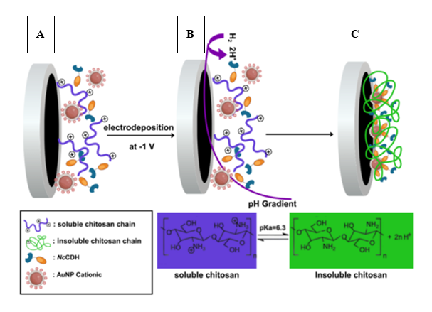
Figure 1 Fabrication of modified-SPCE. Entrapment of NcCDH/Green-AuNPs on the SPCE surface (A) was achieved using chitosan, transitioning from soluble (B) to insoluble (C) form via pH-gradient method.
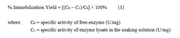
The percentage of the total immobilized enzyme on the SPCE was calculated as % Immobilization Yield as follows:
Lactose detection performance
All electrochemical measurements were performed using a portable potentiostat Galvanoplot GX-102 (Solar Biyoteknoloji, Kordon Kule, Turkey) controlled with GalvanoPlot SUITE software. Unless stated otherwise, all measurements were performed in 100 mM phosphate buffer at pH 6.0 containing 100 mM of KCl. All solutions were purged with nitrogen for 15 min prior to measurements. Three different modified SPCEs (SPCE/NcCDH/Chitosan, SPCE/NcCDH/Chemical-AuNPs/Chitosan, and SPCE/NcCDH/Green-AuNPs/Chitosan) were electrochemically characterized using Cyclic Voltammetry (CV) and Differential Pulse Voltammetry (DPV). The CV measurements were performed at a scan rate of 10 mV/s across a potential window of ‒750 to +750 V to establish baseline current profiles. Subsequent CV analysis with 100 mM lactose solution enabled identification of the oxidation with the net response calculated by subtracting the background currents. DPV measurement was performed at 200 to 700 mV. Calibration curves were created from 3 independent measurements for each point of the curve using varied concentrations of 0 - 100 mM lactose. The calculation of the limit of detection (LOD) was carried out using SigmaPlot 12 (Systat, Chicago, IL, USA) and calculated as the minimum concentration of analyte that can be reliably identified, resulting in a current response greater than the background value plus 3 . Error bars represent the standard deviation of independent measurements obtained from 3 electrodes.
Results and discussion
Extraction of Ocimum americanum leaves
Ocimum americanum leaves contain a spectrum of phytochemicals including phenolics, flavonoids, terpenoids, alkaloids, tannins, saponins, amino acids, and carbohydrates [21,31]. These classes contribute functional groups (e.g., -OH, -C=O, -C=C-, amide, amino, carbonyl-containing moieties) capable of reducing Au3+ to Au0 and simultaneously stabilizing nascent AuNPs via surface binding. The scavenging activity of hydroxyl radical is directly correlated with the antioxidant activity of the phytochemical [32]. In this study, antioxidant activity evaluation was performed by assessing the IC50 value of different preparation methods (Figure 2(A)) and the duration of the extraction using ultrasound-assisted extraction (UAE) method (Figure 2(B)). The antioxidant activity was measured to evaluate the Ocimum americanum leaves’ ability to synthesize nanoparticles. The IC50 value of the extract prepared by dehydrating the leaves has shown the lowest value (163.69 ppm) in comparison to other treatment methods. This value is considered as a moderate activity category [33]. Thus, practically, Ocimum americanum leaves extract is potential reducing agent for the biosynthesis of the gold nanoparticles. The antioxidant properties of the sample depend on the extraction process. Applying drying methods prior to extraction helps to inactivate the enzymes and inhibit the oxidative reaction, thus maintaining the bioactive compounds contained in the sample. Several researchers reported that different drying methods may affect the total phenolic contents, flavonoids, and antioxidant activities of plant extracts [34-36].
Extraction using the UAE probe in various time durations showed that 3-min extraction performed the highest antioxidant activity (IC50 = 83.53), while prolonged extraction up to 10 min showed a decrease in antioxidant activity. This led to the degradation of the phytochemical compound as an effect of excessive exposure to ultrasonic waves [37,38]. Thus, the extract prepared by extracting dry Ocimum americanum leaves using UAE probe for 3 min will be used as a bioreductant for further Green-AuNPs synthesis.
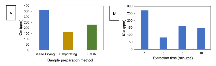
Figure 2 Effect of different preparation conditions of Ocimum americanum leaves extract on the IC50 value. Three different leaves’ preparation methods (A) and 4 different extraction times using ultrasound-assisted extraction (UAE) method (B) were compared.
Characterization of chemically and green-synthesized AuNPs
TEM analysis results showed spherical particles with average sizes of 15.49 ± 2.880 nm for Chemical-AuNPs (Figures 3(A) and 3(C)) and 12.05 ± 4.725 nm for Green-AuNPs (Figures 3(B) and 3(D)). The synthesis of AuNPs using chemical and green reducing agents showed distinct reaction times of 3 and 2.5 min, respectively, as evidenced by color changes from yellow to cherry red (Chemical-AuNPs, Figure 3(E)) and yellow to blue (Green-AuNPs, Figure 3(F)).The UV-Vis spectra results showed a single peak at 521 nm for Chemical-AuNPs, while Green-AuNPs exhibited multiple peaks at 558, 535, and 640 nm, as shown in Figure 3(G). Zeta potential values of ‒60.7 mV (Chemical-AuNPs) and ‒45.4 mV (Green-AuNPs) confirmed good colloidal stability (zeta potential > ±30 mV) in which negative potential values come from the reductor residual as a stabilizing agent [39].
The FT-IR spectrum of AuNPs (Figure 3(H)) chemically synthesized using sodium citrate showed typical absorption bands of carboxylate (-COO-) groups with asymmetric stretching (vas) at 1,516 cm−1 and symmetric stretching (vs) at 1,421 cm−1. These bands indicate a citrate adsorption through carboxylic groups, which is shown by vas and vs vibration that appear around 1,500 - 1,630 cm−1 and 1,305 - 1,415 cm−1, respectively. Typically, citrate will adsorb through oxygen atoms creating a bidentate binding mode, where both oxygen atoms of the carboxylate group coordinate with the gold surface. This confirms that citrate acts as a capping agent through the interaction of its carboxylate groups with Au. A previous study reported that citrate adsorption on gold occurred through both oxygen atoms of the carboxylate group in an η²-COO bridging configuration, with the C–C axis oriented perpendicular to the metal surface [40]. However, in the present study, the presence of both νas and νs bands suggests citrate interaction with Au but does not allow the adsorption configuration to be determined with certainty.
A similar adsorption pattern is observed in AuNPs synthesized with Ocimum americanum extract, where the νas band appears at 1,517 cm⁻¹ and the νs band at 1,422 cm⁻¹ (Figure 3(H)). This suggests that organic compounds in the Ocimum americanum extract, particularly those containing carboxylate or organic acid groups, act similarly to citrate in stabilizing the gold nanoparticles. Therefore, both citrate and natural compounds from Ocimum americanum function as capping agents via –COO⁻ interactions with the Au surface. Chemical-AuNPs displayed monodisperse characteristics dominated by spherical particles, as indicated by the cherry-red colloidal color and single UV-Vis peak [41]. These AuNPs also exhibited higher stability. In contrast, Green-AuNPs showed polydispersity indicated by multiple UV-Vis peaks. Although Green-AuNPs had lower stability compared to Chemical-AuNPs, they maintained adequate colloidal stability. TEM size distribution analysis (Figures 3(A) and 3(B)) revealed that Chemical-AuNPs were larger in size but more uniformly distributed than Green-AuNPs. These size differences are expected to influence electron transfer efficiency in biosensor applications.
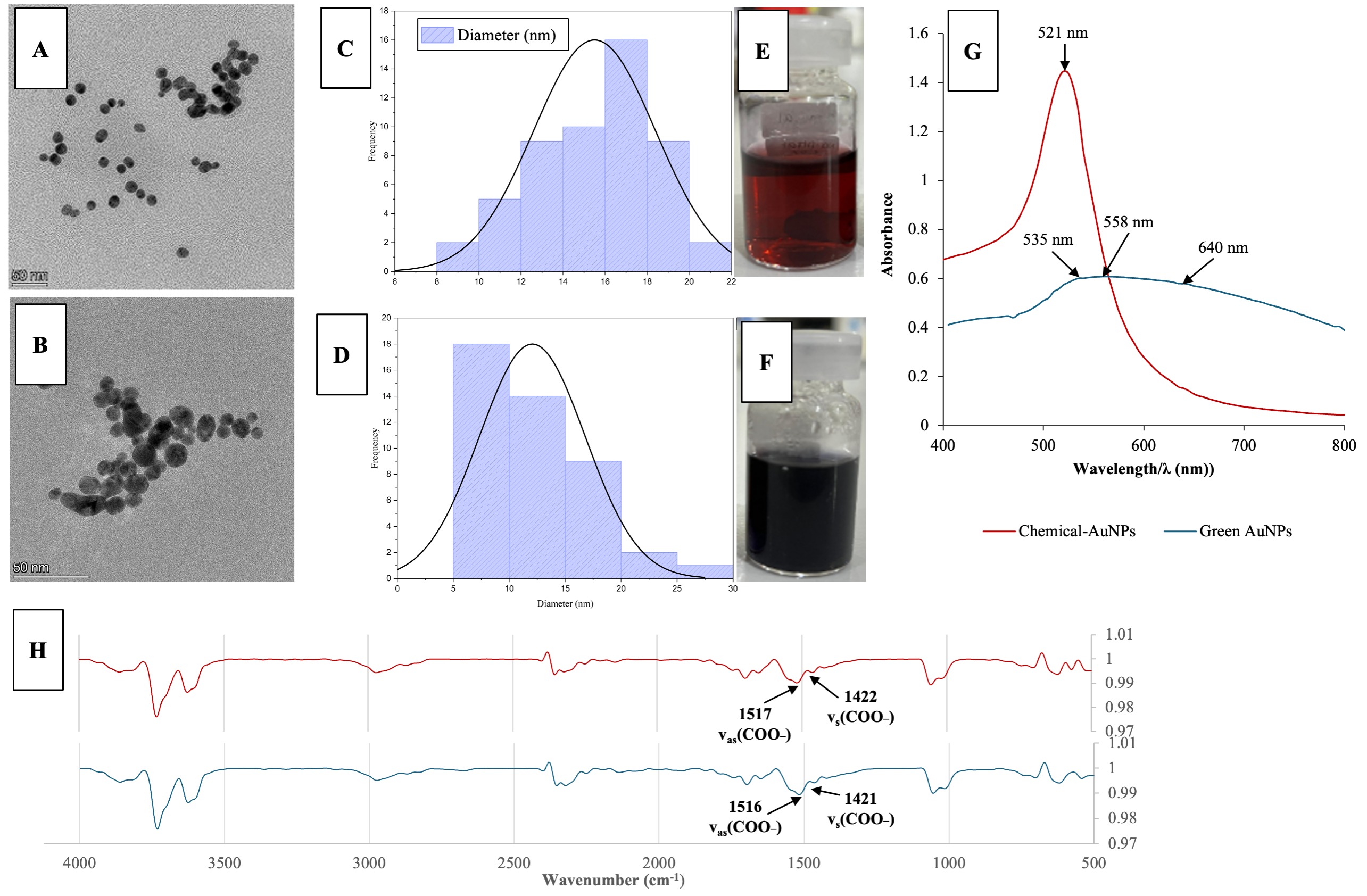
Figure 3 TEM Image Chemical-AuNPs (A) and TEM Image Green-AuNPs (B), Particle Size Distribution of Chemical-AuNPs (C), Particle Size Distribution of Green-AuNPs, Chemical AuNPs Colloid Color (E) Green AuNPs Colloid Color (F), UV-Spectra Curve of Chemical-AuNPs (red line) and Green-AuNPs (blue line) (G) FT-IR of Chemical AuNPs (red) and Green AuNPs (blue) (H).
Effect of chitosan concentration on immobilization of NcCDH/AuNPs onto SPCE
Figure 4(A) shows that 0.5% (v/v) chitosan exhibited the highest immobilization yield of 40.51 ± 3.56%, while 0.1% (v/v) chitosan gave the lowest (19.42 ± 2.96%). Mechanistically, the improvement at 0.2% was attributed to the formation of a stable, porous chitosan film optimal for enzyme entrapment. The decline at 0.3% resulted from increased viscosity disrupting film stability [42] and finally the rise at 0.4% - 0.5% correlated with denser matrix formation. Higher concentrations of chitosan increased the number of positively charged ‒NH3+ groups, allowing electrostatic interaction with the negatively charged enzymes/AuNPs [12]. This was supported by ionotropic crosslinking between chitosan amine groups and the electroactive species [16]. The pH gradient properties of chitosan also facilitate on making the porous hydrogel such that entrapment may occur efficiently. Nevertheless, high concentration may slow down the diffusion of substrate, therefore lowering the activity of the enzymes [42].
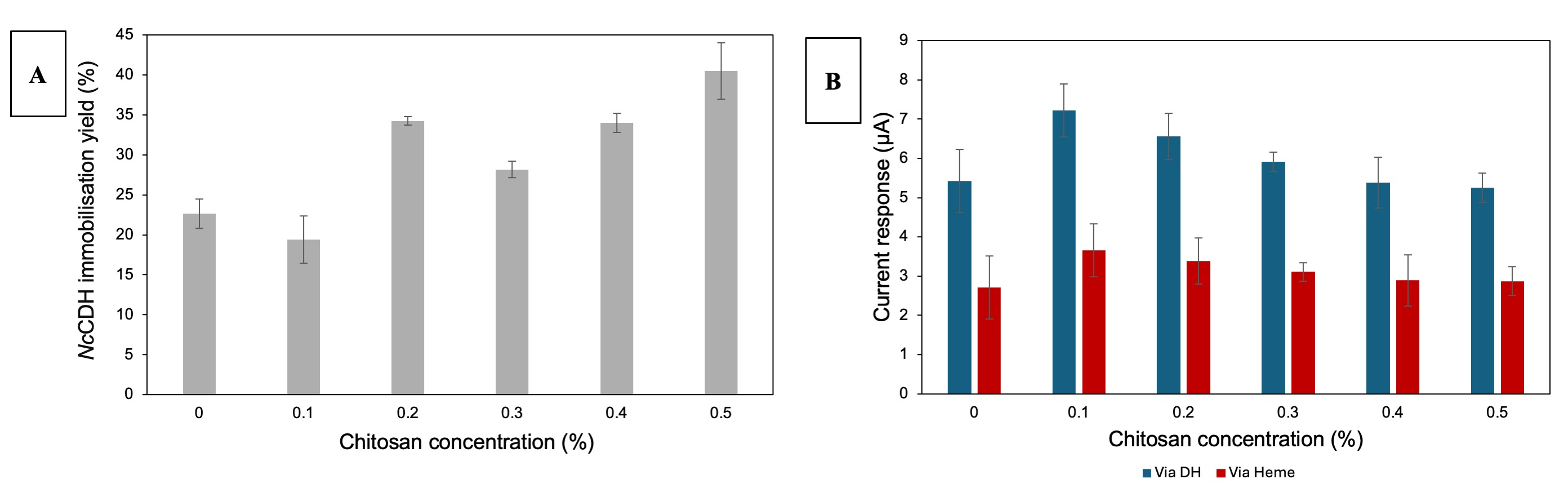
Figure 4 Effect of Chitosan on NcCDH Immobilization Yield (A) and Electrochemical Response (B). The data represent average values from 3 different measurements, and the standard deviation is indicated by error bar.
Electron transfer efficiency was evaluated using cyclic voltammetry (CV) with 100 mM lactose as the substrate and 1 mM DCIP as the redox mediator at a scan rate of 10 mV/s between potentials of ‒750 and 750 mV | Ag vs AgCl (sat. KCl). The net current response was calculated by subtracting the peak current of the blank solution from that of the lactose-containing solution. The modified biosensor showed electron transfer behaviors through multiple peaks in the CV result, establishing 2 electron transfer pathways via flavin adenine dinucleotide (FAD) in the DH domain and heme b in cyt domain. The highest current response was observed at 0.1% chitosan (7.22 ± 0.67 μA), followed by a significant decrease at higher concentrations: 6.56 ± 0.59 μA (0.2%), 5.91 ± 0.24 μA (0.3%), 5.38 ± 0.65 μA (0.4%) and highest decrease was noted at 0.5% chitosan (5.25 ± 0.37 μA) via FAD as shown in Figure 4(B).
This trend is consistent with the findings of [42], who reported that thicker chitosan layers hinder substrate diffusion and thus reduce sensor performance. The decline in DH domain performance at higher chitosan concentrations can be explained through several mechanisms. First, as revealed by [17], increased solution viscosity and the formation of thicker chitosan layers at higher concentrations create substrate diffusion barriers and prolong electron transfer distances. Second, the characteristics of entrapment immobilization methods tend to produce uneven enzyme distribution and orientation. This contributes to prolonged electron transfer distances and reduced electron transfer effectiveness [43].
In contrast, the heme domain exhibited a more stable response profile, with the highest peak value of 7.08 ± 0.82 μA at a 0.1% concentration and limited fluctuations within the range of 6.50 - 6.63 μA for concentrations of 0.2% - 0.5%. The heme domain shows enhanced electron transfer efficiency and stability at higher polymer concentrations [44]. This stability is primarily attributed to strong electrostatic ionotropic crosslinking interactions between the NH₃⁺ groups in chitosan and the negatively charged heme domain [16].
The difference between electron transfer performance via FAD and heme b highlights NcCDH’s electrochemical properties and how its performance is influenced by chitosan concentration. Aligned with CV result, DH domain is more sensitive to changes in the matrix environment compared to the cyt domain, which is more robust due to electrostatic interaction between the heme domain and the chitosan matrix [12]. The superior biosensor performance at 0.1% chitosan is mainly attributed to the formation of a thinner chitosan layer, which minimizes substrate diffusion barriers while maintaining efficient electron transfer across both domains at the electrode. Higher concentration of chitosan generally increases network density, reduce pore size and lengthen diffusion pathways for substrates to access the enzyme. This produces slow diffusion response and lower the apparent reaction rates [45]. The selected concentration of chitosan was based on finding a balance between how well it immobilizes NcCDH and how well it transfers electrons. Higher chitosan concentration (0.5%) led to better immobilization efficiency, but they also made the polymer layer thicker, which can make it harder for electrons to move because of higher diffusion resistance and conductivity. Therefore, the 0.1% chitosan concentration was selected for subsequent experiments.
AuNPs effect on NcCDH/chitosan electrode electrochemistry
Cyclic voltammetry (CV) was conducted to evaluate the effect of gold nanoparticle (AuNP) incorporation on the electrochemical properties of immobilized NcCDH in a chitosan matrix. Three types of SPCEs were fabricated: NcCDH/Chitosan (Control), NcCDH/Chemical-AuNPs/Chitosan, and NcCDH/Green-AuNPs/Chitosan. The immobilized enzyme percentages on the SPCEs were 30.06%, 35.36%, and 34.94%, respectively. NcCDH/Chemical-AuNPs/Chitosan showed the highest percentage due to its monodisperse nature and higher zeta potential compared to NcCDH/Green-AuNPs/Chitosan, though both AuNPs-modified electrodes exhibited higher values than the control without AuNPs. The uniform distribution and surface charge of AuNPs enabled more homogeneous dispersion on the electrode surface, providing a larger and more optimal contact area for NcCDH enzyme binding [18].
Electrochemical properties evaluation using CV revealed multiple peaks indicating electron transfer occurs on both NcCDH domains on different electron transfer pathways. Each electrode produced different buffer values due to DCIP mediator accumulation, so currents were normalized to compare peak currents in the CV curves. The results demonstrated that AuNPs significantly influenced the current response in both domains. Figure 5 presents a bar chart showing the oxidation peak currents for each variation.
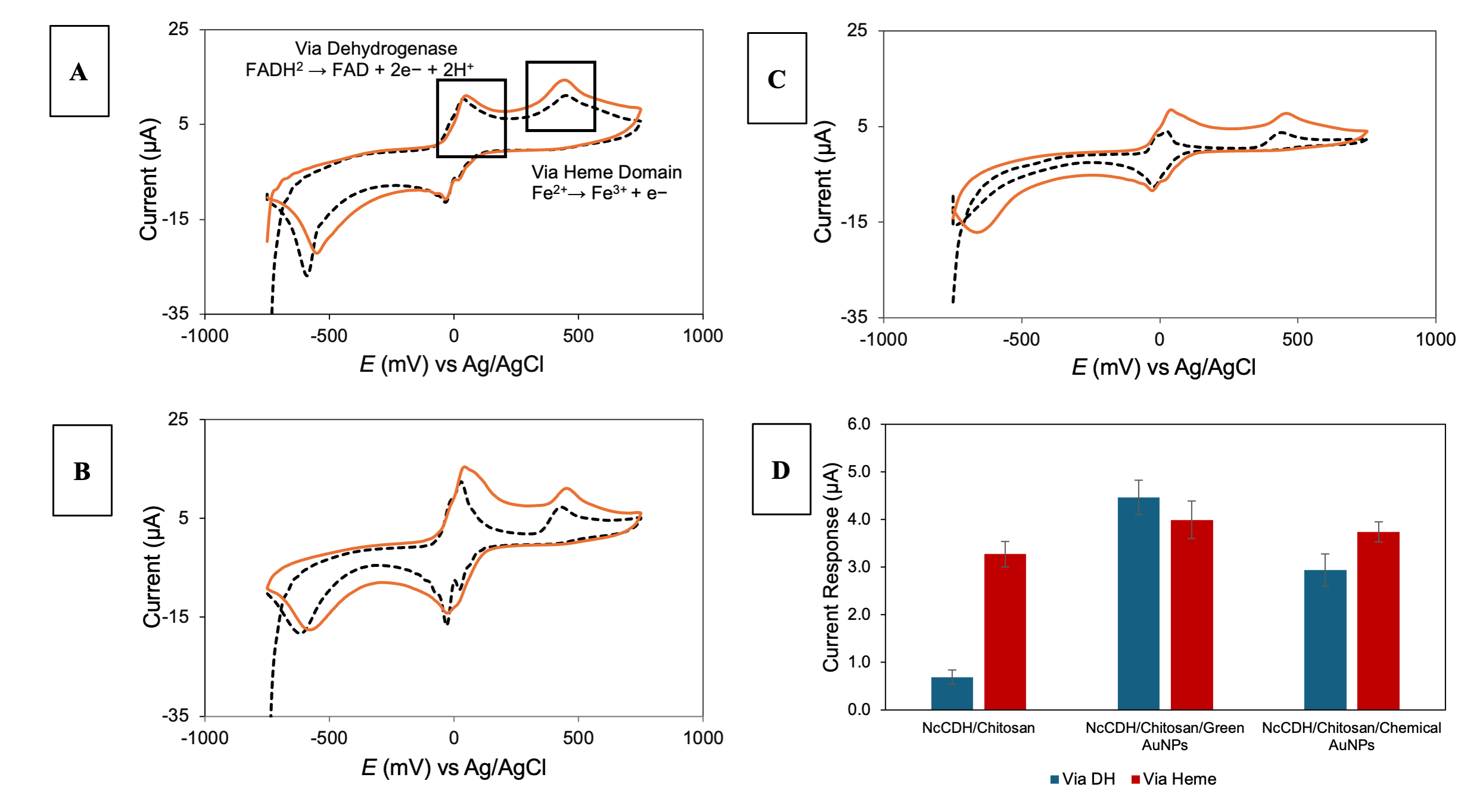
Figure 5 Cyclic voltammogram of NcCDH/Chitosan (A), NcCDH/Green-AuNPs/ Chitosan (B), NcCDH/Chemical-AuNPs/Chitosan (C) in the absence of (dashed black) and in the presence of (orange) 100 mM lactose, peak current response values for NcCDH/Chitosan modified electrodes with and without AuNPs (D). The data represent average values from 3 different measurements, and the standard deviation is indicated by error bar.
The control electrode (NcCDH/Chitosan) generated currents of 0.69 and 3.28 μA (Figures 5(A) and 5(D)), while the Green-AuNPs electrode achieved 4.46 and 3.99 μA (Figures 5(B) and 5(D)), and Chemical-AuNPs electrode generated the highest result of 2.94 and 3.74 μA (Figures 5(C) and 5(D)). Compared to the control, the SPCE/NcCDH/Chemical-AuNPs/Chitosan exhibited current increases of 327.77% and 14.23% via FAD and cyt, respectively. Meanwhile, the SPCE NcCDH/Green-AuNPs/Chitosan showed increases of 549.74% and 14.23%. These results confirm that AuNPs enhance electron transfer efficiency between the enzyme and electrode surface. In the MET system, AuNPs function as electron bridges, which reduce electron hopping distance. AuNPs also increase electrode active surface area and enhanced conductivity though higher conductive molecule density. These properties validate AuNPs’ effectiveness in the MET system [12].
Although both AuNP types improved currents compared to the control, performance differences were observed between synthesis methods. Green-AuNPs yielded higher peak current responses than Chemical-AuNPs. When plant-based reductants are used to make Green-AuNPs, the particles have phytochemical surface functions that help redox enzymes transfer electrons to the electrode more quickly. This makes the electrochemical response and overall biosensor performance better than chemically synthesized AuNPs. The better performance is also due to variations in physicochemical features, especially the smaller particle size, better distribution and better surface stability of Green-AuNPs [18,46]. For Green-AuNPs, phytochemical compounds work as both reducing and capping agents. This makes the nanoparticles smaller but more polydisperse. This smaller size can enlarge the active surface area and shortens the distance that electron have to travel between the redox centers of NcCDH (FAD and heme domains), therefore accelerates electron transfer.
Analytical performance of lactose biosensor
To assess the analytical performance of the fabricated NcCDH/AuNPs/Chitosan biosensor, a various range of lactose concentrations (0, 10, 20, 30, 40, 50, 60, 70, 80, 90, and 100 mM) were applied as depicted in Figure 6. DPV was performed to evaluate the dynamic concentration range as well as the sensitivity and limit of detection (LoD) of the biosensor (Figures 6(A) and 6(C)). The calibration curve was then prepared by plotting the current density (y) vs lactose concentrations (x) (Figures 6(B) and 6(D)). The calibration curve of both biosensors showed the same dynamic range of 10 - 90 mM with correlation equations of y = 2.6689x - 32.066 (R² = 0.9914) for the biosensor incorporated with chemically synthesized AuNPs and y = 2.464x - 22.511 (R² = 0.9957) for the biosensor using green-AuNPs. The lactose sensitivity of NcCDH/Chemical-AuNPs/Chitosan (2.669 μA mM−1 cm-2) was higher than that of NcCDH/Green-AuNPs/Chitosan (2.464 μA mM−1 cm-2). The LoD of NcCDH/Green-AuNPs/Chitosan was found to be lower (8.921 mM) than NcCDH/Chemical-AuNPs/Chitosan (12.015 mM). These detection limits were considered higher than that of biosensors published in other studies, as represented in Table 1. The higher sensitivity observed in the biosensor incorporating Chemical-AuNPs can be due to their more uniform size distribution which facilitate more efficient and consistent enzyme immobilization and electron transfer. On the other hand, Green-AuNPs are smaller which may improve the electron transport, but their phytochemical-derived surface coatings may partially hinder electron transfer pathways. However, the same surface bio-functionalities can act as mediators or help enzymes orient themselves in a way that lowers interfacial resistance and raises the effective electroactive surface area [47,48].
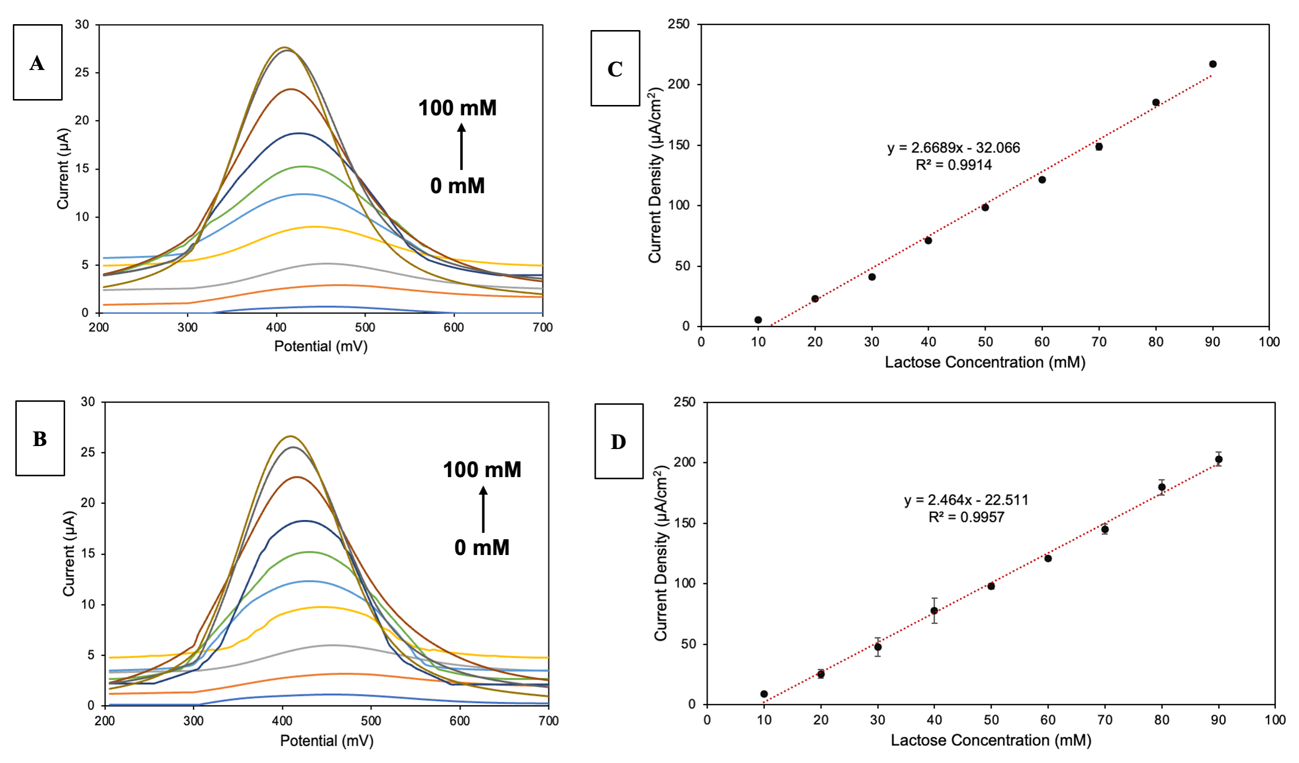
Figure 6 The DPV response and calibration curve of NcCDH/Chemical-AuNPs/Chitosan (A, C) and NcCDH/Green-AuNPs/Chitosan biosensor (B, D) towards 0 - 100 mM lactose in phosphate buffer (100 mM, pH 6.0 containing 100 mM of KCl). The data represent average values from 3 different biosensors measurements, and the standard deviation is indicated by error bar.
Table 1 Comparison of the analytical performance of different electrochemical enzyme-based biosensors for detection of lactose.
| System | Electrochemical technique | Potential of detection (mV) | LoD (mM) | Linear range (mM) | Real sample | Ref. |
|---|---|---|---|---|---|---|
| SPCENcCDH/Green-AuNPs/Chitosan | Voltammetry | 200 - 700 | 8.921 | 10 - 90 | Not applicable | This study |
| MWCNT-RuO2/Gox-β-Gal | Chronoamperometry | 400 | 0.036 | 0.5 - 3.0 | Semi-skim milk | [49] |
| TvCDH/AuNPs/graphite | Amperometry | 250 | 0.35 | 10 - 300 | Not applicable | [48] |
| PsCDH NH2-PD/SWCNT-GC | Amperometry | 200 | 0.5 | 1 - 150 | Not applicable | [50] |
| Pt/Chit/GOxβ-gal/Chit/GA | Constant Potential Coulometry | 650 | 1.29 | 2.47 - 40 | whey permeates | [51] |
| GOx-β-Gal@AuNPs-g-C3N4-MnO2-TiO2/ITO | PEC amperometry | ‒400 | 0.023 | 0.008 - 2.5 | Lactose free milk | [52] |
| CtCDH/AuNPs/BPDT/AuE | FIA amperometry | 250 | 0.3 | 0.5 - 40 | 2% low fat milk, light cream, whipping cream, 80% lactose reduced milk | [18] |
Storage stability
The storage stability of the electrode was evaluated using the same modified electrode over 21 consecutive days. When the modified electrode was not used, the electrode was stored at 4 C under constant humidity in a closed container. The control SPCE without AuNPs showed a current response decrease by about 10% after 2 weeks of storage compared to initial current response and finally declined to 25% by the third week of storage, while NcCDH/Green-AuNPs/Chitosan dropped to only 5.5% after 2 weeks and finally dropped to 10% by day 21.
NcCDH/Chemical-AuNPs/Chitosan also showed comparable storage stability of 9.5% decrease by 21 days of storage. Through this study, we assumed that the AuNPs in the modified electrode were found to be strongly embedded in the NcCDH/Chitosan matrix, assisting the electron transfer. These findings indicate that AuNPs not only enhance electrochemical activity but also maintain the stability and performance of the biosensor.
Possible sensing mechanism
To better illustrate the sensing mechanism of the proposed biosensor, a schematic representation is provided in Figure 7. The sensing mechanism relies on the catalytic function of cellobiose dehydrogenase (NcCDH), which oxidizes lactose at the FAD site within the dehydrogenase (DH) domain, resulting the formation of lactobionate and the reduction of FAD to FADH2. In the cytochrome domain, the electrons are then moved to heme b through internal electron transfer (IET). From this point on, electrons are sent to the electrode either directly (DET) or by a mediated (MET) pathway using 2,6-dichlorophenolindophenol (DCIP). DCIP works as an electron shuttle in the mediated system which takes electrons from the reduced enzyme and is re-oxidized at the electrode surface. This creates a quantifiable current signal that is directly proportional to the lactose concentration.
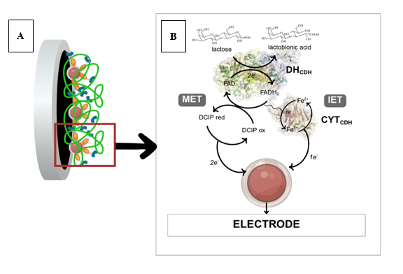
Figure 7 Schematic illustration of SPCE/NcCDH/Green-AuNPs/Chitosan biosensor sensing mechanism toward lactose. Immobilization of NcCDH/Green-AuNPs/Chitosan onto the SPCE (A) and possible electron transfer mechanism from the oxidation of lactose as an analyte to the SPCE surface (B).
Conclusions
This study demonstrated that both chemically and green-synthesized AuNPs can effectively enhance the electrochemical performance of NcCDH-chitosan modified electrodes for lactose detection. While Chemical-AuNPs exhibits mono-dispersity and uniform distribution size, Green-AuNPs yields a higher current response despite its polydispersity and lower negative charge. These different properties are because of the smaller size of the Green-AuNPs particle, therefore increasing the electron transfer and conductivity. The natural active compound in Ocimum americanum extract utilized for green synthesis may enhance electron transfer efficiency, offsetting the challenges associated with particle uniformity. In addition to this analytical performance study, more research into the structure and property relationships of AuNPs-based system is needed to better understand how the shape, surface chemistry, and dispersion of nanoparticles affect electron transfer mechanisms and long-term stability. Regarding the application in dairy products such as milk, the biosensor is incapable of detecting the lactose content for monitoring the lactose-free milk due to its high detection limit but is recommended to be applied in daily rapid assays for checking the lactose content in milk. Nevertheless, the results show that plant-based AuNPs could be an alternative replacement for chemically synthesized nanoparticles in biosensor applications. This is in line with green nanotechnology principles and does not affect the performance.
Acknowledgements
JLL acknowledges UB STARS program (00612/UN10.A0101/B/TU/2025), incorporation with National Research and Innovation Agency (BRIN), Indonesia for facilitating the research. The authors gratefully acknowledge Roland Ludwig and Kwankao Karnpakdee from BOKU University, Vienna 1190, Austria for providing the NcCDH enzyme. WS acknowledges the Research Center for Catalysis, Research Organization for Nanotechnology and Materials (ORNM), National Research and Innovation Agency (BRIN), Indonesia to permit to conduct postdoctoral at the Interdisciplinary Research Center for Hydrogen Technologies and Carbon Management (IRC-HTCM), King Fahd University of Petroleum and Minerals (KFUPM), Dhahran 31261, Saudi Arabia as part of the institutionʼs capacity building.
Declaration of Generative AI in Scientific Writing
The preparation of this manuscript did not use any generative AI tools. No content or data interpretation was executed by generative AI. All authors are responsible for the content and conclusion of this work.
CRediT Author Statement
Sudarma Dita Wijayanti: Conceptualization, Supervision, Writing - review&editing, Project administration; Funding acquisition. Josephine Laurentia Liem: Investigation; Methodology; Formal Analysis; Data Curation, Writing-original draft and editing. Haya Nurhidayah: Investigation. Wirya Sarwana: Data curation; Formal Analysis, Writing - review&editing. Yulizah Hanifah: Data curation; Formal Analysis. Fenty Nurtyastuti Eka Pertiwi: Writing - review. Nur Ida Panca Nugrahini: Writing - review, Supervision. Wisnu Arfian Anditya Sudjarwo: Data Curation, Formal Analysis. All authors read and approved the final manuscript.
References
[1] N Bhalla, P Jolly, N Formisano and P Estrela. Introduction to biosensors. Essays in Biochemistry 2016; 60(1), 1-8.
[2] X Wang, J Zhou and H Wang. Bioreceptors as the key components for electrochemical biosensing in medicine. Cell Reports Physical Science 2024; 5(2), 101801.
[3] SD Wijayanti, L Tsvik and D Haltrich. Recent advances in electrochemical enzyme-based biosensors for food and beverage analysis. Foods 2023; 12(18), 3355.
[4] OY Dudchenko, VM Pyeshkova, OO Soldatkin, B Akata, BO Kasap, AP Soldatkin and SV Dzyadevych. Development of silicalite/glucose oxidase-based biosensor and its application for glucose determination in juices and nectars. Nanoscale Research Letters 2016; 11, 59.
[5] S Dominici, F Marescotti, C Sanmartin, M Macaluso, I Taglieri, F Venturi, A Zinnai and MS Facioni. Lactose: Characteristics, food and drug-related applications, and its possible substitutions in meeting the needs of people with lactose intolerance. Foods 2022; 11(10), 1486.
[6] C Sygmund, D Kracher, S Scheiblbrandner, K Zahma, AKG Felice, W Harreither, R Kittl and R Ludwig. Characterization of the two Neurospora crassa cellobiose dehydrogenases and their connection to oxidative cellulose degradation. Applied and Environmental Microbiology 2012; 78(17), 6161-6171.
[7] G Kovacs, R Ortiz, V Coman, W Harreither, IC Popescu, R Ludwig and L Gorton. Graphite electrodes modified with Neurospora crassa cellobiose dehydrogenase: Comparative electrochemical characterization under direct and mediated electron transfer. Bioelectrochemistry 2012; 88, 84-91.
[8] P Bollella, R Ludwig and L Gorton. Cellobiose dehydrogenase: Insights on the nanostructuration of electrodes for improved development of biosensors and biofuel cells. Applied Materials Today 2017; 9, 319-332.
[9] R Ludwig, W Harreither, F Tasca and L Gorton. Cellobiose dehydrogenase: A versatile catalyst for electrochemical applications. ChemPhysChem 2010; 11(13), 2674-2697.
[10] F Csarman, L Wohlschlager and R Ludwig. Cellobiose dehydrogenase. Elsevier, Netherland, 2020, p. 457-489.
[11] A Giorgianni, A Zenone, L Sützl, F Csarman and R Ludwig. Exploring class III cellobiose dehydrogenase: Sequence analysis and optimized recombinant expression. Microbial Cell Factories 2024; 23, 146.
[12] M Tavahodi, R Ortiz, C Schulz, A Ekhtiari, R Ludwig, B Haghighi and L Gorton. Direct electron transfer of cellobiose dehydrogenase on positively charged polyethyleneimine gold nanoparticles. ChemPlusChem 2016; 82(4), 546-552.
[13] S Ma, CVFP Laurent, M Meneghello, J Tuoriniemi, C Oostenbrink, L Gorton, PN Bartlett and R Ludwig. Direct electron-transfer anisotropy of a site-specifically immobilized cellobiose dehydrogenase. ACS Catalysis 2019; 9(8), 7607-7615.
[14] B Motycka, F Csarman, M Rupp, K Schnabel, G Nagy, K Karnpakdee, S Scheiblbrandner, R Tscheliessnig, C Oostenbrink, M Hammel and R Ludwig. Amino acid residues controlling domain interaction and interdomain electron transfer in cellobiose dehydrogenase. ChemBioChem 2023; 24(22), e202300431.
[15] T Tangkuaram, C Ponchio, T Kangkasomboon, P Katikawong and W Veerasai. Design and development of a highly stable hydrogen peroxide biosensor on screen printed carbon electrode based on horseradish peroxidase bound with gold nanoparticles in the matrix of chitosan. Biosensors and Bioelectronics 2007; 22(9-10), 2071-2078.
[16] H Liu, D Lu, P Li, Y Chen, Y Zhou and T Lu. One-step electrodeposition of chitosan/phosphonate iron(III) hybrids film and its pH-controlled switchable electrocatalytic behavior. Journal of Electroanalytical Chemistry 2014; 717-718, 125-130.
[17] Y Tan, W Deng, C Chen, Q Xie, L Lei, Y Li, Z Fang, M Ma, J Chen and S Yao. Immobilization of enzymes at high load/activity by aqueous electrodeposition of enzyme-tethered chitosan for highly sensitive amperometric biosensing. Biosensors and Bioelectronics 2010; 25(12), 2644-2650.
[18] P Bollella, F Mazzei, G Favero, G Fusco, R Ludwig, L Gorton and R Antiochia. Improved DET communication between cellobiose dehydrogenase and a gold electrode modified with a rigid self-assembled monolayer and green metal nanoparticles: The role of an ordered nanostructuration. Biosensors and Bioelectronics 2017; 88, 196-203.
[19] M Huston, M DeBella, M DiBella and A Gupta. Green synthesis of nanomaterials. Nanomaterials 2021; 11(8), 2130.
[20] MA Qaeed, A Hendi, AS Obaid, AA Thahe, AM Osman, A Ismail, A Mindil, AA Eid, F Aqlan, NMA Osman, A AL-Farga, SM Al-Maaqar and AA Saif. The effect of different aqueous solutions ratios of Ocimum basilicum utilized in AgNPs synthesis on the inhibition of bacterial growth. Scientific Reports 2023; 13, 5866.
[21] BT Aluko, OI Oloyede and AJ Afolayan. Polyphenolic contents and free radical scavenging potential of extracts from leaves of Ocimum americanum L. Pakistan Journal of Biological Sciences 2013; 16(1), 22-30.
[22] AKA Genfi, C Larbie, BO Emikpe, AA Oyagbemi, CK Firempong and CO Adjei. Modulation of oxidative stress and inflammatory cytokines as therapeutic mechanisms of Ocimum americanum L. extract in carbon tetrachloride and acetaminophen-induced toxicity in rats. Journal of Evidence-Based Integrative Medicine 2020; 25, 1-13.
[23] M Shanaida, O Golembiovska, I Jasicka-Misiak, O Oleshchuk, N Beley, I Kernychna and PP Wieczorek. Sedative effect and standardization parameters of herbal medicinal product obtained from the Ocimum americanum L. herb. European Pharmaceutical Journal 2021; 68(1), 1-9.
[24] G Zengin, C Ferrante, DE Gnapi, KI Sinan, G Orlando, L Recinella, A Diuzheva, J Jekő, Z Cziáky, A Chiavaroli, S Leone, L Brunetti, C Picot-Allain, MF Mahomoodally, P Angelini, S Covino, R Venanzoni, B Tirillini and L Menghini. Comprehensive approaches on the chemical constituents and pharmacological properties of flowers and leaves of American basil (Ocimum americanum L.). Food Research International 2019; 125, 108610.
[25] B Islam, D Samir, C Sara and N Janetta. Ocimum basilicum L. leaves extract-mediated green synthesis of MnO NPs: Phytochemical profile, characterization, catalytic and thrombolytic activities. Results in Surfaces and Interfaces 2024; 17, 100284.
[26] N Aggarwal, P Mehta, J Bhambra, A Sharma, D Ali, NK Sharma, S Sharma, G Kumar and N Kumar. Biosynthesis of Ocimum basilicum leaf extract mediated zinc oxide nanoparticles and their cytotoxic activity. Biomass Conversion and Biorefinery 2023; 13, 8373-8380.
[27] DB Manikandan, A Sridhar, RK Sekar, B Perumalsamy, S Veeran, M Arumugam, P Karuppaiah, T Ramasamy. Green fabrication, characterization of silver nanoparticles using aqueous leaf extract of Ocimum americanum (Hoary Basil) and investigation of its in vitro antibacterial, antioxidant, anticancer and photocatalytic reduction. Journal of Environmental Chemical Engineering 2021; 9, 104845.
[28] İ Gulcin and SH Alwasel. DPPH radical scavenging assay. Processes 2023; 11(8), 2248.
[29] TP Patil, AA Vibhute, SL Patil, TD Dongale and AP Tiwari. Green synthesis of gold nanoparticles via Capsicum annum fruit extract: Characterization, antiangiogenic, antioxidant and anti-inflammatory activities. Applied Surface Science Advances 2023; 13, 100372.
[30] AEF Oliveira, AC Pereira, MAC Resende and LF Ferreira. Gold nanoparticles: A didactic step-by-step of synthesis using Turkevich method, mechanisms and characterization. Analytica 2023; 4(2), 250-263.
[31] I Jasicka-Misiak, M Shanaida, N Hudz and PP Wieczorek. Phytochemical and pharmacological evaluation of the residue by-product developed from the Ocimum americanum (Lamiaceae) postdistillation waste. Foods 2021; 10(12), 3063.
[32] ADS Backiam, S Duraisamy, P Karuppaiya, S Balakrishnan, A Sathyan, A Kumarasamy and A Raju. Analysis of the main bioactive compounds from Ocimum basilicum for their antimicrobial and antioxidant activity. Biotechnology and Applied Biochemistry 2023; 70(6), 2038-2051.
[33] A Itam, MS Wati, V Agustin, N Sabri, RA Jumanah and M Efdi. Comparative study of phytochemical, antioxidant, and cytotoxic activities and phenolic content of Syzygium aqueum (Burm. f. Alston f.) extracts growing in West Sumatera Indonesia. The Scientific World Journal 2021; 2021, 5537597.
[34] SB Rad, H Mumivand, S Mollaei and A Khadivi. Effect of drying methods on phenolic compounds and antioxidant activity of Capparis spinosa L. fruits. BMC Plant Biology 2025; 25, 133.
[35] DM Cuong, DK Kim, MK Ediriweera, JE Park, JY Moon and SK Cho. Effects of drying method and extraction solvent on antioxidant and anti-inflammatory activity of Melosira nummuloides bioproducts. Applied Biological Chemistry 2023; 66, 59.
[36] NPE Hikmawanti, S Fatmawati, Z Arifin, N Cahyaningrum and MA Fauzan. The effect of pre-extraction preparation on antioxidant compounds of Sauropus androgynus (L.) Merr. leaves extracts. Pharmaceutical Sciences and Research 2021; 8(3), 128-135.
[37] ASM Sayem, T Ahmed, MUK Mithun, M Rashid and MR Rana. Optimising ultrasound-assisted extraction conditions for maximising phenolic, flavonoid content and antioxidant activity in hog plum peel and seed: A response surface methodology approach. Journal of Agriculture and Food Research 2024; 18, 101312.
[38] MI Prawira-Atmaja and S Puangpraphant. Effects of ultrasound-assisted bath, probe, and extraction time on bioactive compounds and antioxidant activity in white tea (Camellia sinensis) extracts from purple- and green-leaf cultivars. Journal of the Saudi Society of Agricultural Sciences 2025; 24, 10.
[39] MA Ali, TAS Eldin, GME Moghazy, IM Tork and II Omara. Detection of E. coli O157:H7 in feed samples using gold nanoparticles sensor. International Journal of Current Microbiology and Applied Sciences 2014; 3(6), 697-708.
[40] JM Gisbert-González, W Cheuquepán, A Ferre-Vilaplana, E Herrero and JM Feliu. Citrate adsorption on gold: Understanding the shaping mechanism of nanoparticles. Journal of Electroanalytical Chemistry 2020; 875, 114015.
[41] AT Algahiny, OS Elmitwalli, DA Kassim, GR Deen, S Akhtar and FZ Henari. Gum Arabic-assisted green synthesis of gold nanoparticles as fluorescence modulator for potential analytical applications. Exploratory BioMat-X 2024; 1, 190-201.
[42] A Dabhade, S Jayaraman and B Paramasivan. Development of glucose oxidase-chitosan immobilized paper biosensor using screen-printed electrode for amperometric detection of Cr(VI) in water. 3 Biotech 2021; 11, 183.
[43] VP Hitaishi, R Clement, N Bourassin, M Baaden, AD Poulpiquet, S Sacquin-Mora, A Ciaccafava and E Lojou. Controlling redox enzyme orientation at planar electrodes. Catalysts 2018; 8(5), 192.
[44] K Karnpakdee, D Kracher and R Ludwig. Electron transfer of cellobiose dehydrogenase in polyethyleneimine films. ChemElectroChem 2024; 11(8), 202300679.
[45] A Chmayssem, I Shalayel, S Marinesco and A Zebda. Investigation of GOx stability in a chitosan matrix: Applications for enzymatic electrodes. Sensors 2023; 23(1), 465.
[46] DM Radulescu, VA Surdu, A Ficai, D Ficai, AM Grumezescu and E Andronescu. Green synthesis of metal and metal oxide nanoparticles for recent applications. International Journal of Molecular Sciences 2023; 24(20), 15397.
[47] M Popescu and C Ungureanu. Biosensors in food and healthcare industries: Bio-Coatings based on biogenic nanoparticles and biopolymers. Coatings 2023; 13(3), 486.
[48] P Bollella, C Schulz, G Favero, F Mazzei, R Ludwig, L Gorton and R Antiochia. Green synthesis and characterization of gold and silver nanoparticles and their application for development of a third generation lactose biosensor. Electroanalysis 2016; 29(1), 77-86.
[49] S Garehbaghi, V Adam, J Přibyl, L Richtera and AM Ashrafi. An enzyme cascade biosensor based on multiwalled carbon nanotube-RuO2 nanocomposite for selective amperometric determination of lactose in milk samples. Microchemical Journal 2024; 204, 111138.
[50] F Tasca, R Ludwig, L Gorton and R Antiochia. Determination of lactose by a novel third generation biosensor based on a cellobiose dehydrogenase and aryl diazonium modified single wall carbon nanotubes electrode. Sensors and Actuators B: Chemical 2013; 177, 64-69.
[51] G Halpin, S McEntee, C Dwyer, F Lawless and E Dempsey. Lactose biosensor development and deployment in dairy product analysis. Journal of The Electrochemical Society 2022; 169(3), 037528.
[52] B Çakıroğlu, YC Demirci, E Gökgöz and M Özacar. A photoelectrochemical glucose and lactose biosensor consisting of gold nanoparticles, MnO2 and g-C3N4 decorated TiO2. Sensors and Actuators B: Chemical 2019; 282, 282-289.