
Trends
Sci.
2026;
23(11):
13671
Thermostable Amylase from Bacterial Isolate ATF2IV8 of Wae Pellae Hot Spring, Sinjai, Indonesia: Production, Partial Purification and Characterisation
Fitria
Azis1,3,
 ,
Hasnah Natsir2,*,
,
Nur Asmi2,
,
Rugaiyah A. Arfah2,
,
Paulina
Taba2,
,
Abdul Wahid Wahab2,
Amalyah Febryanti3,
,
Maswati
Baharuddin3,
,
Sitti Chadijah3,
,
Iin Novianty3,
and Anita4,
1Magister Student, Department of Chemistry, Faculty of Mathematics and Natural Science, Hasanuddin University, South Sulawesi 90245, Indonesia
2Departement of Chemistry, Faculty of Mathematics and Natural Science, Hasanuddin University,
South Sulawesi 90245, Indonesia
3Department of Chemistry, Faculty of Science and Technology, Universitas Islam Negeri Alauddin Makassar,
South Sulawesi 92118, Indonesia
4Medical Laboratory Technology, Polytechnic Muhammadiyah Makassar, South Sulawesi 90132, Indonesia
(*Corresponding author’s e-mail: [email protected])
Received: 23 February 2026, Revised: 17 April 2026, Accepted: 24 April 2026, Published: 20 June 2026
Abstract
Amylase enzymes isolated from thermophilic bacteria have significant industrial applications due to their thermostability and ability to mantain catalytic activity at high temperatures. This study aimed to identify the bacterial isolate ATF2IV8 from Wae Pellae hot spring, produce amylase, and characterise its biochemical properties. Molecular identification of the ATF2IV8 through 16S rRNA gene sequencing showed a close relationship with Calidifontibacillus sp. The crude enzyme was fractionated using ammonium sulfate (0% - 20%, 20% - 40%, 40% - 60%, 60% - 80%, and 80% - 100% saturation), with fraction F4 showing the highest activity. Dialysis of the F4 increased the specific activity approximately sixfold compared to the crude extract. Biochemical characterisation revealed an optimal temperature of 70 °C, an optimal pH of 7, and an optimal substrate concentration of 5%. The enzyme retained 67.14% of its activity after 120 min at 60 °C and demonstrated significant pH stability under neutral conditions. Kinetic parameters of the amylase were determined by nonlinear regression, yielding a Vmax of 3.128 U/mL and a Km of 1.218% (R² = 0.9714). Enzyme activity was enhanced by Ca²⁺ and Mg²⁺ ions, while inhibited by Fe³⁺ and Zn²⁺. SDS–PAGE analysis revealed a prominent protein band of approximately 53 kDa, which may be associated with amylase activity. These results suggest that the thermostable amylase from Calidifontibacillus sp. ATF2IV8 has further potential as a candidate for industrial biotechnological applications, including high-temperature starch processing.
Keywords: Calidifontibacillus sp., Enzyme characterisation, Hot spring, Thermophilic bacteria, Thermostable amylase, SDS-PAGE, 16S rRNA
Introduction
Amylase stands as one of the most utilized enzymes across modern industrial applications, primarily due to its capacity to break down starch into simpler sugar molecules. Its versatility makes it indispensable in multiple sectors, including food production, fermentation processes, cosmetics
manufacturing, textile processing, paper production, detergent formulation, and bioethanol generation [1-4]. The increasing demand for amylase enzymes has driven research to find new sources with characteristics that meet the demands of industrial processes.
Amylase production is primarily attributed to diverse microorganisms, particularly bacterial and fungal species, which serve as its main biological sources [2,5-7]. However, amylase from mesophilic microorganisms generally suffers from instability at high temperatures [8,9]. Industrial processes typically operate under stringent environmental conditions, characterised by elevated temperatures and precisely controlled pH values, yet amylase continues to demonstrate adequate functionality. Therefore, a thermostable amylase enzyme is needed to remain active and stable under these conditions, thereby increasing production efficiency while reducing the risk of contamination. Recent reviews have highlighted that thermostable microbial amylases, particularly those derived from extremophilic bacteria, have gained increasing attention due to their superior catalytic efficiency, broad pH tolerance, and resistance to harsh industrial conditions, making them highly promising candidates for sustainable bioprocessing applications [4,10,11].
In particular, thermostable α-amylases are highly sought after for industrial starch processing, which requires enzymatic liquefaction at temperatures exceeding 90 °C, as well as for applications in food and beverage production, textile desizing, paper manufacturing, and bioethanol generation — conditions under which conventional mesophilic enzymes rapidly lose activity [11]. Compared with commercial amylase preparations, enzymes derived from extremophilic bacteria offer several competitive advantages, including superior thermal stability, activity across a broad pH range, and resistance to harsh industrial conditions [12]. Polyextremophilic α-amylases from various extremophilic sources have demonstrated the capacity to function under multiple simultaneous stress conditions [13], and thermostable amylases from hot spring isolates have been reported to maintain high catalytic activity across a broad temperature range [14].
Thermophilic microorganisms are a potential source of thermostable amylase because their extreme natural habitats have developed enzymes that are resistant to heat and other extreme conditions [15]. Hot springs are a prime habitat for thermophilic microbes, rich in biodiversity. Exploring bacteria from these environments is crucial not only for increasing knowledge of the ecology of extreme microorganisms but also for discovering new enzymes with potential for broader industrial applications.
Indonesia has numerous hot springs associated with geothermal activity [16]. However, to date, exploration of thermophilic microorganisms from hot springs in Indonesia remains relatively limited. The exploration of thermophilic microorganisms from Indonesia is particularly relevant for advancing the development of thermostable enzymes that enable energy-efficient and environmentally sustainable industrial bioprocesses. This opens up a significant opportunity to discover new enzyme-producing isolates with superior properties, while simultaneously contributing to the development of local biological resources for biotechnological applications [17,18].
The Wae Pellae hot spring in South Sulawesi was selected as the sampling site based on several strategic considerations. During sampling, in situ measurements showed a temperature of 55 °C and a pH of 7.1 — conditions highly suitable for the growth of thermophilic bacteria capable of producing thermostable enzymes. This hot spring offers high bioprospecting potential as a source of thermophilic microorganisms that remain largely uncharacterised. Several previous studies have reported the potential of extreme microorganisms as sources of thermostable amylase. Arfah et al. [19] obtained thermophilic bacteria capable of producing amylase from the Lejja hot springs in South Sulawesi and subsequently undertook detailed characterisation of the resulting enzyme. Ardhi et al. [20] identified thermophilic amylase-synthesising bacteria from the Bukit Gadang geothermal springs located in West Sumatra. Similarly, Fazal et al. [21] recovered amylase-producing bacterial isolates from hot springs in Sabah, Malaysia. Comparable findings have been documented in the literature, including the isolation of amylase-producing bacteria from geothermal springs in India [22] and from thermophilic soil environments in Ethiopia [3]. Another study showed that Bacillus methylotrophicus DCS1 can produce amylase that is stable under alkaline conditions and is also an applicable biosurfactant in detergents [23]. These findings substantiate the significance of thermophilic microorganisms as valuable biological sources for enzymes exhibiting enhanced properties suitable for industrial utilisation [24].
Amylolytic bacteria have been widely isolated from hot spring environments. In this study, we focused on isolate ATF2IV8 obtained from the Wae Pellae hot spring (South Sulawesi) to produce and characterise its amylase enzyme. Characterisation included determining optimum temperature and pH, assessing temperature and pH stability, measuring enzyme kinetic parameters (Km and Vmax), and evaluating the effect of metal ions on enzyme activity. The results of this study are expected to provide new information about the potential of amylase enzymes from thermophilic bacteria in Indonesia, particularly from the Wae Pellae hot spring. Additionally, it aims to enrich data related to the diversity and prospects of utilising thermostable enzymes for various industrial applications. Compared to previous reports on thermophilic amylase-producing bacteria from Lejja, Bukit Gadang, and other geothermal sites, isolate ATF2IV8 obtained from Wae Pellae hot spring exhibits a distinct genetic profile. The Wae Pellae site exhibits specific environmental conditions, including a moderate temperature (55 °C) and a neutral pH (7.1), which were measured directly during sampling. These conditions differentiate it from several other hot springs previously reported in Indonesia and the surrounding region. These unique physicochemical conditions are presumed to influence the adaptive characteristics of isolate ATF2IV8, thus potentially producing an amylase enzyme with stability and responsiveness profiles toward environmental factors that differ from those of other thermophilic isolates. Therefore, comprehensive research on isolated ATF2IV8 can provide new insights regarding variations in biochemical properties of amylase from local thermophilic strains and open opportunities for discovering biocatalysts with characteristics suitable for industrial and environmental applications.
Materials and methods
Materials
This study used materials consisting of ATF2IV8 isolates isolated from Wae Pellae Sinjai hot water samples, distilled water (Waterone Onemed), starch (Merck), nutrient agar (Merck), nutrient broth (Merck), Geneaid Presto™ Mini gDNA bacteria kit, magnesium sulfate (Merck), sodium chloride (Merck), calcium chloride (Merck), potassium dihydrogen phosphate (Merck), magnesium chloride (Merck), iron (III) chloride (Merck), zinc (II) chloride (Merck), glucose (Merck), 3,5-Dinitrosalicylic acid (Merck), sodium potassium tartrate (Merck), sodium hydroxide (Merck), sodium sulfite (Merck), phosphate buffer, bovine serum albumin (Merck), sodium carbonate (Merck), copper sulfate pentahydrate (Merck), Folin-Ciocalteu (Merck), ammonium sulfate (Merck), cellophane membrane, acrylamide (Himedia), bis acrylamide (Himedia), APS (Merck), comassie brilliant blue R-250 (Merck), TEMED (Merck), sodium dodecyl sulfate, glycine (Merck), sodium dodecyl sulfate and tris (Merck), glycerol (Merck), 2-mercaptoethanol (Merck), bromphenol blue (Merck), glacial acetic acid (Merck), methanol (Merck), and blue prestained protein marker (Jena Bioscience).
Bacterial isolation and rejuvenation
The ATF2IV8 strain, an amylolytic bacterial isolate successfully recovered from the Wae Pellae geothermal springs in Sinjai, South Sulawesi, was cultured on a starch-containing agar medium (nutrient agar 2.75 g; starch 1.00 g; NaCl 0.05 g; KH₂PO₄ 0.01 g; MgSO₄.7H₂O 0.05 g; CaCl₂.2H₂O 0.015 g) and maintained at an incubation temperature of 40 °C.
Molecular characterisation and taxonomic identification of bacterial isolate ATF2IV8
Molecular identification was performed using the 16S rRNA gene amplification method. The amylolytic bacterial isolate was first rejuvenated on nutrient broth (NB) medium. Genomic DNA was then extracted using standard bacterial DNA extraction procedures. The 16S rRNA gene was subsequently amplified by polymerase chain reaction (PCR), and the amplification products were analysed by agarose gel electrophoresis [25]. A single DNA band of approximately 1,500 bp was observed, indicating successful amplification. The PCR product was then used for sequencing.
Genomic DNA was subsequently extracted using the Geneaid Presto™ Mini gDNA Bacteria Kit, in accordance with the manufacturer's protocol [26]. Gram-positive bacterial cells (approximately 1×10⁹ cells) were transferred into a 1.5 mL microcentrifuge tube and pelleted by centrifugation at 13,900 rpm for 1 min, after which the supernatant was discarded. The cell pellet was resuspended in 200 µL of buffer GA, and 200 µL of lysozyme was added, mixed thoroughly by vortexing, and incubated at 37 °C for 30 min with intermittent inversion every 10 min. Subsequently, 20 µL of proteinase K and 200 µL of buffer GB were added, mixed, and incubated at 70 °C for 10 min with periodic mixing. After lysis, 200 µL of 96% - 100% ethanol was added and vortexed. The lysate was transferred to a CB3 spin column and centrifuged at 12,000 rpm for 30 s; the filtrate was discarded. The column was sequentially washed with 500 µL of buffer GD and 600 µL of buffer PW, each followed by centrifugation at 12,000 rpm for 30 s. An additional centrifugation at 12,000 rpm for 2 min was performed to dry the membrane completely. The spin column was transferred to a sterile 1.5 mL microcentrifuge tube, and DNA was eluted with 50 - 200 µL of TE buffer after incubation at room temperature (15 - 25 °C) for 2 - 5 min, followed by centrifugation at 12,000 rpm for 2 min. The eluted genomic DNA was collected as the supernatant for subsequent analysis.
Amplification was performed on extracted DNA samples. The PCR reaction mixture was prepared by combining 8 µL of 1×MyTaq HS Red Mix, 1 µL of forward primer 63F (5′-CAGGCCTAACACATGCAAGTC-3′), 1 µL of reverse primer 1387R (5′-GGGGGGTGTGTACAAGGC-3′), and 2 µL of ddH₂O. Subsequently, 12 µL of this mixture was transferred into a PCR tube, followed by the addition of 3 µL of DNA extract. The mixture underwent homogenisation via vortex agitation. Subsequently, the tubes were subjected to PCR amplification in a thermocycler for 35 cycles. The thermal cycling protocol consisted of the following parameters: Initial denaturation at 95 °C for 5 min, followed by denaturation at 95 °C for 1 min, primer annealing at 55 °C for 45 s, and extension at 72 °C for 1 min, with a final extension step at 72 °C for 10 min [27].
The sequencing data were processed and subjected to comparative analysis against GenBank database sequences utilising the nucleotide BLAST programme accessible at https://blast.ncbi.nlm.nih.gov/Blast.cgi?PROGRAM=blastx&PAGE_TYPE=BlastSearch&LINK_LOC=blasthome [28]. Phylogenetic reconstruction of the 16S rRNA gene sequences was performed using MEGA version 11 software [29].
Inoculum preparation
The composition of the inoculum medium was the same as that of the rejuvenation medium, except that the nutrient agar was replaced with 0.8% nutrient broth. The sterilized medium (121 °C, 15 min, 2 atm) was then aseptically inoculated with the ATF2IV8 isolate and incubated in a shaker incubator (150 rpm, 40 °C, 24 h) to obtain an active inoculum culture. To measure the growth curve, samples were taken every 12 h from 0 to 120 h. Optical density measurements at a wavelength of 600 nm and amylase enzymatic activity determinations were performed on all sampling specimens utilising a visible light spectrophotometer [19].
Amylase enzyme production
The amylase enzyme was produced using a liquid fermentation medium containing starch at the same concentration as the inoculum medium. The medium was inoculated at 15% (v/v) with an actively metabolising culture and subjected to aerobic cultivation in a temperature-controlled shaker incubator at 40 °C for the established optimum fermentation time. Following incubation, the culture supernatant was obtained via refrigerated centrifugal separation at 4,500 rpm for 15 min (4 °C), and this crude extract was retained for subsequent enzymatic assays [19].
Partial purification of amylase enzyme
Amylase enzyme purification was accomplished through sequential ammonium sulphate precipitation at differential saturation concentrations: F1 (0% - 20%), F2 (20% - 40%), F3 (40% - 60%), F4 (60% - 80%), and F5 (80% - 100%). Ammonium sulphate was progressively introduced to the crude enzyme preparation to achieve 0% - 20% saturation whilst maintaining gentle agitation via magnetic stirrer under refrigerated conditions. Following 24 h storage at 4 °C, the mixture underwent cold centrifugation at 13,000 rpm for 30 min. The resulting protein precipitate constituted fraction 1, whilst the supernatant was reserved for subsequent fractionation (20% - 40%). This procedure was systematically repeated until all fractions (F1 - F5) were obtained. Each recovered precipitate was reconstituted in 10 mL of 50 mM phosphate buffer (pH 7) and assayed for enzymatic activity. The fraction exhibiting maximum activity was selected for further purification via dialysis [30]. Dialysis was carried out using a cellophane membrane [31]. The most enzymatically active fraction underwent dialysis using cellophane membrane tubing against 50 mM phosphate buffer (pH 7) under refrigerated conditions with continuous gentle stirring. The buffer was replaced every 4 h [27].
Determination of amylase enzyme activity
Amylase enzymatic activity was quantified using the 3,5-dinitrosalicylic acid (DNS) method as described by Miller [32]. A stock glucose standard solution (1 mg/mL) was prepared. This solution was diluted to concentrations from 0.10 to 0.35 mg/mL [33]. The DNS reagent was made by mixing 3,5-dinitrosalicylate (2.5 g), sodium hydroxide (2.5 g), sodium sulphite (2.5 g), and sodium potassium tartrate (45.5 g) in distilled water. The final volume was adjusted to 250 mL. For the assay, 2 mL of sample, standard reference, or blank control was mixed with 2 mL of DNS reagent. The mixture was heated in water at 100 °C for 10 min, then cooled to room temperature. Absorbance was measured at 540 nm using a spectrophotometer. The concentration of reducing sugars was calculated from a calibration curve with glucose standards. Enzyme preparations were first incubated in phosphate buffer (pH 7) at 40 °C for 30 min before the DNS reagent was added.
Determination of protein concentration using the Lowry method
Protein concentration was quantified utilising the Lowry assay method with bovine serum albumin (BSA) employed as the calibration standard. A series of protein standards was prepared at concentrations ranging from 0.1 to 1.0 mg/mL in distilled water. The Lowry reagent system comprised two components: Lowry A (Folin-Ciocalteu reagent combined with distilled water in a 1:1 volumetric ratio) and Lowry B (a mixture of 2% (w/v) sodium carbonate in 0.1 N sodium hydroxide, 1% (w/v) copper sulphate pentahydrate, and 2% (w/v) sodium potassium tartrate combined in a 100:1:1 ratio). Lowry B was introduced to blank control, standard, and sample tubes, which were then incubated for 15 min at ambient temperature. Subsequently, Lowry A was added, the mixture was immediately homogenised, and samples were incubated for 30 min. The volumetric proportions of test solution, Lowry B, and Lowry A were maintained at 1:8:1. Spectrophotometric measurement of absorbance was performed at 650 nm, utilizing a visible light spectrophotometer [34].
Determination of protein molecular weight
Molecular weight assessment of proteins using sodium dodecyl sulphate polyacrylamide gel electrophoresis (SDS-PAGE) [35]. The procedure began with the preparation of a polyacrylamide gel, consisting of a 4% stacking gel and a 12.5% resolving gel. Next, a polyacrylamide-based electrophoresis buffer and the protein sample were prepared. Following this, the proteins were separated by electrophoresis under constant current and voltage. After separation, the gel was stained with Coomassie Brilliant Blue R-250 and destained until the protein bands became clearly visible.
Characterisation methods
Effect of temperature on amylase activity
One millilitre of amylase enzyme extract was combined with 1 mL of 50 mM phosphate buffer (pH 7) and 1 mL of 1% (w/v) starch substrate solution. The reaction mixture was subjected to thermal incubation at temperatures of 30, 40, 50, 60, 70, and 80 °C for a period of 30 min. The enzymatic reaction was terminated by adding 3 mL of DNS reagent. This was followed by heating in a boiling water bath for 10 min, cooling to room temperature, and measuring absorbance spectrophotometrically at 540 nm using a UV–Vis spectrophotometer [36]. Enzyme activity was expressed in units per millilitre (U/mL), defined operationally as the enzyme quantity catalyzing the liberation of 1 µmol of reducing sugar per minute of reaction time.

where AE represents the amylase enzyme activity expressed in units per millilitre (U/mL), P is the amount of product formed (μmol or equivalent), DF is the dilution factor, t is the reaction time (minutes), and Venzyme is the volume of enzyme used in the reaction (mL).
Effect of pH on amylase activity
One millilitre of crude enzyme extract was combined with 1 mL of 50 mM phosphate buffer adjusted to pH values of 5, 6, 7, and 8, followed by the addition of 1 mL of 1% (w/v) starch solution as the enzyme substrate. The reaction mixture was incubated at the predetermined optimum temperature for 30 min. The enzymatic reaction was terminated by adding 3 mL of DNS reagent, followed by heating in water for 10 min, cooling to ambient temperature, and measuring absorbance spectrophotometrically at a 540 nm wavelength [36].
Effect of substrate concentration on amylase activity
One millilitre of crude enzyme extract was combined with 1 mL of 50 mM phosphate buffer adjusted to the predetermined optimum pH, followed by sequential addition of starch substrate solutions at varying concentrations: 0.5% (w/v), 1.0% (w/v), 2.0% (w/v), 4.0% (w/v), and 5.0% (w/v). The reaction mixture was incubated at the experimentally determined optimum temperature for 30 min. The enzymatic reaction was terminated by adding 3 mL of DNS reagent, followed by heating in water for 10 min, cooling to ambient temperature, and measuring absorbance at 540 nm using a UV–Vis spectrophotometer [36].
Determination of amylase thermal stability
Ten millilitres of crude enzyme extract was combined with 10 mL of 50 mM phosphate buffer adjusted to the predetermined optimum pH and incubated at the experimentally determined optimum temperature. Aliquots of 2 mL were withdrawn at regular intervals of 0, 30, 60, 90, and 120 min and subsequently analysed for enzymatic activity employing standard assay conditions. An identical procedure was performed at the temperature condition exhibiting the second-highest enzymatic activity, thereby facilitating comparative evaluation of enzyme thermal stability across two distinct temperature environments [37].
Determination of amylase pH stability
Ten millilitres of crude enzyme extract was combined with 10 mL of 50 mM phosphate buffer adjusted to the predetermined optimum pH and incubated at a constant temperature. Aliquots of 2 mL were withdrawn at regular intervals of 0, 30, 60, 90, and 120 min and subsequently analysed for enzymatic activity employing standard assay conditions. An identical procedure was performed at the pH condition exhibiting the second-highest enzymatic activity, thereby facilitating comparative evaluation of enzyme pH stability across two distinct buffer pH environments [37].
Effect of metal ions on amylase activity
One mL of crude enzyme solution was mixed with 700 µL of 50 mM phosphate buffer (optimum pH) and 300 µL of metal ion solution. The mixture was incubated at the optimum temperature for 30 min. After incubation, 1 mL of 1.0% (w/v) starch substrate solution was added, followed by further incubation at the optimum temperature for another 30 min. The reaction was stopped by adding 3 mL of DNS reagent, the mixture was vortexed until homogeneous, and then heated in boiling water for 10 min. After that, the solution was cooled on ice. Enzyme activity was measured using a UV–VIS spectrophotometer at a maximum wavelength of 540 nm. The metal salts tested were CaCl₂, MgSO₄, FeCl₃, and ZnSO₄, each at concentrations of 10 mM and 50 mM. Controls were carried out without the addition of metal ions, which were considered to represent 100% enzyme activity [38].
The concentrations of 10 and 50 mM represent moderate and high levels of metal ion exposure, respectively. The 10 mM concentration was used to assess activation effects, as certain metal ions may act as cofactors enhancing enzyme activity at this level. In contrast, the 50 mM concentration was employed to evaluate potential inhibitory effects resulting from excess metal ions, which could disrupt the enzyme’s structure or active site. This concentration range was selected based on previous studies investigating the roles of metal ions as activators or inhibitors of thermophilic amylase.
Statistical evaluation
One-way ANOVA was employed to determine the statistical significance of treatment effects on amylase enzyme activity. Results are expressed as mean ± standard deviation (SD) from three independent replicates (n = 3). Statistical significance of temperature, pH, substrate concentration, and metal ions on enzyme activity was evaluated using one-way ANOVA at the 95% confidence level (p < 0.05), followed by Tukey’s honestly significant difference (HSD) test to identify significant differences among treatments, with significance accepted at p < 0.05.
Results and discussion
Molecular identification of the ATF2IV8 bacterial isolate
Genomic DNA was isolated using standard bacterial DNA extraction procedures and verified by agarose gel electrophoresis prior to PCR amplification. The 16S rRNA gene was then amplified via PCR, yielding a single DNA fragment of approximately 1,500 bp as visualised on agarose gel (Figure 1). The presence of this single, discrete band confirmed specific amplification, and the PCR product was deemed suitable for sequencing.
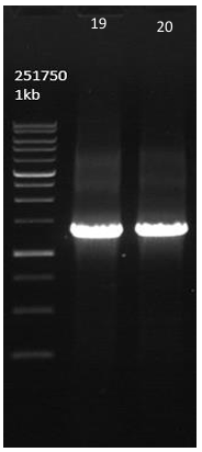
Figure 1 Agarose gel electrophoresis of 16S rRNA polymerase chain reaction result. M: DNA marker; Lane 19: PCR product of isolate ATF2IC showing band at ~1500 bp; Lane 20: PCR product of isolate ATF2IV8 showing band at ~1500 bp. Both PCR products passed quality control for sequencing.
Homology analysis using the NCBI GenBank database identified isolate ATF2IV8 as a member of the genus Calidifontibacillus. BLAST findings indicated that the 16S rRNA sequence of ATF2IV8 exhibited 94.85% similarity with Calidifontibacillus erzurumensis strain P2. A phylogenetic analysis of the 16S rRNA gene, performed by the neighbour-joining method in MEGA 11 software, is illustrated in Figure 2. The high sequence identity from the BLAST results and the consistent topology of the phylogenetic tree confirm that isolate ATF2IV8 belongs to the genus Calidifontibacillus. This genus comprises thermophilic bacteria known for producing hydrolytic enzymes, including amylase.
However, the phylogenetic analysis revealed that ATF2IV8 clustered with C. erzurumensis strain P2 (bootstrap 81%) but formed a distinct clade with a genetic distance of 2.437. Notably, the BLAST similarity value (94.85%) is below the 97% species delineation threshold [39], and together with its separate phylogenetic position, suggests that ATF2IV8 represents a distinct member within the genus Calidifontibacillus. This approximately 5% genetic divergence may reflect specific adaptation to the unique geothermal environment of the Wae Pellae hot spring. Collectively, these findings indicate that ATF2IV8 is an evolutionarily significant isolate with potential as a source of thermostable amylase exhibiting unique properties. However, definitive species-level identification requires further polyphasic taxonomic analysis, including genomic (e.g., Average Nucleotide Identity and whole-genome sequencing), phenotypic, and chemotaxonomic characterisation.
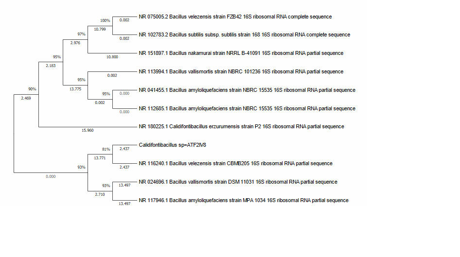

Figure 2 Phylogenetic tree of isolate ATF2IV8 based on 16S rRNA gene sequences, constructed using the Neighbour-Joining method with 1,000 bootstrap replicates (MEGA11). Bootstrap values (%) are shown at branch nodes and branch lengths represent the number of substitutions per site.
The genus Calidifontibacillus is known as a group of thermophilic bacteria typically native to hot spring ecosystems and related to the Bacillaceae family. The type species, C. erzurumensis P2ᵀ, was first established through genome-based classification of Ilica-Erzurum hot spring isolates and has since been separated from the Bacillus sensu lato group (transferring Bacillus azotoformans and B. oryziterrae to Calidifontibacillus) [40]. Culture data sources (BacDive) also confirmed the thermophilic nature of C. erzurumensis P2ᵀ and its origin in hot environments, consistent with our findings in Wae Pellae hot springs [41]. Relevant to this study, members of Calidifontibacillus have been reported to produce hydrolytic enzymes (including amylase), thus having the potential to be producers of biocatalysts that are stable at high temperatures. Recent reports on soil/rhizosphere microbial communities have identified C. erzurumensis as a producer of amylase, cellulase, and protease, strengthening its potential for high-temperature industrial processes [42]. Furthermore, recent studies on close relatives within the Bacillales have demonstrated the optimisation of production and characterisation of thermostable amylases for food/bioprocess applications. This positions Calidifontibacillus as a current producer of thermostable enzymes [43-45]. These findings reinforce the merit of exploring ATF2IV8 amylase as a thermostable amylase for applications under challenging temperature/pH process conditions.
Production of amylase enzyme
Comprehensive characteristics of amylase enzyme production were carried out by monitoring microorganism growth and enzyme activity during the fermentation period. Observed parameters included optical density as an indicator of cell biomass and amylase enzyme activity expressed in units per millilitre (U/mL). The relationship between these 2 parameters during the fermentation period is shown in Figure 3.
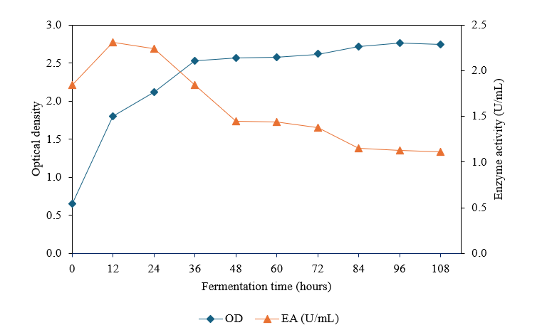
Figure 3 Correlation of biomass growth and amylase production during fermentation of Calidifontibacillus sp. ATF2IV8 at 1% starch concentration, pH 7, 40 °C, and 180 rpm agitation. Biomass growth was monitored by optical density (OD) at 600 nm and amylase activity was determined using the DNS method.
Based on the analysis of the fermentation profiles obtained, it can be observed that there is a complex correlation between the growth phase of the microorganisms and the enzyme production pattern. Understanding this dynamic is key to determining the timing of amylase enzyme production. Amylase production in this study showed peak activity during the log-stationary transition phase (around 12 - 24 h), then decreased even though biomass continued to increase. Several studies report that extracellular amylase activity reaches its maximum value in the first 12 - 24 h of cultivation. For example, Bacillus licheniformis BT5.9 showed peak activity at 12 h [46], while Bacillus subtilis J12 strains reported a peak at 24 h [47], thus supporting the enzyme production process within the 12 - 24 h timeframe.
Partial purification of amylase enzyme
Partial purification is a crucial step in enzyme characterisation because it produces enzymes with a higher degree of purity. This study involved the partial purification of the amylase enzyme through ammonium sulphate fractionation, followed by dialysis. Fractionation with ammonium sulphate at varying saturation levels (0% - 20%, 20% - 40%, 40% - 60%, 60% - 80%, 80% - 100%) yielded fractions F1 through F5, with F4 exhibiting the highest activity (Figure 4). Additionally, the desalting of the F4 fraction via dialysis led to a notable enhancement in specific activity, approximately sixfold relative to the crude extract, as presented in Table 1. This increase was primarily due to the removal of low-molecular-mass inhibitors and excess salts that inhibit enzyme activity in the crude extract.
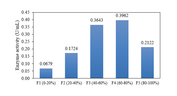
Figure 4 Effect of ammonium sulfate fractionation on amylase activity. Enzyme fractions F1 - F5 correspond to 0% - 20%, 20% - 40%, 40% - 60%, 60% - 80%, and 80% - 100% saturation, respectively.
Table 1 Overview of amylase enzyme purification.
| Enzyme | Volume (mL) | Amylase activity (U/mL) | Amylase activity total (Unit) | Protein (mg/mL) | Protein Total (mg) | Specific activity (U/mg) | Persen Yield (%) | Fold purification (x) |
|---|---|---|---|---|---|---|---|---|
| Crude extract | 500 | 1.2999 | 649.9500 | 2.0354 | 1,017.7000 | 0.6386 | 100.00 | 1.00 |
| Dialysate (F4) | 79 | 0.4203 | 33.2037 | 0.1104 | 8.7216 | 3.8071 | 5.11 | 5.96 |
Determination of protein molecular weight
Determining the molecular weight of an enzyme is a crucial step in characterisation because it provides information about the purity and identity of the resulting protein. Amylase enzymes generally have a range of molecular weights that vary depending on their source [48]. The molecular weight of the amylase enzyme was ascertained utilising the sodium dodecyl sulfate-polyacrylamide gel electrophoresis (SDS-PAGE) technique as per Laemmli (1970). SDS-PAGE analysis is used as a standard method to estimate protein subunit size and compare protein band profiles with known molecular markers. The results of protein molecular weight determination using Bio-Rad Mini-PROTEAN® Tetra Cell electrophoresis are shown in Figure 5. In this study, the blue prestained protein marker (Jena Bioscience) was used.
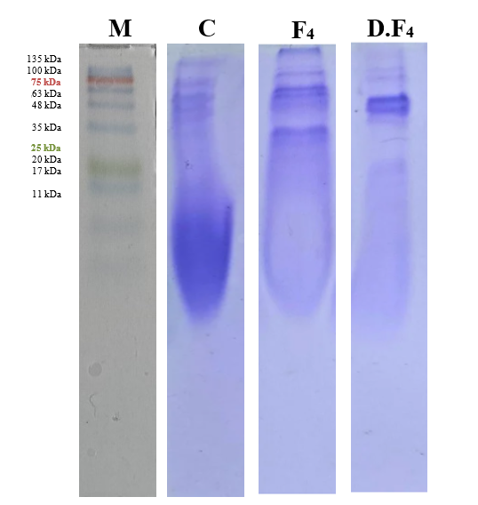
Figure 5 SDS-PAGE of amylase. Lane M: marker; C: crude extract; F4: 60% - 80% ammonium sulfate fraction; D.F4: dialysed fraction 4. Distinct bands (72, 69, 53, and 47 kDa) in D.F4 indicate partial purification.
SDS-PAGE analysis was conducted to estimate the molecular weight of the amylase enzyme obtained during the partial purification process. In the crude extract (C), the protein profile appeared as a diffuse smear without a clearly dominant band, indicating the presence of multiple proteins. Following ammonium sulphate fractionation (F4), a slight reduction in the number of protein bands was observed, suggesting partial enrichment.
Dialysis of the F4 fraction (D.F4) resulted in a simpler banding pattern with several distinguishable protein bands, among which a band of approximately 53 kDa appeared more prominent. The increased intensity of this band coincided with the observed increase in specific enzyme activity following dialysis, suggesting a possible association with amylase activity. This observation is consistent with previous reports indicating that amylases from Bacillus spp. and marine actinomycetes typically exhibit molecular weights in the range of 50 - 60 kDa [49,50].
In addition, minor bands at approximately 47 kDa and 69 kDa were detected in the D.F4 fraction. The 47 kDa band may represent a proteolytic degradation product or a co-purified protein, whereas the 69 kDa band could correspond to an aggregated form of the enzyme or a non-specific contaminant. However, the presence of multiple bands indicates that the enzyme preparation was not fully purified. Further purification using chromatographic techniques, such as ion exchange or gel filtration, was not performed in this study; therefore, the assignment of the ~53 kDa band as the amylase enzyme remains tentative and requires further confirmation through advanced purification or activity-based assays, such as zymography.
Characterisation
Effect of temperature on amylase enzyme activity
Temperature exerted a statistically significant influence on amylase enzymatic activity. The temperature optimum for the amylase enzyme derived from this bacterial isolate was determined to be 70 °C, at which the enzyme exhibited maximal activity of 1.4771 ± 0.0017 U/mL (Figure 6). Enzyme activity decreased beyond this temperature. At different temperatures, the isolateʼs amylase activity differed significantly (p ≤ 0.05).
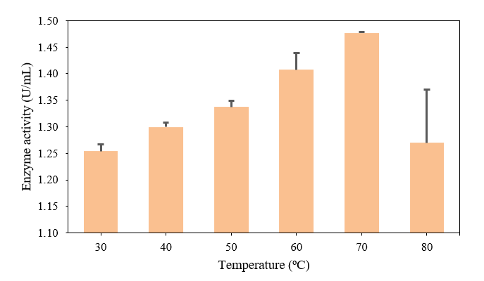
Figure 6 Effect of temperature on amylase activity of isolate ATF2IV8 at 1% starch, pH 7.0, and 30 min incubation in 50 mM phosphate buffer. Maximum activity was obtained at 70 °C using the DNS method. Data are presented as mean ± SD (n = 3).
Temperature emerged as a significant determinant of amylase enzymatic activity. The temperature at which maximum enzyme activity was achieved was 70 °C, a finding that is consistent with previously documented reports in the literature indicating that amylase enzymes isolated from thermophilic bacterial sources typically function optimally between 50 - 75 °C. Significantly decreased activity at higher temperatures is attributed to structural damage to the enzyme [2,32,51]. This finding is further corroborated by Burhanoğlu et al. [52], who reported a thermostable α-amylase from Geobacillus sp. GS33 with optimum activity at 60 °C, and by Fincan et al. [51], who reported a thermostable α-amylase from Bacillus licheniformis So-B3 isolated from a thermal spring with an optimum temperature of 70°. Conversely, Istia’nah [53] and Abo-Kamer [54] reported that maximum amylase activity from Bacillus isolated from Cosmos sulphureus Cav. and soil sources occurred at a lower temperature range, namely 35 - 40 °C. The difference in results between this study and previous studies is likely due to differences in the source of bacterial isolation and variations in the strains used.
Effect of pH on amylase enzyme activity
The influence of pH on amylase enzymatic activity was investigated by subjecting cultures to incubation across a pH spectrum ranging from 5 to 8. Results indicated that elevation of pH corresponded with increased enzyme activity. The pH optimum for enzymatic activity of this isolate was established within the range of 6 - 7, with maximal activity observed at pH 7. Conversely, enzyme activity demonstrated a decline at pH 8. Amylase enzymatic activity exhibited statistically significant differences (p ≤ 0.05) across the tested pH values.
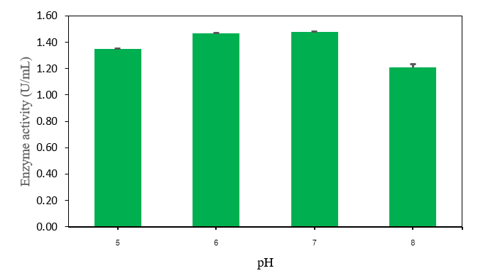
Figure 7 Effect of pH on amylase activity of isolate ATF2IV8 at 70 °C, a substrate concentration of 1% starch, and 30 min incubation in 50 mM phosphate buffer. Maximum activity was observed at pH 7.0 using the DNS method. Data are presented as mean ± SD (n = 3).
pH represents another significant factor influencing amylase enzymatic activity, bearing important implications for the practical application of this enzyme. During characterisation to establish the pH optimum, the amylase demonstrated catalytic activity across the pH range of 5 - 8. Enzymatic activity progressively increased from pH 5 and attained optimal performance within the pH range of 6 - 7, corresponding to an activity value of approximately 1.4771 ± 0.0017 U/mL. After passing the optimum pH, enzyme activity decreased drastically at pH 8, with an activity of approximately 1.2082 U/mL ± 0.0282. The highest activity at neutral to slightly acidic pH (pH 6 - 7) indicates that the amylase produced by this isolate is more stable and efficient under these conditions. The reduction in enzymatic activity observed beyond pH 7 may be attributable to alterations in the electrostatic charge distribution at the enzymeʼs active site and the substrate molecule, which consequently disrupts the enzyme-substrate complex formation and diminishes catalytic efficiency. This observation is consistent with previously published research demonstrating that amylase enzymes derived from thermophilic bacteria characteristically exhibit optimal performance under neutral to slightly alkaline pH conditions. However, there are variations depending on the source of the isolate and the type of strain [2,55]. Elevated pH optima for amylase activity were documented in the literature within the pH range of 9 - 10 by Simair et al. [56], whose investigations utilised the Bacillus BCC 01-50 bacterial strain.
Effect of substrate concentration on amylase enzyme activity
One factor influencing amylase enzyme activity is substrate concentration [57]. The substrate used in this study was starch. Table 2 shows that enzyme activity increased with increasing substrate concentration. At a starch concentration of 0.5%, enzyme activity was still relatively low, at 1.0517 ± 0.0036 U/mL. However, when the substrate concentration was increased to 1%, enzyme activity increased to 1.3825 ± 0.0400 U/mL. This increasing pattern continued up to a substrate concentration of 5%, which produced the highest activity of 2.5645 ± 0.0285 U/mL.
Table 2 Effect of Substrate Concentration on Amylase Activity of Isolate ATF2IV8 at 70 °C, pH 7.0, and 30-minute incubation. Data are presented as mean ± SD (n = 3).
| Substrate concentration (%) | Enzyme activity (U/mL) |
|---|---|
| 0.5 | 1.0517 ± 0.0036 |
| 1.0 | 1.3825 ± 0.0400 |
| 2.0 | 1.7882 ± 0.0215 |
| 4.0 | 2.4363 ± 0.0511 |
| 5.0 | 2.5645 ± 0.0285 |
At low substrate concentrations, most enzymes cannot interact optimally due to limited substrate availability. As substrate concentration increases, more active sites can bind to the enzyme, resulting in a significant increase in enzyme activity until saturation is reached. At a concentration of 5%, the rate of activity increase begins to slow at higher substrate concentrations. This indicates that the enzyme is approaching saturation, where nearly all active sites are occupied by substrate molecules.
This response pattern aligns with the enzyme kinetics theory proposed by Michaelis and Menten, which remains relevant and continues to be used in modern enzyme research [58]. The relationship between substrate concentration and enzyme reaction rate follows a hyperbolic curve, not a straight line. These enzyme activity data at various substrate concentrations then serve as the basis for further analysis in determining kinetic parameters.
The determination of the kinetic parameters Km and Vmax in the amylase enzyme was nonlinear regression fitting to the Michaelis-Menten equation (GraphPad Prism), as recommended for accurate kinetic parameter estimation [59]. The Lineweaver–Burk plot (Figure 8) was also constructed for visual representation and comparison purposes [60]. In this procedure, the relationship between the concentration of starch substrate [S] and the enzyme reaction rate (V) was visualised in the form of a plot as shown in Figure 8. The linear pattern formed corresponds to the characteristics of the Lineweaver-Burk graph, so that the calculation of kinetic parameters can be determined accurately [60].
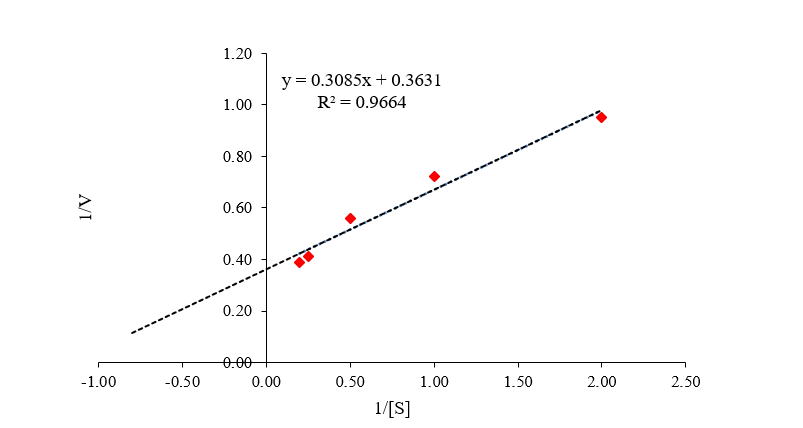
Figure 8 Lineweaver-Burk plot of amylase from Calidifontibacillus sp. ATF2IV8 using soluble starch as substrate at 70 °C and pH 7.0. The reciprocal plot of 1/V versus 1/[S] was used to determine the kinetic parameters Km and Vmax from the linear regression equation.
Lineweaver–Burk equation

From the linear relationship obtained between the two variables, the following equation was derived:

Based on equation 3, the Vmax value is 2.7541 U/mL and the Km value is 0.8496%.
Nonlinear regression analysis yielded a Vmax of 3.128 U/mL (95% CI: 2.528 - 4.102) and a Km of 1.218% (95% CI: 0.5948 - 2.499), with R² = 0.9714. For comparison, the Lineweaver-Burk plot (Figure 8) yielded values of Vmax = 2.7541 U/mL and Km = 0.8496%, confirming consistency between both approaches. The Km parameter reflects the substrate concentration required to produce 50% of the enzymeʼs maximum activity. The Michaelis-Menten curve in Figure 9 illustrates the relationship between substrate concentration and the amylase enzyme reaction rate. The experimental measurements demonstrate a characteristic pattern wherein the enzymatic reaction rate progressively increases with rising substrate concentration until reaching saturation conditions. The curve in Figure 9 represents the Michaelis-Menten model based on kinetic parameters obtained from nonlinear regression, namely a Vmax of 3.128 U/mL (95% CI: 2.528 - 4.102) and a Km of 1.218% (95% CI: 0.5948 - 2.499), which is consistent with the Lineweaver - Burk plot calculation. Thus, the Michaelis - Menten curve can be used to visualise enzyme efficiency and the substrate concentration required to achieve half-maximum activity (Km).
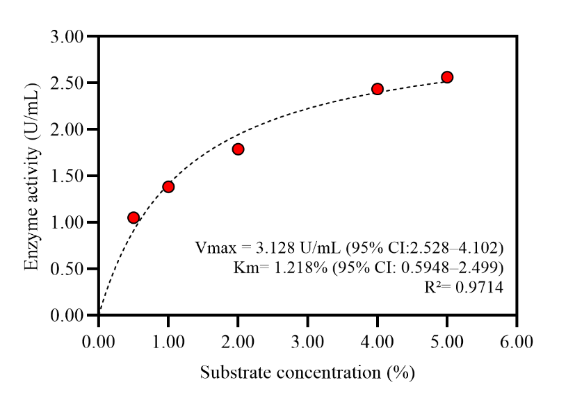
Figure 9 Michaelis-Menten plot of enzyme activity at varying substrate concentrations (GraphPad Prism nonlinear regression).
The Km value of 1.218% (equivalent to 12.18 mg/mL) obtained in this study is higher than those reported for some thermophilic bacterial amylases, including those from Geobacillus icigianus (Km = 2.17 mg/mL, Vmax = 4.16 U/mL) [61] and Bacillus subtilis KIBGE HAS (Km = 2.68 mg/mL, Vmax = 1,773 U/mL) [62]. This suggests that the enzyme may have a lower substrate affinity compared to these amylases; however, the relatively high Vmax of 3.128 U/mL at 70 °C indicates that the enzyme retains strong catalytic activity under thermophilic conditions, which may compensate for the lower substrate binding affinity in high-substrate environments. Like other thermophilic α-amylases, the enzyme from Calidifontibacillus sp. ATF2IV8 is presumed to act via a retaining mechanism involving the hydrolysis of α-1,4-glycosidic bonds in starch, consistent with the classical double-displacement mechanism reported for bacterial α-amylases [9,63].
Temperature stability
Temperature stability is an important characteristic determining the use of enzymes in industrial processes. The enzyme obtained in this study was tested for stability at 60 and 70 °C. These temperatures were selected based on the highest activity at the optimum temperature. A comparison of enzyme stability at these two temperatures is shown in Figure 10.
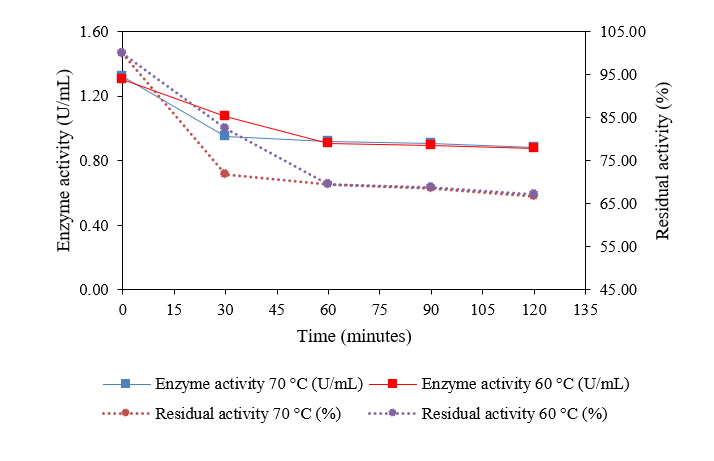
Figure 10 Thermal stability of amylase from isolate ATF2IV8 at 60 and 70 °C under a substrate concentration of 1% starch, pH 7.0, and 30-minute incubation in 50 mM phosphate buffer. Enzyme activity was determined using the DNS method.
At 60 °C, the amylase enzyme exhibited fairly good stability characteristics. Initial enzyme activity was recorded at approximately 1.299 U/mL and gradually decreased during the incubation period. In the first 30 min, the decline in activity was relatively slow, but after that the rate of decline accelerated. Analysis of residual enzyme activity expressed as a percentage revealed that following 120 min of incubation at 60 °C, the enzyme maintained approximately 67% of its original activity. These results indicate that the amylase at 60 °C in this study had a comparable, or even higher, level of stability than the amylase from the Bacillus methylotrophicus DCS1 strain [23], which at 50 °C only retained 53% of its activity after 1 h of incubation, although still slightly lower than its stability at 40 °C (73%). This finding is in line with other reports showing that amylase from several other isolates was able to maintain more than 66% of its initial activity in the temperature range of 55 - 80 °C for 50 min, and even specific isolates, such as M13, remained active up to 80.94% after 60 min at 80 °C [3]. Furthermore, research on Bacillus subtilis RK6 also demonstrated high stability, with the enzyme retaining 94%, 82%, 60%, and 51% of its activity after heating for 30 min at 60 °C, 70 °C, 80 °C, and 90 °C, respectively [14]. This confirms that amylases from various microbial sources generally exhibit good stability at medium to high temperatures, although with variations depending on the species and environmental conditions.
The thermal stability of amylase from the ATF2IV8 isolate can be attributed to several molecular factors that enhance its structural integrity compared with mesophilic enzymes. These factors include a compact molecular structure, stronger hydrophobic interactions, and an extensive network of hydrogen and disulphide bonds, which are typical features of extremozymes. The evolutionary adaptations of such enzymes enable them to function efficiently under extreme environmental conditions, thereby contributing to their remarkable stability [64]. These characteristics are consistent with those reported for thermostable enzymes derived from extremophilic microorganisms, which are known to possess unique structural and thermodynamic properties that confer resistance to thermal denaturation [12]. Microbial α-amylases exhibiting such properties have been widely applied in various industrial processes including starch liquefaction, food processing, and biofuel production [11]. Polyextremophilic α-amylases in particular have been reported to maintain catalytic efficiency across a broad range of temperatures and environmental conditions [13], and thermostable amylases from hot spring isolates reflect the evolutionary adaptation of their molecular architecture to extreme thermal conditions [14].
pH stability
pH stability constitutes a critical parameter in the comprehensive characterisation of enzymatic systems, including amylase preparations, owing to the significant influence of pH on both the three-dimensional structural conformation of the enzyme and its catalytic performance. Amylase, which hydrolyses glycosidic bonds in starch molecules, exhibits activity that is highly dependent on environmental pH conditions. In this study, pH stability measurements were conducted at pH 6 and pH 7, as shown in Figure 11.
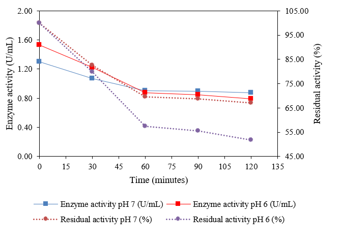
Figure 11 pH stability of amylase from isolate ATF2IV8 at pH 6 and 7 under a substrate concentration of 1% starch, a temperature of 60 °C, and a 30-minute incubation in 50 mM phosphate buffer. Enzyme activity was determined using the DNS method.
At pH 6, the initial enzyme activity was recorded at 1.5296 U/mL and decreased to 0.7903 U/mL after 120 min. This decrease indicates that although pH 6 provides higher initial activity, the enzymeʼs stability over time is relatively lower compared to pH 7. Conversely, at pH 7, the initial enzyme activity was 1.2999 U/mL and decreased to 0.8728 U/mL after 120 min. The residual activity at this pH decreased from 100% to 67.14%, indicating that the enzyme is more stable at pH 7 than at pH 6, despite its slightly lower initial activity. This indicates that pH 7 favours the structural stability of the amylase enzyme over more extended incubation periods. These findings are consistent with previously published research by Agustien et al. [65], who documented enzyme stability of up to 80% maintained over a 10-hour incubation period at pH 6 - 7. Additionally, Lim et al. [66] reported that α-amylase derived from Bacillus sp. demonstrated substantial stability under neutral to mildly alkaline pH conditions.
Overall, the enzymatic characterisation indicates that the amylase exhibits optimal activity at elevated temperature and neutral pH, with moderate stability under these conditions. The combined effects of temperature and pH suggest that the enzyme is functionally adapted to thermophilic environments, although its stability profile indicates that further optimisation may be required for sustained performance. These findings highlight the potential of the isolate as a source of thermostable amylase, while emphasising the need for further validation under application-relevant conditions.
Effect of metal ions on amylase enzyme activity
Metal ions are external factors that can significantly influence enzyme activity, either as activators or inhibitors. In the context of amylase enzymology, numerous metal ions are documented in the scientific literature to augment enzyme activity through mechanisms involving stabilisation of the enzyme-substrate complex structure or functioning as essential catalytic cofactors. The effects of various metal ions on amylase enzymatic activity determined in this investigation are presented graphically in Figure 12.
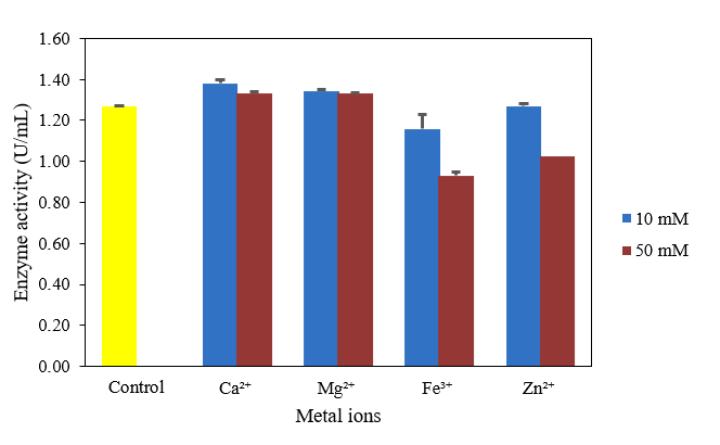
Figure 12 Effect of metal ions on amylase activity of isolate ATF2IV8 at 1% starch concentration, 60 °C, and pH 7.0. Enzyme activity was determined by the DNS method after 30 min of incubation. Data are presented as mean ± SD (n = 3).
The results showed that the addition of metal ions had varying effects on amylase enzyme activity, depending on the ion type and concentration. Ca²⁺ and Mg²⁺ ions at a concentration of 10 mM significantly increased enzyme activity by 1.3810 ± 0.0184 U/mL and 1.3438 ± 0.0049 U/mL, respectively. However, at a concentration of 50 mM, activity decreased to 1.3317 ± 0.0098 U/mL for both. This decrease was relatively small, indicating that Ca²⁺ and Mg²⁺ continued to support enzyme stability even at higher concentrations. This is in line with the findings of Ejike et al. [67], who reported that Ca²⁺ and Mg²⁺ act as important cofactors in maintaining the active conformation of the amylase enzyme from Lactobacillus plantarum. This is supported by research [68], which reported that amylase shows a selective response to the metal ion Ca²⁺. Mechanistically, stability by Ca²⁺ is largely associated with its role in maintaining the three-dimensional structure of the enzyme. Calcium ions were found at the interface of domains A and B of α-amylase, contributing to maintaining the structural consistency of the enzyme and preventing thermal denaturation [69]. At the structural level, Ca²⁺ ions coordinate with negatively charged amino acid residues in the protein, stabilising the inter-domain and intra-domain regions of the enzyme. This coordination maintains the integrity of the active site and prevents unfolding at elevated temperatures, which is particularly important for thermophilic amylases operating at 70 °C. Furthermore, Ca²⁺ ions have been shown to significantly increase the maximum velocity, enzyme half-life, and catalytic efficiency of α-amylases, consistent with the activation observed in this study [70].
In contrast, Fe³⁺ and Zn²⁺ ions showed a decrease in amylase enzyme activity. This decrease indicates a potential inhibitory effect, possibly through disruption of the active site or interaction with important residues in the enzyme structure. At the structural level, Zn²⁺ has been shown to alter the secondary structure and dynamic characteristics of α-amylase, leading to conformational changes that reduce catalytic activity. This inhibitory effect may be attributed to competition with Ca²⁺ for metal-binding sites at the domain interface, displacing essential calcium ions and disrupting the active conformation of the enzyme [71]. A study by Zhang et al. [72] also showed that the metal ions Zn²⁺ and Fe³⁺ can interfere with amylase enzyme activity. Other studies have shown that Fe³⁺ can actually act as an activator or inhibitor depending on the enzyme source. For example, amylase from Streptomyces griseus is stimulated by Fe³⁺ and Mg²⁺ [73]. This suggests that the influence of metal ions is highly dependent on the enzyme type and its molecular structure. These results are in line with the concept that α-amylase belongs to the group of metalloenzymes, where the presence of specific metal ions is crucial for maintaining the active conformation of the enzyme and increasing its catalytic efficiency.
Statistical analysis
One-way ANOVA analysis was used to determine the effect of temperature, pH, substrate concentration, and metal ions on enzyme activity. The analysis results showed that all parameters had a significant effect (p < 0.05) on enzyme activity (Table 3).
Table 3 ANOVA results for enzyme activity.
| Parameter | F | p - value | F crit | Significance |
|---|---|---|---|---|
| Temperature | 11.7885 | 0.000271605 | 3.1059 | Significant |
| pH | 233.2737 | 3.99755E-08 | 4.0662 | Significant |
| Substrate concentration | 1168.1914 | 2.6599E-13 | 3.4780 | Significant |
| Metal ions | 71.5752 | 3.0681E-07 | 3.2296 | Significant |
As one-way ANOVA revealed statistically significant differences among the treatment groups (p < 0.05), Tukey’s post hoc test was subsequently applied to determine which treatment levels differed significantly. The results of Tukey’s test for each parameter are presented in Table 4.
Table 4 Effect of temperature, pH, substrate concentration, and metal ions on enzyme activity based on Tukey’s HSD test.
| Parameter | Treatment | Enzyme Activity (U/mL) | Tukey |
|---|---|---|---|
| Temperature (°C) | 30 | 1.2543 ± 0.0125 | a |
| 40 | 1.2999 ± 0.0078 | ab | |
| 50 | 1.3374 ± 0.0113 | ab | |
| 60 | 1.4073 ± 0.0314 | bc | |
| 70 | 1.4771 ± 0.0017 | c | |
| 80 | 1.2699 ± 0.1001 | a | |
| pH | 5 | 1.3507 ± 0.0017 | b |
| 6 | 1.4667 ± 0.0035 | c | |
| 7 | 1.4771 ± 0.0017 | c | |
| 8 | 1.2082 ± 0.0282 | a | |
| Substrate (%) | 0.5 | 1.0517 ± 0.0036 | a |
| 1 | 1.3825 ± 0.0400 | b | |
| 2 | 1.7882 ± 0.0215 | c | |
| 4 | 2.4363 ± 0.0511 | d | |
| 5 | 2.5645 ± 0.0285 | e | |
| Metal Ions | Control | 1.2711 ± 0.0024 | c |
| Ca²⁺ (10 mM) | 1.3810 ± 0.0184 | d | |
| Ca²⁺ (50 mM) | 1.3317 ± 0.0098 | cd | |
| Mg²⁺ (10 mM) | 1.3438 ± 0.0049 | cd | |
| Mg²⁺ (50 mM) | 1.3317 ± 0.0098 | cd | |
| Fe³⁺ (10 mM) | 1.1577 ± 0.0722 | b | |
| Fe³⁺ (50 mM) | 0.9317 ± 0.0024 | a | |
| Zn²⁺ (10 mM) | 1.2693 ± 0.0122 | c | |
| Zn²⁺ (50 mM) | 1.0226 ± 0.0159 | a |
Values are expressed as mean ± standard deviation (n = 3).
Means followed by different lowercase letters within each factor differ significantly at p < 0.05 (Tukeyʼs HSD test).
Letter groupings are independent per factor and must not be compared across factors.
As shown in Table 4, enzyme activity was significantly influenced by all experimental factors tested. Temperature treatment showed a gradual increase in enzyme activity from 30 to 70 °C, with the highest activity observed at 70 °C (1.4771 ± 0.0017 U/mL). According to Tukey’s HSD grouping, the activity at 70 °C differed significantly from 30 °C and 80 °C, while no significant difference was observed compared with 60 °C, indicating that the enzyme reached its optimum catalytic performance near 70 °C.
The pH treatment also significantly affected enzyme activity, with the highest activity recorded at pH 7 (1.4771 ± 0.0017 U/mL). However, no significant difference was found between pH 6 and pH 7, suggesting that the enzyme remained highly active within a narrow neutral pH range, whereas activity decreased significantly under alkaline conditions (pH 8). A consistent increase in enzyme activity was observed with increasing substrate concentration, reaching the highest value at 5% substrate concentration (2.5645 ± 0.0285 U/mL). Each concentration level showed distinct Tukey grouping, indicating significant differences among all treatments. Among metal ion treatments, Ca²⁺ at 10 mM produced the highest stimulatory effect on enzyme activity, while Fe³⁺ at 50 mM caused the strongest inhibitory effect. The grouping results further indicated that Ca²⁺ and Mg²⁺ treatments generally maintained higher enzyme activity than Fe³⁺ and Zn²⁺ treatments.
Conclusions
This study demonstrates that the thermophilic bacterial isolate ATF2IV8, obtained from the Wae Pellae hot springs, was identified as Calidifontibacillus sp. with 94.85% similarity based on molecular analysis. This isolate produced a thermostable extracellular amylase enzyme, with optimum activity at 70 °C and pH 7. Stability tests showed that the enzyme retained more than half of its activity at elevated temperatures and under neutral pH conditions. Moreover, enzyme activity was enhanced in the presence of Ca²⁺ and Mg²⁺ ions, but inhibited by Fe³⁺ and Zn²⁺ ions. Partial purification through ammonium sulphate fractionation followed by dialysis increased the enzyme purity by approximately six-fold compared with the crude extract. SDS-PAGE analysis revealed a prominent protein band of approximately 53 kDa, which may be associated with amylase activity and is consistent with the reported molecular weight range of bacterial amylases. These findings suggest that the thermostable amylase from Calidifontibacillus sp. has further potential as a candidate for industrial biotechnological applications, including starch processing.
Acknowledgements
This research was supported by the Beasiswa Indonesia Bangkit, funded by the Lembaga Pengelola Dana Pendidikan (LPDP) as the funding sponsor in collaboration with the Kementerian Agama Republik Indonesia.
Declaration of Generative AI in Scientific Writing
The authors declare that artificial intelligence tools, including Chat GPT (Open AI), Google Translate, QuillBot, and Grammarly, were used to help correct language structure, grammar, and clarity in the manuscript. All content and interpretations were critically reviewed and approved by the authors.
CRediT Author Statement
Fitria Azis: Conceptualisation, Methodology, Investigation, Resources, Writing - original draft. Hasnah Natsir: Conceptualisation, Methodology, Writing - reviewing and editing, Supervision. Nur Asmi: Conceptualisation, Writing - reviewing and editing. Rugaiyah A. Arfah: Methodology, Writing - reviewing and editing. Paulina Taba: Methodology, Writing - reviewing and editing. Abdul Wahid Wahab: Supervision. Amalyah Febryanti: Software, Data curation, Writing- original draft. Maswati Baharuddin: Methodology, Visualisation. Sitti Chadijah: Methodology, Formal analysis. Iin Novianty: Formal analysis, Validation. Anita: Writing - reviewing and editing, Software.
[1] S Mondal, K Mondal, SK Halder, N Thakur and KC Mondal. Microbial amylase: Old but still at the forefront of all major industrial enzymes. Biocatalysis and Agricultural Biotechnology 2022; 45, 102509.
[2] JS Paul, N Gupta, E Beliya, S Tiwari and SK Jadhav. Aspects and recent trends in microbial α-amylase: A Review. Applied Biochemistry and Biotechnology 2021; 193, 2649-2698.
[3] SN Yassin, TM Jiru and M Indracanti. Screening and characterization of thermostable amylase-producing bacteria isolated from soil samples of Afdera, Afar Region, and molecular detection of amylase-coding gene. International Journal of Microbiology 2021; 2021, 5592885.
[4] AG Patil, K Khan, S Aishwarya, S Padyana, R Huchegowda, KR Reddy, R Pais, H Alrafas, R Dsouza, J Madhavi, AN Yadav, AV Raghu and F Zameer. Fungal amylases and their industrial applications. Springer, Cham, 2021, p. 407-434.
[5] BA Kikani and SP Singh. Amylases from thermophilic bacteria: Structure and function relationship. Critical Reviews in Biotechnology 2022; 42(3), 325-341.
[6] SA Shaikh, DR Khot and GR Pathade. Isolation and screening of amylase producing bacteria from soil and production of amylase. Ecology, Environment and Conservation 2023; 29, 227-231.
[7] A Suresh, D Shravan Ramgopal, K Panchamoorthy Gopinath, J Arun, P SundarRajan and A Bhatnagar. Recent advancements in the synthesis of novel thermostable biocatalysts and their applications in commercially important chemoenzymatic conversion processes. Bioresource Technology 2021; 323, 124558.
[8] BE Far, Y Ahmadi, AY Khosroushahi and A Dilmaghani. Microbial alpha-amylase production: Progress, challenges and perspectives. Advanced Pharmaceutical Bulletin 2020; 10(3), 350-358.
[9] MA Farooq, S Ali, A Hassan, HM Tahir, S Mumtaz and S Mumtaz. Biosynthesis and industrial applications of α-amylase: A review. Archives of Microbiology 2021; 203, 1281-1292.
[10] M Shad, N Hussain, M Usman, MW Akhtar and M Sajjad. Exploration of computational approaches to predict the structural features and recent trends in α‐amylase production for industrial. Biotechnology and Bioengineering 2023; 120(8), 2092-2116.
[11] PM de Souza and P de Oliveira e Magalhães. Application of microbial α-amilase in industry-A review. Brazilian Journal of Microbiology 2010; 41, 850-861.
[12] S Elleuche, C Schröder, K Sahm and G Antranikian. Extremozymes-biocatalysts with unique properties from extremophilic microorganisms. Current Opinion in Biotechnology 2014; 29, 116-123.
[13] S Kumar, J Grewal, A Sadaf, R Hemamalini and SK Khare. Halophiles as a source of polyextremophilic α-amylase for industrial applications. AIMS Microbiology 2016; 2(1), 1-26.
[14] S Kiran, A Singh, C Prabha, S Kumari and S Kumari. Isolation and characterization of thermostable amylase producing bacteria from hot springs of Bihar, India. International Journal of Pharma Medicine and Biological Sciences 2018; 7(2), 28-34.
[15] S Sharma, S Vaid, B Bhat, S Singh and BK Bajaj. Thermostable enzymes for industrial biotechnology. Elsevier, Netherlands, 2019, p. 469-495.
[16] ANM Anugrah, SW Hastuti, K Nafista, AK Ilham and D Nandiwardhana. A review: Study of potential direct use geothermal in Indonesia. In: Proceedings of the 10th ITB International Geothermal Workshop 2021 Bandung Institute of Technology, Bandung, Indonesia. 2021.
[17] D Wulandari, A Budiharjo, AAK Putri and RHB Setiarto. Diversity of thermophilic bacteria isolated from extreme environments in Indonesia: A perspective in biotechnology applications. Journal of Tropical Biodiversity and Biotechnology 2025; 10(3), 19548.
[18] A Budiharjo, D Wulandari, J Shabrina, RA Mawarni, AR Maulana, Nurhayati, Wijanarka, L Hartajanie and Lindayani. Bioprospecting and molecular identification of amylase and cellulase producing thermophilic bacteria from sediment of Nglimut Hot Springs, Kendal Regency. Journal of Tropical Biodiversity and Biotechnology 2024; 9(3), 86756.
[19] RA Arfah, A Ahmad, MN Djide, M Anis and M Zakir. Production, optimization and characterization of amylase enzyme isolated from termofil bacteria Bacillus sp. RSAII-1b from Lejja Hot Spring South Sulawesi. American Journal of Biomedical and Life Sciences 2015; 3(6), 115-119.
[20] A Ardhi, AN Sidauruk, N Suraya, NW Pratiwi, U Pato and Saryono. Molecular identification of amylase-producing thermophilic bacteria isolated from Bukit Gadang Hot Spring, West Sumatra, Indonesia. Biodiversitas Journal of Biological Diversity 2020; 21(3), 994-1000.
[21] BZ Fazal, C Budiman, Z Amin and CMWV Ling. Screening, isolation, and characterization of amylase-producing bacteria from Poring Hot Spring Sabah, Malaysia. Biodiversitas 2022; 23(6), 2807-2815.
[22] S Soy, U Lakra, P Prakash, P Suravajhala, VK Nigam, SR Sharma and N Bayal. Exploring microbial diversity in hot springs of Surajkund, India through 16S rRNA analysis and thermozyme characterization from endogenous isolates. Scientific Reports 2023; 13, 14221.
[23] N Hmidet, N Jemil and M Nasri. Simultaneous production of alkaline amylase and biosurfactant by Bacillus methylotrophicus DCS1: Application as detergent additive. Biodegradation 2019; 30, 247-258.
[24] S Arbab, H Ullah, MIU Khan, MNK Khattak, J Zhang, K Li and IU Hassan. Diversity and distribution of thermophilic microorganisms and their applications in biotechnology. Journal of Basic Microbiology 2022, 62(2), 95-108.
[25] A Rodiansyah, AF Mahmudah, MM Ulfah, U Rohmawati, D Listyorini, EA Suyono and S Prabaningtyas. Identification of potential bacteria on several lakes in East Java Indonesia based on 16S rRna sequence analysis. HAYATI Journal of Biosciences 2021; 28(2), 136-143.
[26] Geneaid. Presto TM Mini gDNA Bacteria Kit. Geneaid Biotech Ltd, Taipei, Taiwan, 2017.
[27] MA Rakaz, MO Hussien and HM Ibrahim. Isolation, extraction, purification, and molecular characterization for thermostable α‐amylase from locally isolated Bacillus species in Sudan. Biochemistry Research International 2021; 2021, 6670380.
[28] T Govil, M Paste, D Samanta, A David, KM Goh, X Li, DR Salem and RK Sani. Metagenomics and culture dependent insights into the distribution of firmicutes across two different sample types located in the Black Hills Region of South Dakota, USA. Microorganisms 2021; 9(1), 113.
[29] R Ortega-Villar, A Escalante, F Astudillo-Melgar, L Lizárraga-Mendiola, GA Vázquez-Rodríguez, ME Hidalgo-Lara and C Coronel-Olivares. Isolation and characterization of thermophilic bacteria from a hot spring in the State of Hidalgo, Mexico, and geochemical analysis of the thermal water. Microorganisms 2024; 12(6), 1066.
[30] ALN Aminin, Y Milarsih and NS Mulyani. Isolasi dan karakterisasi amilase termostabil dari Geobacillus dYtae-14. Greensphere: Journal of Environmental Chemistry 2022; 2(2), 1-6.
[31] A Davenport. The changing face of dialyzer membranes and dialyzers. Seminars in Dialysis 2025; 38(1), 25-34.
[32] MJ Msarah, I Ibrahim, AA Hamid and WS Aqma. Optimisation and production of alpha amylase from thermophilic Bacillus spp. and its application in food waste biodegradation. Heliyon 2020; 6(6), 04183.
[33] F Azis, S Si and M Si. Teknik dasar analisis biomolekul. Eureka Media Aksara, Indonesia, 2024.
[34] ST Sagu, L Zimmermann, E Landgräber, T Homann, G Huschek, H Özpinar, FJ Schweigert and HM Rawel. Comprehensive characterization and relative quantification of α-amylase/trypsin inhibitors from wheat cultivars by targeted HPLC-MS/MS. Foods 2020; 9(10), 1448.
[35] B Yao, C Jin, Y Guan, Z Chang, Q Liu and H Gao. Purification and characteristics of β-amylase from soybean whey wastewater. Catalysts 2024; 14(12), 909.
[36] M Baharuddin, N Alfina, A Febryanti, F Azis and W Wahyuningsih. Karakterisasi enzim amilase isolat bakteri R2M larva kumbang sagu dari Luwu Utara. Chimica et Natura Acta 2022; 10(2), 81-87.
[37] AM Saeed, EH El-shatoury and HAE Sayed. Statistical factorial designs for optimum production of thermostable α-amylase by the degradative bacterium Parageobacillus thermoglucosidasius Pharon1 isolated from Sinai, Egypt. Journal of Genetic Engineering and Biotechnology 2021; 19(1), 24.
[38] JA Kim, MJ Kim, JH Yim, IC Kim, J Rhee and SJ Han. Isolation and characterization of low-temperature and high-salinity amylase from Halomonas sp. KS41843. Fermentation 2025; 11(8), 465.
[39] HJ Gwak and M Rho. Data-driven modeling for species-level taxonomic assignment from 16S rRNA: Application to human microbiomes. Frontiers in Microbiology 2020; 11, 570825.
[40] A Adiguzel, H Ay, MO Baltaci, S Akbulut, S Albayrak and MA Omeroglu. Genome-based classification of Calidifontibacillus erzurumensis gen. Nov., sp. nov., isolated from a hot spring in Turkey, with reclassification of Bacillus azotoformans as Calidifontibacillus azotoformans comb. nov. and Bacillus oryziterrae as Calidifontibacillus oryziterrae comb. nov. International Journal of Systematic and Evolutionary Microbiology 2020; 70(12), 6418-6427.
[41] Leibniz Institute DSMZ. Calidifontibacillus erzurumensis (DSM 107530, P2). Leibniz Institute DSMZ, Braunschweig, Germany, 2026.
[42] SM Samal, D Singh, RN Kharwar and RS Upadhaya. Characterization of plant growth promoting Rhizobacteria and identify their potentiality to manage wilt disease of pigeonpea inciting by Fusarium udum. SSRN 2020. http://dx.doi.org/10.2139/ssrn.4768642
[43] A Kholikov, K Vokhidov, A Murtozoyev, ZS Tóth, GN Nagy, BG Vértessy and A Makhsumkhanov. Characterization of a thermostable α-amylase from Bacillus licheniformis 104.K for industrial applications. Microorganisms 2025; 13(8), 1757.
[44] KM Hossain, U Khan, SMM Rahman and MS Khan. Potential antimicrobial and fruit juice clarification activity of amylase enzyme from Bacillus strains. Biotechnology Reports 2024; 44, 00861.
[45] S Harirchi, T Sar, M Ramezani, H Aliyu, Z Etemadifar, SA Nojoumi, F Yazdian, MK Awasthi and MJ Taherzadeh. Bacillales: From taxonomy to biotechnological and industrial perspectives. Microorganisms 2022; 10(12), 2355.
[46] D Ibrahim, HL Zhu, N Yusof, Isnaeni and LS Hong. Bacillus licheniformis BT5.9 isolated from Changar Hot Spring, Malang, Indonesia, as a potential producer of thermostable α-amylase. Tropical Life Sciences Research 2013; 24(1), 71-84.
[47] MK Mahfudz, S Jaikhan, K Phirom-On and J Apiraksakorn. Cost-effective strategy and feasibility for amylase production from okara by Bacillus subtilis J12. Fermentation 2024; 10(11), 561.
[48] H Zhang, F Zhang, F Wu, L Guo and X Xu. Purification and characterization of endogenous α-amylase from glutinous rice flour. Foods 2025; 14(10), 1679.
[49] B Enez. Purification and characterization of thermostable α-amylase from soil bacterium Bacillus sp. Protein and Peptide Letters 2021; 28(12), 1372-1378.
[50] MH Al-Agamy, MR Alhuzani, MS Kelany and MM Hamed. Production and partial characterization of α-amylase enzyme from marine Actinomycetes. BioMed Research International 2021; 2021, 5289848.
[51] SA Fincan, S Özdemir, A Karakaya, B Enez, SD Mustafov, MS Ulutaş and F Şen. Purification and characterization of thermostable α-amylase produced from Bacillus licheniformis So-B3 and its potential in hydrolyzing raw starch. Life Sciences 2021; 264, 118639.
[52] T Burhanoğlu, Y Sürmeli and G Şanlı-Mohamed. Identification and characterization of novel thermostable α-amylase from Geobacillus sp. GS33. International Journal of Biological Macromolecules 2020; 164, 578-585.
[53] D Istia’Nah, U Utami and A Barizi. Karakterisasi enzim amilase dari bakteri Bacillus megaterium pada variasi suhu, pH dan konsentrasi substrat. Jurnal Riset Biologi dan Aplikasinya 2020; 2(1), 11-17.
[54] AM Abo-Kamer, IS Abd-El-Salam, FA Mostafa, AERA Mustafa and LA Al-Madboly. A promising microbial α-amylase production, and purification from Bacillus cereus and its assessment as antibiofilm agent against Pseudomonas aeruginosa pathogen. Microbial Cell Factories 2023; 22, 141.
[55] I Ullah, MS Khan, SS Khan, W Ahmad, L Zheng, SUA Shah, M Ullah and A Iqbal. Identification and characterization of thermophilic amylase producing bacterial isolates from the Brick Kiln Soil. Saudi Journal of Biological Sciences 2021; 28(1), 970-979.
[56] AA Simair, I Khushk, AS Qureshi, MA Bhutto, HA Chaudhry, KA Ansari and C Lu. Amylase production from thermophilic Bacillus sp. BCC 021-50 isolated from a marine environment. Fermentation 2017; 3(2), 25.
[57] L VanDee, A Teague, T East, KR Jacinto, M Carter, J Totty, PD Adams and D Muhoza. A cost-effective enzyme kinetics and inhibition model for biochemistry education and research. Biochemistry and Molecular Biology Education 2024; 52(5), 588-598.
[58] B Choi, GA Rempala and JK Kim. Beyond the Michaelis-Menten equation: Accurate and efficient estimation of enzyme kinetic parameters. Scientific Reports 2017; 7, 17018.
[59] RA Copeland. Evaluation of enzyme inhibitors in drug discovery: A guide for medicinal chemists and pharmacologists. Wiley, New Jersey, 2005.
[60] P Tallapragada, R Dikshit, A Jadhav and U Sarah. Partial purification and characterization of amylase enzyme under solid state fermentation from Monascus sanguineus. Journal of Genetic Engineering and Biotechnology 2017; 15(1), 95-101.
[61] S Soy, VK Nigam and SR Sharma. Enhanced production and biochemical characterization of a thermostable amylase from thermophilic bacterium Geobacillus icigianus BITSNS038. Journal of Taibah University for Science 2021; 15(1), 730-745.
[62] S Bano, SAU Qader, A Aman, MN Syed and A Azhar. Purification and characterization of novel α-Amylase from Bacillus subtilis KIBGE HAS. AAPS PharmSciTech 2011; 12(1), 255-261.
[63] Š Janeček and B Svensson. How many α-amylase GH families are there in the CAZy database? Amylase 2022; 6(10), 1-10.
[64] MF ur Rehman, A Shaeer, AI Batool and M Aslam. Chapter 2-Structure-Function relationship of extremozymes. Academic Press, Massachusetts, 2022, p. 9-30.
[65] A Agustien, D Bendrianis, F Alamsyah, M Muqarramah and M Jannah. Enhanced amylase activity by modulating abiotic factors and enzyme stability in the thermophilic bacterial isolates. On Line Journal of Biological Sciences 2024; 24(2), 295-301.
[66] SJ Lim, SN Hazwani-Oslan and SN Oslan. Purification and characterisation of thermostable α-amylases from microbial sources. BioResources 2020; 15(1), 2005-2029.
[67] EN Ejike, OK Achi and E Nwachukwu. Optimization of carbon and nitrogen sources and effect of metal ions on amylase production by lactic acid bacteria strains isolated from fermented starchy foods. International Journal of Microbiology and Applied Sciences 2024; 3(3), 47-52.
[68] OE Owolabi, OO Olaniyi and FA Akinyosoye. Catalytic properties of purified alpha amylase from Aspergillus flavus cultivated on low-cost agricultural substrate. Revista Facultad Nacional de Agronomía Medellín 2023; 76(1), 10213-10225.
[69] N Jaiswal and P Jaiswal. Thermostable α-amylases and laccases: Paving the way for sustainable industrial applications. Processes 2024; 12(7), 1341.
[70] E Abedi, H Torabizadeh and L Orden. Enhancement of alpha-amylase’s stability and catalytic efficiency after modifying enzyme structure using calcium and ultrasound. Food and Bioprocess Technology 2024; 17, 1546-1562.
[71] SM Liao, NK Shen, G Liang, B Lu, ZL Lu, LX Peng, F Zhou, LQ Du, YT Wei, GP Zhou and RB Huang. Inhibition of α-amylase activity by Zn2+: Insights from spectroscopy and molecular dynamics simulations. Medicinal Chemistry 2019; 15(5), 510-520.
[72] Z Zhang, R Ma, Y Tao, Z Wang and Y Yang. Effects of Fe and Zn alone and combined treatment on Triticum aestivum L. seed germination. BMC Plant Biology 2025; 25, 430.
[73] B Divya, S Shiburaj and NS Pradeep. Purification and characterization of thermostable and calcium independant α-amylase from Streptomyces griseus TBG19NRA1. Indian Journal of Applied and Pure Biology 2021; 36(1), 85-102.