
Trends
Sci.
2026;
23(11):
13531
Single Clove Garlic Extract Attenuates Dexamethasone-Induced C2C12 Myotube Atrophy through Akt-Related Signalling
Putri Elok Septiana Dewi1,2, Piyaporn Surinlert3,4, Tanaporn Hengpratom2,
Thanvarin Thitiphatphuvanon5, Sri Rahayu Lestari6 and Chittipong Tipbunjong2,*
1Applied Biology Department, Faculty of Health, Science, and Psychology, Universitas Sunan Gresik,
East Java 61124, Indonesia
2Department of Anatomy, Division of Health and Applied Sciences, Faculty of Science, Prince of Songkla University, Songkhla 90110, Thailand
3Chulabhorn International College of Medicine, Thammasat University, Pathum-Thani 12120, Thailand
4Thammasat University Research Unit in Synthesis and Applications of Graphene, Thammasat University,
Pathum-Thani 12120, Thailand
5Faculty of Medicine, Kasetsart University, Bangkok 10900, Thailand
6Department of Biology, Faculty of Mathematics and Natural Science, Universitas Negari Malang,
East Java 65145, Indonesia
(*Corresponding author’s e-mail: [email protected])
Received: 2 February 2026, Revised: 16 April 2026, Accepted: 26 April 2026, Published: 1 June 2026
Abstract
The number of older adults with skeletal muscle loss or sarcopenia is gradually increasing. In response, the interest in exploring novel drugs and supplements to attenuate muscle atrophy is growing. This research aimed to investigate the effect of single clove garlic extract (SGE) on muscle cell proliferation, differentiation, and atrophy. SGE was prepared using the maceration method. C2C12 skeletal myoblasts were treated with SGE before being subjected to MTT assay and flow cytometry to determine cell viability and proliferation. Myoblast differentiation was induced to form multinucleated myotubes, which were then treated with 10 µM dexamethasone (DEX) for 24 h to induce atrophy, in the presence or absence of SGE. After treatment, genes and proteins related to myotube atrophy were quantified using real-time PCR, immunostaining, and Western blot. Treatment with SGE (5 - 40 µg/mL) had no effect on cell viability, proliferation, or differentiation of C2C12 myoblasts. However, in myotubes treated with DEX, diameter and the expression of myosin heavy chain (MHC), MyoD, and myogenin proteins were all significantly reduced; pretreatment with SGE significantly attenuated these effects. SGE pretreatment also significantly increased the phosphorylation of Akt, P70S6K, and β-catenin, and significantly suppressed the expression of atrogin-1 compared to DEX treatment alone. Furthermore, the genes PGC-1α, NRF-1, and TFAM were significantly down-regulated in myotubes treated with DEX compared to control. Pretreatment with SGE significantly alleviated this effect. Thus, this study shows that SGE prevents muscle protein degradation and muscle atrophy induced by DEX via Akt protein phosphorylation and subsequent suppression of the atrogin-1 protein.
Keywords: C2C12 myoblasts, Muscle atrophy, Myogenesis, Myotube, Sarcopenia, Garlic extract, Skeletal muscle
Introduction
Skeletal muscle is a specialised tissue responsible for body movement and metabolism. Healthy skeletal muscle maintains mass by balancing protein synthesis
and protein degradation. Various conditions, including ageing, lead to a gradual loss of this muscle mass [1,2]. Muscle atrophy in older people, or sarcopenia, is characterised by reduced muscle mass, and strength, and physical functions which contribute to disability in this population. This problem places a heavy economic burden on society. Two biomarkers of muscle wasting have been identified in sarcopenia: increases in the proteins muscle atrophy F-box (atrogin-1) and muscle RING finger-1 (MuRF-1) [3]. Both atrogin-1 and MuRF-1 promote ubiquitin-mediated protein degradation, leading to skeletal muscle protein loss. Therefore, an increase in either atrogin-1 or MuRF-1 protein exacerbates skeletal muscle atrophy [4]. With growing ageing populations, interest is rising in the exploration of new interventions and supplements to prevent muscle atrophy and increase muscle protein synthesis.
Garlic (Allium sativum) is a widely used herb in food preparation, and is also common in traditional medicine in Asian and other countries for the treatment of various diseases [5]. The major active compounds in garlic are reported to be organosulfurs, such as diallyl thiosulfonate (allicin), diallyl sulfide (DAS), diallyl disulfide (DADS), diallyl trisulfide (DATS), E/Z-ajoene, S-allyl-cysteine (SAC), and S-allyl-cysteine sulfoxide (alliin) [6]. For nutraceutical usages, single clove garlic extract (SGE) is preferable due to its 5 - 6-fold higher content of bioactive compounds than multi-clove garlic [7]. Single clove garlic contains diverse bioactive compounds, including organic sulfides, saponins, phenolic compounds, and polysaccharides [6]. Numerous studies have demonstrated multimodal biological functions of garlic extract, including anti-oxidant, anti-cancer, anti-inflammatory, anti-diabetes, anti-obesity, anti-thrombotic, and anti-atherosclerotic effects, alongside decreased blood lipids, blood glucose modulation, cardiovascular disease treatment, and stimulation of the immune system [6,8].
Age-related cellular oxidative stress has been linked to the atrophy of skeletal muscle [1,2]. Given the anti-oxidant and other varied biological activities of SGE [6,8], research has suggested its potential for preventing muscle atrophy. Previous research reports that oral administration of garlic supplement upregulated muscle mitochondrial biosynthesis in skeletal muscle after exercise [9] and prevented skeletal muscle damage by reducing inflammation and apoptosis [10]. In addition, it has been reported to alleviate skeletal muscle loss and enhance myogenic differentiation in obese subjects via the modulation of Akt/mTOR/p70S6K signalling [11]. Moreover, a specific compound present in garlic extract has also been reported to exert a beneficial effect for the treatment and prevention of muscle atrophy. Saponins are reported to protect against H2O2 and inhibit myoblast growth and DNA damage by scavenging intracellular reactive oxygen species (ROS) [12]. DATS further demonstrated a protective effect against oxidative stress-induced DNA damage and apoptosis in skeletal muscle myoblast cells through ROS scavenging [13]. Additionally, Z-ajoene has been shown to ameliorate muscle atrophy by regulating myokine secretion in the JAK/STAT3 and SMADs/forkhead box O (FoxO) signalling pathways, aiding the preservation of muscle mass. It also functions to degrade myoprotein under muscle wasting and stimulate myogenesis [14]. Moreover, garlic extract has been reported to inhibit the expression of the protein FoxO [14], which acts as the master regulator of muscle atrophy in ageing through the activation of the ubiquitin proteasome system, which ultimately leads to muscle protein degradation [15]. Inhibition of the activity of FoxO may reduce the expression of atrogin-1 and MuRF-1, thus preventing muscle atrophy [16].
We hypothesise that the numerous biologically active compounds found in SGE—each with beneficial individual effects on muscle cells—may exert a synergistic therapeutic effect for the prevention and treatment of muscle atrophy and sarcopenia in older adults. Therefore, the present study aimed to explore the synergistic effect and possible mechanisms of SGE on muscle cell proliferation, differentiation, and myotube atrophy prevention.
Materials and methods
Single clove garlic extract preparation
Single clove garlic (Allium sativum L.) was collected from Temanggung, Central Java, Indonesia. SGE was prepared using the maceration method with an ethanol solvent, following previous reports with little modification [17-19]. This extraction protocol has been reported to extract the organosulfur compounds validated by Fourier transform infrared (FTIR) spectroscopy and high-performance liquid chromatography (HPLC). Briefly, 1 kg of mashed single clove garlic was soaked in 70% ethanol at a ratio of 1:3. It was placed in a water bath at 37 °C for 24 h with constant shaking at 100 rpm. The solution was then passed through filter paper. This step was repeated 3 times with new 70% ethanol. The 70% ethanol containing active compounds was evaporated using a rotary evaporator. The extraction yield was 27.5% (w/w). The SGE was kept at 4 °C until use, at which point it was reconstituted in dimethyl sulfoxide (DMSO) at 40 mg/mL as a stock solution. To create the working solution, the stock solution was diluted in cell culture medium and then filtered through a sterile 0.2 µm syringe filter. The final concentration of DMSO in the culture medium was 0.1%. An identical solution of 0.1% DMSO in cell culture medium without the active SGE compounds was used as a control solution.
C2C12 myoblast cell culture
A C2C12 myoblast cell line was purchased from the American Type Culture Collection (ATCC). It was maintained in growth medium (GM) composed of Dulbecco’s modified Eagle medium (DMEM) supplemented with 10% fetal bovine serum (FBS) and 1% penicillin-streptomycin solutions (penicillin 100 U/mL, streptomycin 100 µg/mL) at 37 °C in a humidified 5% CO2 incubator. Cell passages 10 - 15 were used. To study the cytotoxic and proliferative effects of SGE, C2C12 myoblasts were seeded at 1×10⁴ cells/well in a 96-well plate and cultured in low serum medium or GM containing SGE at 0 - 40 µg/mL for 24, 48, and 72 h [20,21]. Cell morphology was observed and photographed. For myotube formation, C2C12 myoblasts were seeded at 3×10⁵ cells/well in a 6-well plate. To induce myotube differentiation, confluent cells were switched to a differentiation medium (DM) composed of DMEM supplemented with 2% horse serum (HS) for 3 days [20,21]. The myotubes were pretreated with SGE at 0 - 40 µg/mL for 2 h before 10 µM dexamethasone (DEX) was added directly to the medium. Treatment with 10 µM DEX for 24 h has been reported to induce myotube atrophy [22]. The myotubes were further cultured under these conditions for 24 h. Myotube morphology was observed and photographed. To test for mycoplasma contamination, routine Hoechst staining was performed every month.
MTT assay
The treated cells were incubated with GM containing 0.5 mg/mL 3-(4,5-dimethylthiazol-2-yl)-2,5- diphenyltetrazolium bromide (MTT) for 3 h. Thereafter, the solution was discarded and replaced with 100 µL DMSO. Absorbance was measured at 570 nm using a microplate reader [20,21]. Cell viability and proliferation were normalised to a control group and reported as a percentage of the control value.
Nuclear staining
The treated cells were fixed with ice-cold methanol at −20 °C for 10 min. After several washes and rehydration with phosphate buffered saline (PBS), the cells were incubated with DAPI (4′,6-diamidino-2-phenylindole) diluted in PBS for 30 min with constant agitation. Thereafter, the solution was discarded, and the cells were washed again with PBS. Nuclear density images were captured using a fluorescence microscope [20,21].
Flow cytometry
The treated cells were trypsinised and fixed in 70% ice-cold ethanol for 10 min. After several washes with PBS, the cell pellet was incubated with ribonuclease A (50 μg/mL) diluted in PBS for 30 min at 37 °C. The cells were then immediately placed on ice for 10 min. Propidium iodide (0.5 mg/mL) was directly added, and the reaction was kept in the dark for 15 min [20,21]. The cell cycle stage was determined using a BD FACSCanto flow cytometer (BD Biosciences, USA).
Immunofluorescence staining and myotube diameter measurement
The treated cells were fixed with ice-cold methanol at −20 °C for 10 min. The fixed cells were washed and rehydrated with PBS. After permeabilisation and blocking with 5% normal goat serum at room temperature (RT) for 1 h, a mouse monoclonal anti-MHC antibody was applied and incubated overnight at 4 °C. After several washes, the cells were incubated with goat anti-mouse IgG conjugated to Alexa Fluor 488 fluorescent dye for 1 h at RT. Images were captured using a fluorescence microscope [18,19]. Myotube diameter was measured using ImageJ. Myotube diameters were averaged over 6 fields per treatment, with at least ten myotubes per field [22]. In order to blind the measurement, 3 observers were assigned to perform measurements.
RNA isolation and real-time polymerase chain reaction (PCR)
The treated cells were subjected to total RNA isolation using Trizol. The RNA was treated with DNase to remove contamination before conversion to cDNA using a cDNA synthesis kit. Real-time PCR was performed using the Luna qPCR master mix, and relative gene expression was calculated using the comparative Ct method (2−ΔΔCt) normalised to GAPDH [20,21]. The oligonucleotide primers used in this experiment were nuclear respiratory factor 1 (NRF-1) Fw: GGCAACAGTAGCCACATTGGCT, Rw: GTCTGGATGGTCATTTCACCGC; peroxisome proliferator-activated receptor gamma coactivator 1-alpha (PGC-1α) Fw: GAATCAAGCCACTACAGACACCG, Rw: CATCCCTCTTGAGCCTTTCGTG; transcription factor A mitochondrial (TFAM) Fw: GAGGCAAAGGATGATTCGGCTC, Rw: CGAATCCTATCATCTTTAGCAAGC; glyceraldehyde-3-phosphate dehydrogenase (GAPDH) Fw: TCAATGAAGGGGTCGTTGAT, Rw: CGTCCCGTAGACAAAATGGT [23].
Protein extraction and Western blot analysis
The treated cells were subjected to protein extraction using a radioimmunoprecipitation assay buffer containing protease and phosphatase inhibitors. Equal amounts of proteins were separated using sodium dodecyl sulfate-polyacrylamide gel electrophoresis, and were then transferred to a polyvinylidene fluoride membrane. The membrane was blocked for non-specific binding with 5% goat serum for 1 h before incubation with the desired primary antibody on a rocking table overnight at 4 °C. After washing, the membrane was incubated with an appropriate horseradish peroxidase (HRP)-conjugated secondary antibody at RT for 1 h. The protein signal was visualised using the HRP-substrate by Alliance Q9 advanced chemiluminescence imaging. Band intensity was measured using ImageJ. To measure the phosphorylation of proteins, the band intensity of phosphorylated protein was normalised to total protein and then to tubulin, which served as a loading control. The intensity of each treatment group was then normalised to the control group and reported as fold-change [20,21]. Throughout the experiment, all blots were exposed in automatic mode without any saturation, which confirmed the exposure as linear.
Statistical analysis
For each experiment, at least 3 independent experiments were performed with 3 technical replicates. Data were presented as mean ± s.e.m. Statistical analysis was conducted by one-way analysis of variance (ANOVA) followed by post-hoc analysis with Tukey’s test using the GraphPad Prism. The data exhibited homogeneity of variance. p-values < 0.05 were considered statistically significant.
Results and discussion
Non-toxic effects of SGE on C2C12 myoblast cells
To determine a possible toxic effect of SGE, C2C12 myoblasts were treated with SGE at different concentrations in low serum media for 24, 48, and 72 h. MTT assay revealed that treatment for these durations with SGE at 5 - 40 µg/mL had no effect on C2C12 myoblast cell viability compared to the non-treated control (Figure 1(A)). In addition, myoblast cells cultured in low serum media containing 5 - 40 µg/mL SGE for up to 72 h showed no detrimental cell morphology effects. Myoblast cells remained normal, showing flat, star-shaped, or fusiform morphology (Figure 1(B)). These findings show that SGE has no cytotoxic effect on C2C12 myoblast viability. This result is consistent with previous reports that garlic saponins are non-toxic to C2C12 myoblasts [12]. Similarly, SGE is also reported to show no effect on the viability of cardiomyocytes [24], neuronal cells [25], epithelial cells [26], or osteoblasts [27]. In addition, the unchanged myoblast morphology is another indicator of the non-toxicity of SGE. Myoblasts exposed to toxic substances have been reported to express long cytoplasmic extensions, showing a round shape, and detaching from the culture dish [20,21]. Notably, the most biologically active compound in garlic is allicin (S-(2-propenyl)-2-propene-1-sulfinothioate) [28], which has been reported to show positive effects on cell viability [29]. It has also been shown to have potent anti-cancer and anti-oxidant properties, via the activation of the Nrf2 signalling pathway [30]. Collectively, these findings suggest that SGE is safe and appropriate for development as a drug or supplement.
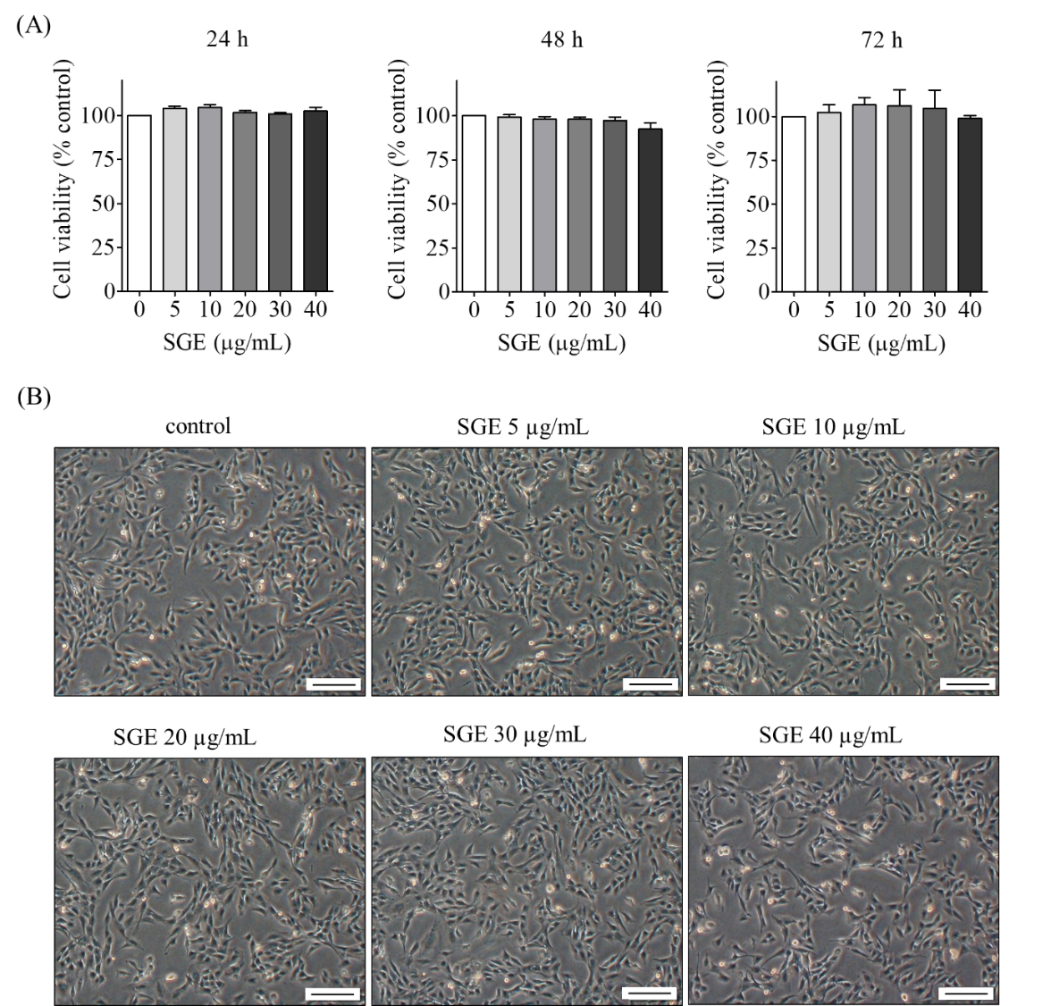
Figure 1 Non-cytotoxic effects of single clove garlic extract (SGE) on C2C12 myoblasts. Subconfluent C2C12 myoblasts were cultured in low serum media containing SGE at the indicated concentrations for 24, 48, and 72 h. The treated cells were subjected to MTT assay (A) and morphology photography at 72 h (B). Scale bar = 200 µm.
SGE did not affect C2C12 myoblast cell proliferation
To evaluate the effects of SGE on cell proliferation, C2C12 myoblasts were treated with SGE at different concentrations (5 - 40 µg/mL) in growth media for 24, 48 and 72 h. MTT assay revealed no significant increase in cell proliferation of C2C12 myoblasts treated with SGE at any concentration compared to non-treated controls at 24, 48 and 72 h (Figure 2(A)). In addition, no differences were observed in the total number of nuclei per area after 72 h treatment (Figure 2(B)), indicating equal myoblast growth rates. We further examined the effect of SGE on cell cycle distribution by flow cytometry. The percentage of cell distribution in G0/G1, S, and G2/M phases of SGE-treated groups at all doses was not significantly different compared to non-treated controls (Figures 2(C) and 2(D)). Although SGE contains numerous beneficial compounds, none have been reported to stimulate proliferation of myoblasts or other cells. Allicin has been reported to have potent anti-proliferative properties in zebrafish [30]. Meanwhile, mouse fibroblasts treated with allicin from garlic extract exhibited disrupted microtubule properties, leading to the inhibition of cell proliferation [31]. In addition, diallyl polysulfides, which are derivatives of allicin, have been shown to inhibit cell proliferation by stimulated cell-cycle arrest in HCT116 human colon cancer cells [32]. Several forms of garlic, from both water and alcohol extracts, have also been shown to have antiproliferative activity in H520, H1975, and A549 lung cancer cell lines [33]. However, our data indicated no adverse effect of SGE on C2C12 myoblast proliferation. This discrepancy is possibly due to different cell types having different sensitivities and physiological responses to garlic extract.
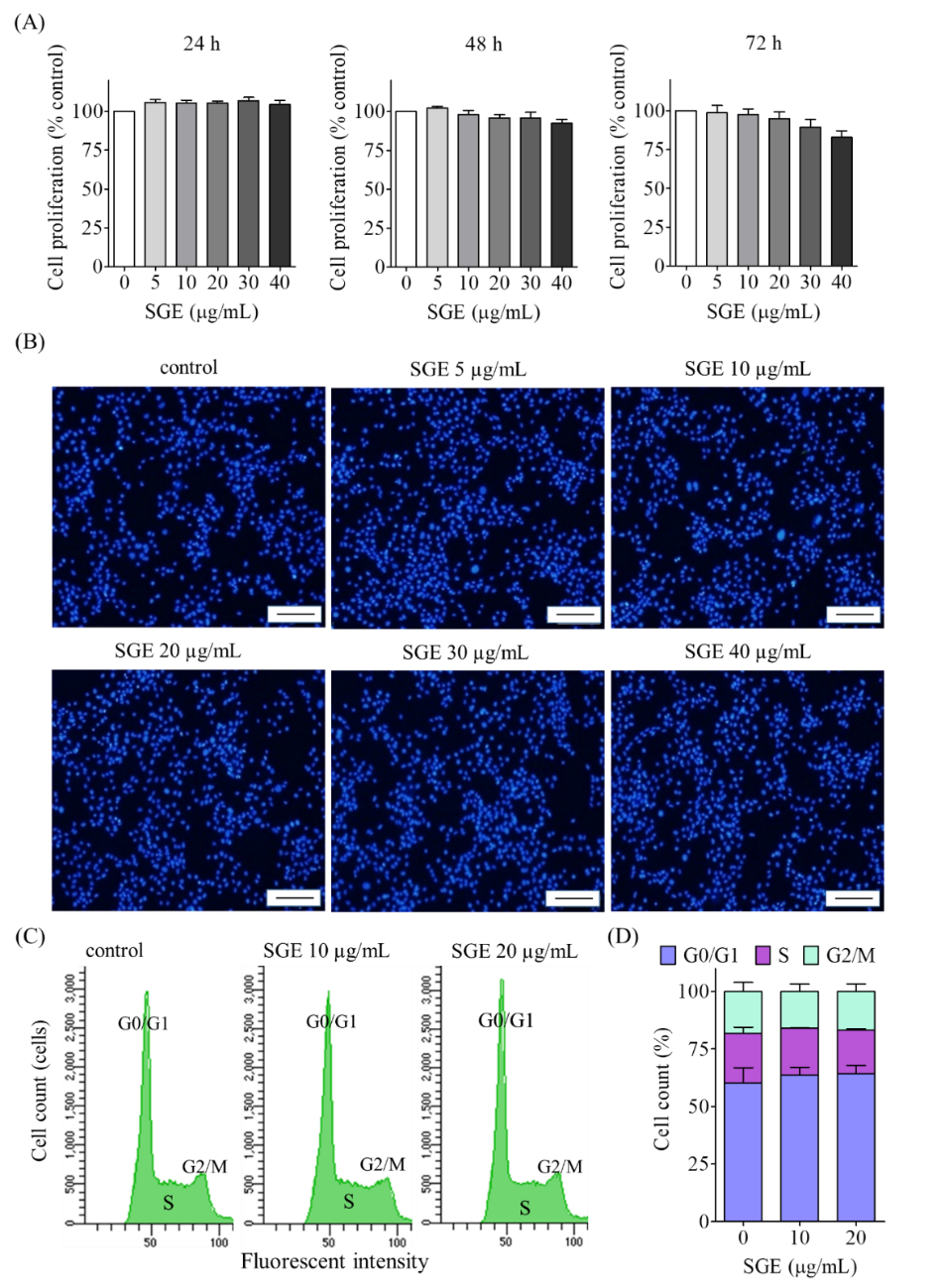
Figure 2 Effects of single clove garlic extract (SGE) on C2C12 myoblast cell proliferation. Subconfluent C2C12 myoblasts were cultured in growth media containing SGE at 5 - 40 µg/mL for 24, 48, and 72 h. The treated cells were subjected to MTT assay (A), cell density observation (B), and cell cycle determination (C). The percentage of cell cycle distributions was calculated (D). Scale bar = 200 µm.
Effect of SGE on C2C12 myoblast cell differentiation
To assess whether SGE enhanced myoblast cell differentiation, confluent C2C12 myoblasts were cultured in DM containing SGE at 5 - 40 µg/mL for 72 h. Phase contrast microscopy and MHC immunostaining revealed numerous elongated and multinucleated myotubes at all doses of SGE treatment. However, there were no significant differences in either the number or
size of myotubes for any dose of SGE compared to the non-treated control (Figure 3(A)). In addition, levels of MHC, MyoD, and myogenin proteins were not significantly different between SGE-treated and non-treated control groups (Figures 3(B) - 3(E)). Similar to myoblast proliferation, SGE did not enhance myoblast differentiation. This is in contrast to previous reports, in which garlic extract has been shown to enhance PC12 nerve cell morphological differentiation with neurite outgrowth [34] and induce morphological differentiation of MCF7 breast cancer cells via up-regulation of eIF2-α phosphorylation [35]. Meanwhile, garlic extract decreased 3T3-L1 adipocyte differentiation through activation of AMP-activated protein kinase (AMPK) [36]. These varying results may indicate cell type-specific effects. However, garlic extract has been reported to possess estrogenic activity [37], and phytoestrogen has been reported to enhance myoblast differentiation [38-40]. Thus, the correlation between estrogenic activity in garlic extract and myoblast differentiation requires further investigation.
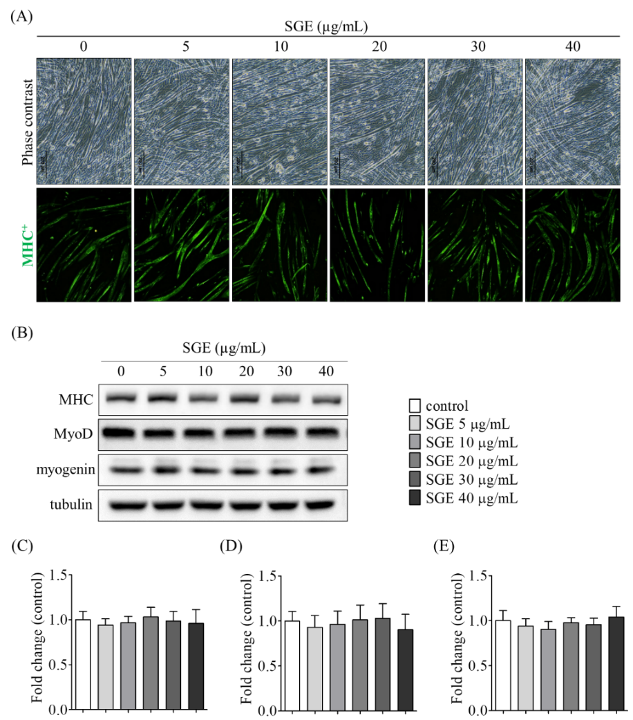
Figure 3 Effects of single clove garlic extract (SGE) on C2C12 myoblast differentiation. Confluent C2C12 myoblasts were cultured in differentiation media containing SGE at 0 - 40 µg/mL for 72 h. The differentiated myotubes were stained with anti-myosin heavy chain (MHC, green) and photographed (A) or subjected to Western blot with anti-MHC, anti-MyoD, and anti-myogenin antibodies (B). The band intensities of MHC (C), MyoD (D), and myogenin (E) were measured. Scale bar = 200 µm.
SGE protects myotubes against DEX-induced atrophy
DEX is commonly used to induce muscle atrophy in vitro through a similar mechanism to that observed in sarcopenia. We therefore aimed to determine the protective effect of SGE against DEX-induced myotube atrophy. The endpoint markers of myotube atrophy in this experiment were decreased MHC protein expression, decreased myotube size, and increased atrogin-1 protein expression. Multinucleated myotubes were treated with DEX (10 µM) in the presence or absence of SGE for 24 h. Visual inspection of immunofluorescence staining revealed that DEX treatment produced smaller myotubes than control conditions, but this effect was reversed by pretreatment with SGE (Figure 4(A)). Measurement confirmed that DEX-treated myotubes were significantly smaller than controls, and that SGE pretreatment at 20 - 40 µg/mL prevented significant DEX-induced myotube atrophy (Figure 4(B)). In addition, the levels of MHC, MyoD, and myogenin—important proteins for myotube growth and development—were altered, consistent with the myotube size, being significantly decreased after DEX treatment compared to the control group. Pretreatment with SGE at 10 - 40 µg/mL significantly alleviated the decreased expression of these proteins (Figures 4(C) - 4(F)). It is noteworthy that our findings are the first report about the preventive effect of SGE on DEX-induced myotube atrophy. Prolonged administration of DEX, a corticosteroid, produces muscle atrophy by increasing the activity of 2 major muscle-specific ubiquitin ligases, atrogin-1 and MuRF-1, via the upregulation of the FoxO transcription factor [41,42]. It also induces cellular oxidative stress and mitochondrial dysfunction, further contributing to muscle atrophy [24,43]. Our findings are congruent with the previous reports on natural extracts of Valeriana fauriei, which possess anti-oxidant activity and can prevent myotube atrophy [44]. Thus, prevention of DEX-induced myotube atrophy may arise from the anti-oxidative activity of SGE [33,45].
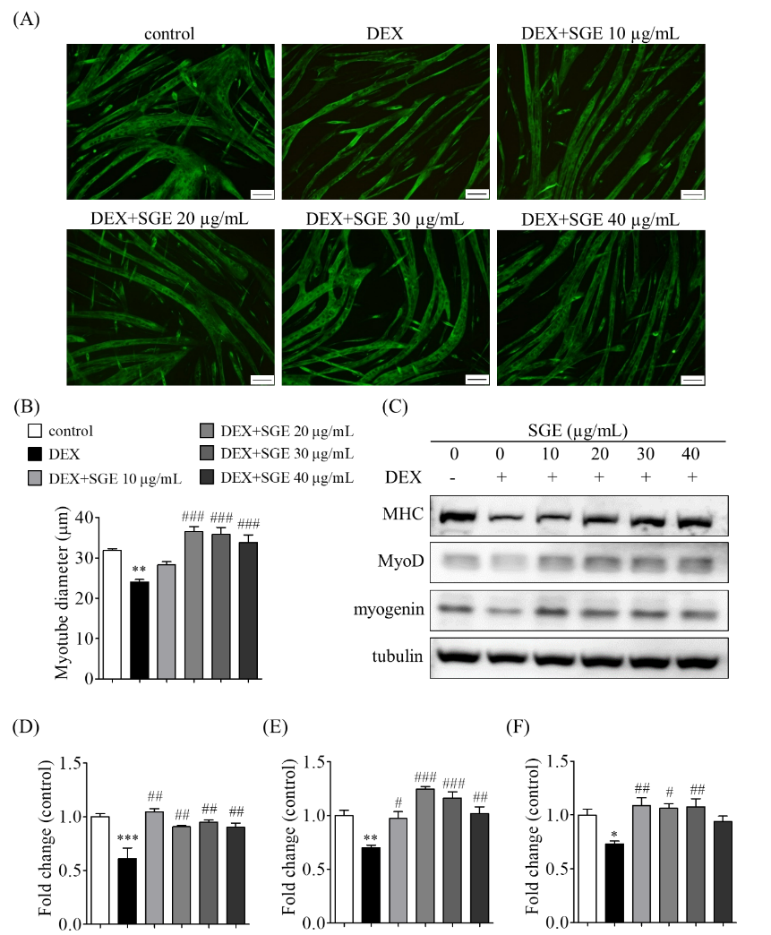
Figure 4 Single clove garlic extract (SGE) protects myotubes against dexamethasone (DEX)-induced atrophy. Confluent C2C12 myoblasts were cultured in differentiation media for 72 h. The differentiated myotubes were pretreated with SGE (10 - 40 µg/mL) for 2 h before being exposed to DEX for 24 h. The treated myotubes were stained with anti-MHC antibody (green) (A), and myotube size was measured (B). The treated myotubes were subjected to Western blot with anti-MHC, anti-MyoD, and anti-myogenin antibodies (C). The band intensities of MHC (D), MyoD (E), and myogenin (F) were measured. *p < 0.05, **p < 0.01, ***p < 0.001 compared to control, and #p < 0.05, ##p < 0.01, ###p < 0.001 compared to the DEX-treated group. Scale bar = 100 µm.
SGE alleviates DEX-induced myotube atrophy associated with increased Akt phosphorylation
To determine possible mechanisms of the protective effect of SGE against DEX-induced myotube atrophy, we further investigated the expression of the Akt protein and its downstream signalling molecules; phosphorylation of Akt is reported to be involved with both suppression of muscle protein degradation and stimulation of muscle protein synthesis. Western blot revealed significantly increased expression of atrogin-1 in DEX-treated myotubes compared to the control group. In addition, DEX induced significant decreases in the expression of the signalling molecules p-Akt, β-catenin, and p-P70S6K. Meanwhile, pretreatment with 20 µg/mL SGE significantly mitigated the downregulation of these proteins by DEX (Figures 5(A) - 5(E)). These results were consistent with myotube morphology and size (Figures 5(F) and 5(G)). In addition, our findings indicated that SGE stimulated the phosphorylation of Akt and P70S6K and suppressed atrogin-1 expression related to DEX-induced muscle atrophy. These results are similar to previous reports on various extracts—including flavonoid dihydromyricetin [46], fucoxanthine [47], ginseng [22], V. fauriei [44], Salvia plebeian, and rosemarinic acid [48]—which can activate p-Akt, p-mTOR, and p-P70S6K, and exhibit beneficial effects in attenuating DEX-induced muscle atrophy. Garlic extract has previously been linked to the expression of phosphorylated Akt [49]. Activation of Akt has been reported to inhibit FoxO [50], which stimulates the expression of MuRF-1 and atrogin-1, two proteins responsible for muscle protein degradation and subsequent muscle atrophy [51]. Treatment of skeletal muscle with DEX has been reported to induce proteasomal proteolysis via MuRF-1 and atrogin-1 expression [52]. Our results show that SGE prevented DEX-induced muscle atrophy by suppressing the expression of atrogin-1, at least in part, through Akt-related signalling. Meanwhile, p‑P70S6K and p-mTOR have been reported to be downstream mediators of Akt phosphorylation in skeletal muscle cells [53]. Activation of Akt triggers the mTOR signalling cascade, which stimulates protein synthesis and leads to the prevention of muscle atrophy [54]. Phosphorylation of mTOR also inhibits protein degradation-induced skeletal muscle atrophy in mice by attenuating autophagy-related pathways [52]. In addition, increased β-catenin is also important for the maintenance of muscle structure [55]. Apart from mTOR, phosphorylation of P70S6K in skeletal muscle cells has been shown to play an essential role in the regulation of protein translation, which is necessary for muscle protein synthesis [53,56]. Activation of P70S6K in skeletal muscle has also been reported as essential for skeletal muscle mass maintenance [56]. Altogether, these results suggest that prevention of DEX-induced myotube atrophy by SGE may act through the activation of Akt/P70S6K signalling pathways.
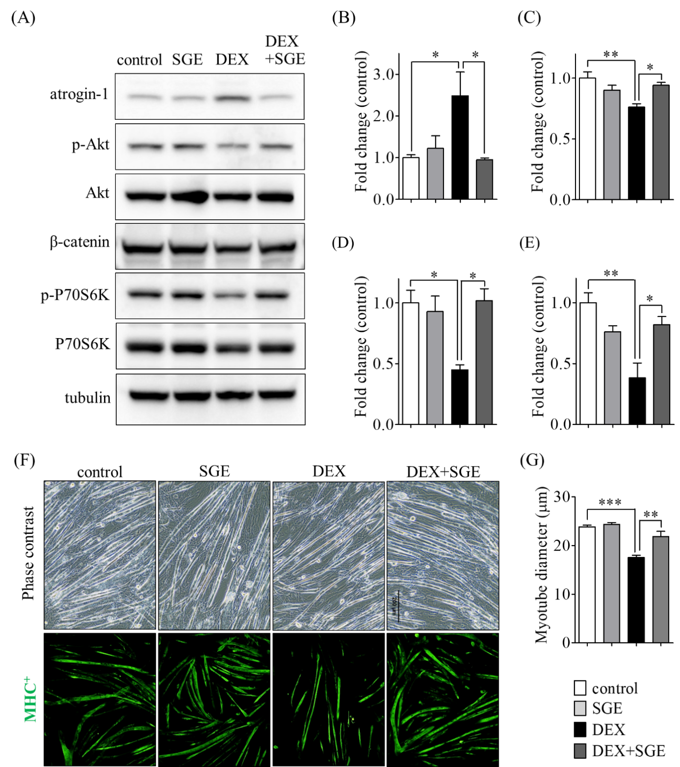
Figure 5 Single clove garlic extract (SGE) alleviates dexamethasone (DEX)-induced myotube atrophy through Akt-related signalling. Differentiated myotubes were pretreated with SGE (20 µg/mL) for 2 h before exposure to DEX for 24 h. The treated myotubes were subjected to Western blot (A). The band intensities of atrogin-1 (B), p-Akt (C), β-catenin (D), and p-P70S6K (E) proteins were measured. The treated myotubes were stained with anti-MHC antibody (green) (F), and diameters were measured (G). *p < 0.05, **p < 0.01, ***p < 0.001 compared to the corresponding group. Scale bar = 200 µm.
SGE modulates mitochondrial biogenesis gene expression in DEX-induced myotube atrophy
As mitochondria play an important role in muscle physiology and homeostasis, disruption of their function can contribute to skeletal muscle atrophy. We evaluated the modulation of the PGC-1α, NRF-1, and TFAM genes, which are implicated in mitochondrial function and muscle atrophy. These genes are also reported to be involved with the expression of phosphorylated proteins involved with muscle atrophy. Real-time PCR revealed that differentiated myotubes treated with DEX exhibited significant downregulation of PGC-1α, NRF-1, and TFAM expression compared to controls. Treatment with SGE alone had no effect on the expression of these genes. However, pretreatment with SGE before 24 h-exposure to DEX significantly attenuated their DEX-induced downregulation (Figure 6). The modulation of myotube PGC-1α, NRF-1, and TFAM levels by SGE provides new insights for the prevention and treatment of muscle loss and atrophy. PGC-1α, NRF-1, and TFAM are involved with mitochondrial biogenesis and function [57]. SGE has been previously reported to stimulate the expression of PGC-1α and NRF-1 [11], which leads to the expression of TFAM [58]. Expression of these genes has been linked to the ablation of FoxO-mediated atrophy in skeletal muscle [59]. In addition, PGC-1α has also been reported to prevent muscle fibre atrophy through the suppression of atrogin-1 and MuRF-1 expression [11,59]. In addition, the upregulation of phosphorylated Akt and P70S6K has been reported to occur through PGC-1α/TFAM signalling pathways [46]. Hence, the modulation of these genes by SGE may ultimately lead to Akt and P70S6K phosphorylation in myotubes treated with DEX, protecting them from atrophy.
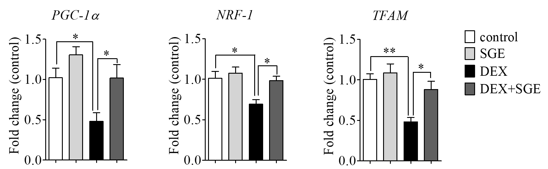
Figure 6 Single clove garlic extract (SGE) pretreatment prevents dexamethasone (DEX)-induced downregulation of PGC-1α, NRF-1, and TFAM genes. Confluent C2C12 myoblasts were cultured in differentiation media for 72 h. The differentiated myotubes were pretreated with SGE before exposure to DEX for 24 h. The treated myotubes underwent real-time PCR analysis. *p < 0.05 and **p < 0.01 compared to the corresponding group.
Conclusions
To the best of our knowledge, these findings provide new information, providing mechanistic insight into the preventive effect of SGE on DEX-induced myotube atrophy. These effects may act through by the upregulation of the Akt/β-catenin and Akt/P70S6K signalling pathways and of mitochondrial biogenesis genes. These results suggest that SGE has potential as a supplement for the prevention of muscle loss and atrophy in older people. However, this experiment was performed in vitro, which does not reflect the full physiology and response of an organism. Further studies using in vivo models and clinical trials are required. In addition, the extracted chemical constituents of SGE should be standardised for comparison across batches and studies.
Acknowledgements
This research was financial supported by Prince of Songkla University, Thailand Grant no. SCI6202017N and Thammasat University, Thailand Research Fund, Contract No TUFT 18/2567, Thammasat University, Thailand. Partially supported by the Thailand Science Research and Innovation (TSRI) Fundamental Fund, fiscal year 2026, Thammasat University, Thailand to P.S. is gratefully acknowledged. We would like to thank Prof. Dr. Koji Ikegami, Hiroshima University, Japan for the technical support in Western blot.
Declaration of Generative AI in Scientific Writing
No content was generated by AI. The authors take full responsibility for the content and conclusions of this work.
CRediT Author Statement
Putri Elok Septiana Dewi: Investigation; Writing-original draft preparation. Piyaporn Surinlert: Conceptualization; Methodology: Software; Validation; Formal analysis; Investigation; Resources; Data curation: Writing-original draft preparation; Writing-review and editing; Project administration; Funding acquisition. Tanaporn Hengpratom: Software; Investigation; Writing-original draft preparation. Thanvarin Thitiphatphuvanon: Investigation; Writing-original draft preparation. Sri Rahayu Lestari: Investigation; Supervision. Chittipong Tipbunjong: Conceptualization; Methodology; Software; Validation; Formal analysis; Investigation, Resources; Data curation; Writing-original draft preparation; Writing-review and editing; Visualization; Supervision; Project administration; Funding acquisition. Putri Elok Septiana Dewi and Piyaporn Surinlert have equal contribution as co-first authors.
References
[1] E Volpi, R Nazemi and S Fujita. Muscle tissue changes with aging. Current Opinion in Clinical Nutrition & Metabolic Care 2004; 7(4), 405-410.
[2] BC Clark. Neuromuscular changes with aging and sarcopenia. Journal of Frailty & Aging 2019; 8(1), 7-9.
[3] JP Gumucio and CL Mendias. Atrogin-1, MuRF-1, and sarcopenia. Endocrine 2013; 43(1), 12-21.
[4] SC Bodine and LM Baehr. Skeletal muscle atrophy and the E3 ubiquitin ligases MuRF1 and MAFbx/atrogin-1. American Journal of Physiology-Endocrinology and Metabolism 2014; 307(6), 469-484.
[5] P Mikaili, S Maadirad, M Moloudizargari, S Aghajanshakeri and S Sarahroodi. Therapeutic uses and pharmacological properties of garlic, shallot, and their biologically active compounds. Iranian Journal of Basic Medical Sciences 2013; 16(10), 1031-1048.
[6] A Shang, SY Cao, XY Xu, RY Gan, GY Tang, H Corke, V Mavumengwana and HB Li. Bioactive compounds and biological functions of garlic (Allium sativum L.). Foods 2019; 8(7), 246.
[7] Y Susanti, M Adriani and A Adi. Effect of single clove garlic extract (Allium Sativum Linn) on blood sugar levels, malondialdehyde, insulin levels and insulin resistance (experiments in rats (Rattus Novergicus) induced by streptozotocin. STRADA Jurnal Ilmiah Kesehatan 2020; 9(2), 954-963.
[8] X Wang. Drug metabolism and pharmacokinetics of organosulfur compounds from garlic. Journal of Drug Metabolism & Toxicology 2013; 04(5), 159.
[9] IS Cheng, JP Tsao, JR Bernard, TW Tsai, CC Chang and SF Liao. Oral post-exercise garlic extract supplementation enhances glycogen replenishment but does not up-regulate mitochondria biogenesis mRNA expression in human-exercised skeletal muscle. Journal of the International Society of Sports Nutrition 2024; 21(1), 2336095.
[10] NA Abd El-Mottaleb, GS Mahmoud, EA Negm and FMA Maksoud. Garlic antagonizes skeletal muscle ischemia reperfusion injury through regulating inflammation, apoptosis and desmin expression in adult male rats. International Journal of Physiology, Pathophysiology and Pharmacology 2019; 11(4), 126.
[11] J Chae, E Lee, SM Oh, HW Ryu, S Kim and JO Nam. Aged black garlic (Allium sativum L.) and aged black elephant garlic (Allium ampeloprasum L.) alleviate obesity and attenuate obesity-induced muscle atrophy in diet-induced obese C57BL/6 mice. Biomedicine & Pharmacotherapy 2023; 163, 114810.
[12] JS Kang, SO Kim, GY Kim, HJ Hwang, BW Kim, YC Chang, WJ Kim, CM Kim, YH Yoo and YH Choi. An exploration of the antioxidant effects of garlic saponins in mouse-derived C2C12 myoblasts. International Journal of Molecular Medicine 2016; 37(1), 149-156.
[13] JS Kang, GY Kim, BW Kim and YH Choi. Antioxidative effects of diallyl trisulfide on hydrogen peroxide-induced cytotoxicity through regulation of nuclear factor-E2-related factor-mediated thioredoxin reductase 1 expression in C2C12 skeletal muscle myoblast cells. General Physiology and Biophysics 2017; 36(2), 129-139.
[14] H Lee, JW Heo, AR Kim, M Kweon, S Nam, JS Lim, MK Sung, SE Kim and JH Ryu. Z-ajoene from crushed garlic alleviates cancer-induced skeletal muscle atrophy. Nutrients 2019; 11(11), 2724.
[15] Y Yoshioka, Y Kubota, Y Samukawa, Y Yamashita and H Ashida. Glabridin inhibits dexamethasone-induced muscle atrophy. Archives of Biochemistry and Biophysics 2019; 664, 157-166.
[16] D Lee and AL Goldberg. SIRT1 protein, by blocking the activities of transcription factors FoxO1 and FoxO3, inhibits muscle atrophy and promotes muscle growth. Journal of Biological Chemistry 2013; 288(42), 30515-30526.
[17] A El-Baroroh, Y Annisa, S Lestari, H Susanto, A Gofur and S Sunaryono. Efficacy of single love garlic extract on self nanoemulsifying drug delivery system (SCGE-SNEDDS) on cytokine expression in vitro. Indonesian Journal of Pharmacy 2024; 35(4), 690-701.
[18] SR Lestari, PES Dewi, SI Maslikah, A Gofur, Y Annisa, NANN Malek, H Susanto, H Rachmawati and R Irwanto. Novel self-nanoemulsifying drug delivery system (SNEDDS) of single garlic extract: Bio-accessibility, cytotoxicity, and anti-inflammatory studies. Malaysian Journal of Fundamental and Applied Sciences 2022; 18(6), 674-683.
[19] SR Lestari, A Ghofur, SI Maslikah, AN Rahma, DN Aisyah, IN Mufidah, ND Rifqi, N Prastita, DS Miasih and AR El Baroroh. Profiling of single garlic extract microencapsulation: Characterization, antioxidant activity, and release kinetic. Journal of Tropical Biodiversity & Biotechnology 2023; 08(3), jtbb79072.
[20] P Surinlert, N Kongthong, M Watthanard, T Sae-Lao, P Sookbangnop, C Pholpramool and C Tipbunjong. Styrene oxide caused cell cycle arrest and abolished myogenic differentiation of C2C12 myoblasts. Journal of Toxicology 2020; 2020, 1807126.
[21] C Tipbunjong, T Thitiphatphuvanon, C Pholpramool and P Surinlert. Bisphenol-A abrogates proliferation and differentiation of C2C12 mouse myoblasts via downregulation of phospho-P65 NF-κB signaling pathway. Journal of Toxicology 2024; 2024, 3840950.
[22] R Kim, H Kim, M Im, SK Park, HJ Han, S An, JS Kang, SJ Lee and GU Bae. BST204 protects dexamethasone-induced myotube atrophy through the upregulation of myotube formation and mitochondrial function. International Journal of Environmental Research and Public Health 2021; 18(5), 2367.
[23] KS Park, H Kim, HJ Kim, KI Lee, SY Lee and J Kim. Paeoniflorin alleviates skeletal muscle atrophy in ovariectomized mice through the ERα/NRF1 mitochondrial biogenesis pathway. Pharmaceuticals 2022; 15(4), 390.
[24] XL Louis, R Murphy, SJ Thandapilly, L Yu and T Netticadan. Garlic extracts prevent oxidative stress, hypertrophy and apoptosis in cardiomyocytes: A role for nitric oxide and hydrogen sulfide. BMC Complementary and Alternative Medicine 2012; 12(1), 140.
[25] F Agahi, R Penalva-Olcina, G Font, A Juan-García and C Juan. Effects of Voghiera garlic extracts in neuronal human cell line against zearalenone's derivates and beauvericin. Food and Chemical Toxicology 2022; 162, 112905.
[26] M Ohtani and T Nishimura. Sulfur-containing amino acids in aged garlic extract inhibit inflammation in human gingival epithelial cells by suppressing intercellular adhesion molecule-1 expression and IL-6 secretion. Biomedical Reports 2020; 12(3), 99-108.
[27] S Bose, SF Robertson and AA Vu. Garlic extract enhances bioceramic bone scaffolds through upregulating ALP & BGLAP expression in hMSC-monocyte co-culture. Biomaterials Advances 2023; 154, 213662.
[28] GB El-Saber, AM Beshbishy, LG Wasef, YHA Elewa, AA Al-Sagan, MEA El-Hack, AE Taha, YM Abd-Elhakim and HP Devkota. Chemical constituents and pharmacological activities of garlic (Allium sativum L.): A review. Nutrients 2020; 12(3), 872.
[29] Y Guo, P Huang, W Wang, J Wu, J Du, J Li, J Gao, H Zhu, J Gao and Y Zheng. Positive effects of allicin on cytotoxicity, antioxidative status, and immunity in “Eriocheir sinensis” hepatopancreatic cells against oxidative stress-induced injury. Antioxidants 2026; 15(1), 93.
[30] J Ravindra, Y Ug, DP Nanjappa, K Kalladka, R Dhakal, A Chakraborty and G Chakraborty. Allicin extracted from Allium sativum shows potent anti-cancer and antioxidant properties in zebrafish. Biomedicine & Pharmacotherapy 2023; 169, 115854.
[31] M Prager-Khoutorsky, I Goncharov, A Rabinkov, D Mirelman, B Geiger and AD Bershadsky. Allicin inhibits cell polarization, migration and division via its direct effect on microtubules. Cell Motility and the Cytoskeleton 2007; 64(5), 321-337.
[32] JD Song, SK Lee, KM Kim, SE Park, SJ Park, KH Kim, SC Ahn and YC Park. Molecular mechanism of diallyl disulfide in cell cycle arrest and apoptosis in HCT‐116 colon cancer cells. Journal of Biochemical and Molecular Toxicology 2009; 23(1), 71-79.
[33] Z Farhat, T Scheving, DS Aga, PA Hershberger, JL Freudenheim, RH Blair, MJ Mammen and L Mu. Antioxidant and antiproliferative activities of several garlic forms. Nutrients 2023; 15(19), 4099.
[34] K Kohda, K Itoh, H Goda, K Samejima, T Fukuuchi, N Morihara, K Imamura, Y Kodera and T Oka. Enhancing effect of aged garlic extract on induction of morphological differentiation with neurite outgrowth in NGF-treated PC12 cells. Pharmacology & Pharmacy 2012; 3(1), 35-41.
[35] S Modem, SE Dicarlo and TR Reddy. Fresh garlic extract induces growth arrest and morphological differentiation of MCF7 breast cancer cells. Genes Cancer 2012; 3(2), 177-186.
[36] O Balogun and HW Kang. Garlic scape (Allium sativum L.) extract decreases adipogenesis by activating AMK-activated protein kinase during the differentiation in 3T3-L1 adipocytes. Journal of Medicinal Food 2022; 25(1), 24-32.
[37] W Mazur. Phytoestrogen content in foods. Baillière's Clinical Endocrinology and Metabolism 1998; 12(4), 729-742.
[38] C Tipbunjong, Y Kitiyanant, G Chaturapanich, N Sornkaew, A Suksamrarn, N Kitiyanant, K Esser and C Pholpramool. Natural diarylheptanoid compounds from Curcuma comosa Roxb. promote differentiation of mouse myoblasts C2C12 cells selectively via ER alpha receptors. Medicinal Chemistry Research 2017; 26(2), 359-370.
[39] Q Yan, Z Fei, M Li, J Zhou, G Du and X Guan. Naringenin promotes myotube formation and maturation for cultured meat production. Foods 2022; 11(23), 3755.
[40] I Soundharrajan, DH Kim, P Kuppusamy and KC Choi. Modulation of osteogenic and myogenic differentiation by a phytoestrogen formononetin via p38MAPK-dependent JAK-STAT and Smad-1/5/8 signaling pathways in mouse myogenic progenitor cells. Scientific Reports 2019; 9(1), 9307.
[41] WE Mitch and AL Goldberg. Mechanisms of muscle wasting. The role of the ubiquitin-proteasome pathway. New England Journal of Medicine 1996; 335(25), 1897-1905.
[42] SC Bodine, E Latres, S Baumhueter, VK Lai, L Nunez, BA Clarke, WT Poueymirou, FJ Panaro, E Na, K Dharmarajan, ZQ Pan, DM Valenzuela, TM DeChiara, TN Stitt, GD Yancopoulos and DJ Glass. Identification of ubiquitin ligases required for skeletal muscle atrophy. Science 2001; 294(5547), 1704-1708.
[43] E Balboa, F Saavedra, LA Cea, V Ramírez, R Escamilla, AA Vargas, T Regueira and JC Sáez. Vitamin E blocks connexin hemichannels and prevents deleterious effects of glucocorticoid treatment on skeletal muscles. International Journal of Molecular Sciences 2020; 21(11), 4094.
[44] YI Kim, H Lee, FS Nirmala, HD Seo, TY Ha, CH Jung and J Ahn. Antioxidant activity of Valeriana fauriei protects against dexamethasone-induced muscle atrophy. Oxidative Medicine and Cellular Longevity 2022; 2022, 3645431.
[45] HJ Jang, HJ Lee, DK Yoon, DS Ji, JH Kim and CH Lee. Antioxidant and antimicrobial activities of fresh garlic and aged garlic by-products extracted with different solvents. Food Science and Biotechnology 2018; 27(1), 219-225.
[46] Y Huang, K Chen, Q Ren, L Yi, J Zhu, Q Zhang and M Mi. Dihydromyricetin attenuates dexamethasone-induced muscle atrophy by improving mitochondrial function via the PGC-1α pathway. Cellular Physiology and Biochemistry 2018; 49(2), 758-779.
[47] L Zhiyin, C Jinliang, C Qiunan, Y Yunfei and X Qian. Fucoxanthin rescues dexamethasone induced C2C12 myotubes atrophy. Biomedicine & Pharmacotherapy 2021; 139, 111590.
[48] JY Kim, HM Kim, JH Kim, S Guo, DH Lee, GM Lim, W Kim and CY Kim. Salvia plebeia R.Br. and rosmarinic acid attenuate dexamethasone-induced muscle atrophy in C2C12 myotubes. International Journal of Molecular Sciences 2023; 24(3), 1876.
[49] M Dong, G Yang, H Liu, X Liu, S Lin, D Sun and Y Wang. Aged black garlic extract inhibits HT29 colon cancer cell growth via the PI3K/Akt signaling pathway. Biomedical Reports 2014; 2(2), 250-254.
[50] A Brunet, A Bonni, MJ Zigmond, MZ Lin, P Juo, LS Hu, MJ Anderson, KC Arden, J Blenis and ME Greenberg. Akt promotes cell survival by phosphorylating and inhibiting a Forkhead transcription factor. Cell 1999; 96(6), 857-868.
[51] C Mammucari, G Milan, V Romanello, E Masiero, R Rudolf, PD Piccolo, SJ Burden, RD Lisi, C Sandri, J Zhao, AL Goldberg, S Schiaffino and M Sandri. FoxO3 controls autophagy in skeletal muscle in vivo. Cell Metabolism 2007; 6(6), 458-471.
[52] H Lee, Y Eo, SY Kim and Y Lim. Guava leaf extract attenuated muscle proteolysis in dexamethasone induced muscle atrophic mice via ubiquitin proteasome system, mTOR-autophagy, and apoptosis pathway. Nutrition Research 2024; 127, 97-107.
[53] C Tipbunjong, P Khuituan, Y Kitiyanant, A Suksamrarn and C Pholpramool. Diarylheptanoid 1-(4-hydroxyphenyl)-7-phenyl-(6E)-6-hepten-3-one enhances C2C12 myoblast differentiation by targeting membrane estrogen receptors and activates Akt-mTOR and p38 MAPK-NF-κB signaling axes. Journal of Natural Medicines 2019; 73(4), 735-744.
[54] RA Saxton and DM Sabatini. mTOR signaling in growth, metabolism, and disease. Cell 2017; 168(6), 960-976.
[55] A Suzuki, A Scruggs and J Iwata. The temporal specific role of WNT/β-catenin signaling during myogenesis. Journal of Natural Sciences 2015; 1(8), 143.
[56] NE Zanchi and AH Lancha. Mechanical stimuli of skeletal muscle: Implications on mTOR/p70s6k and protein synthesis. European Journal of Applied Physiology 2008; 102(3), 253-263.
[57] OA Shelbayeh, T Arroum, S Morris and KB Busch. PGC-1α is a master regulator of mitochondrial lifecycle and ROS stress response. Antioxidants 2023; 12(5), 1075.
[58] DP Kelly and RC Scarpulla. Transcriptional regulatory circuits controlling mitochondrial biogenesis and function. Genes & Development 2004; 18(4), 357-368.
[59] M Sandri, J Lin, C Handschin, W Yang, ZP Arany, SH Lecker, AL Goldberg and BM Spiegelman. PGC-1alpha protects skeletal muscle from atrophy by suppressing FoxO3 action and atrophy-specific gene transcription. Proceedings of the National Academy of Sciences of the United States of America 2006; 103(44), 16260-16265.