Trends
Sci.
2026;
23(8):
12711
Development and Validation of A C57BL/6 Mouse Model of Androgenetic Alopecia Through Testosterone Propionate Induction
Adniana Nareswari1,2,*, Didik Gunawan Tamtomo1, Harijono Kariosentono1,2,
Ratih Puspita Febrinasari3, Muhammad Eko Irawanto1,2, Brian Wasita1,4,
Novan Adi Setyawan1,4 and Paramasari Dirgahayu1,5
1Doctoral Program of Medical Sciences, Faculty of Medicine, Universitas Sebelas Maret,
Jawa Tengah 57126, Indonesia
2Department of Dermatology and Venereology, Faculty of Medicine, Universitas Sebelas Maret,
Jawa Tengah 57126, Indonesia
3Department of Pharmacology, Faculty of Medicine, Universitas Sebelas Maret, Jawa Tengah 57126, Indonesia
4Department of Anatomical Pathology, Faculty of Medicine, Universitas Sebelas Maret, Jawa Tengah 57126, Indonesia
5Department of Parasitology, Faculty of Medicine, Universitas Sebelas Maret, Jawa Tengah 57126, Indonesia
(*Corresponding author’s e-mail: [email protected])
Received: 19 November 2025, Revised: 27 December 2025, Accepted: 2 January 2026, Published: 15 March 2026
Abstract
Androgenetic alopecia (AGA) is the most prevalent form of progressive hair loss, primarily influenced by genetic and androgenic factors, particularly dihydrotestosterone. Existing therapeutic options show limited efficacy, emphasizing the need for reliable and representative animal models. This study established a testosterone propionate (TP)-induced C57BL/6 mouse model of AGA and evaluated the associated clinical, histopathological, and molecular alterations within the Wnt/β-catenin pathway. Twenty-one male C57BL/6 mice were randomized into seven groups (n = 3 per group): One control and six groups receiving subcutaneous testosterone propionate at 0.5, 1.0, or 1.5 mg/day for 7 or 14 days consecutive days. Hair regrowth, dermal thickness, follicle density, and β-catenin expression were analyzed using standardized morphometric and immunohistochemical analyses. Data with normal distribution were assessed using one-way ANOVA, while non-parametric data were evaluated with the Kruskal-Wallis test, followed by LSD or Dunn’s post hoc analysis. Statistical significance was set at p < 0.05. Low-dose, short-term treatment (0.5 mg/day for 7 days) produced only mild dermal thinning and partial follicular reduction, whereas higher doses and prolonged exposure induced progressive inhibition of hair regrowth. The regimen of 1.5 mg/day for 14 days resulted in complete growth arrest, pronounced dermal thinning, follicular depletion, and marked β-catenin suppression (p < 0.001, 0.028, and 0.017, respectively). Post hoc analysis confirmed significant reductions in dermal thickness (groups receiving 1.0 - 1.5 mg/day for 7 - 14 days), follicle density (1.5 mg/day for 7 days and 1.0 - 1.5 mg/day for 14 days), and β-catenin expression (1.5 mg/day for 7 and 14 days). Despite the small sample size, this study demonstrates that subcutaneous TP administration at 1.5 mg/day for 14 days successfully induces androgenetic alopecia in C57BL/6. This model provides a robust, reproducible, and physiologically relevant platform for mechanistic exploration and preclinical testing of therapeutic agents tergeting Wnt/β-catenin signaling.
Keywords: Androgenetic alopecia, Testosterone propionate, C57BL/6 mice, Dermal thickness, Hair follicle density, Wnt/β-catenin signaling, Animal model
Introduction
Androgenetic alopecia (AGA), also known as male- or female-pattern baldness, is the most common progressive hair loss, affecting about 50% of men and women worldwide, with prevalence increasing with age [1]. In men, it typically begins with temporal recession forming an “M” shape and extends to the vertex, while in women it presents as diffuse thinning over the crown without frontal recession [2]. Although AGA is not life-threatening, its impact on quality of life is substantial, as it often triggers psychological distress including anxiety, depression, and decreased self-esteem [3].
The pathogenesis of involves genetic predisposition and androgenic influence, with dihydrotestosterone (DHT) as the key mediator. DHT, converted from testosterone via 5α-reductase, binds androgen receptors with higher affinity than testosterone [4,5]. Elevated activity of 5α-reductase type 2 (SRD5A2) is found in balding follicles, while individuals with congenital deficiency do not develop AGA, underscoring DHT’s central role [6]. The DHT-receptor complex in dermal papilla cells induces apoptosis, suppresses proliferation, prolongs telogen, and causes follicular miniaturization, converting terminal hairs into vellus hairs and reducing the anagen-telogen ratio [7].
Current treatments, such as topical minoxidil and oral finasteride show variable efficacy and adverse effects, underscoring the need for safer and more effective therapies [8]. The development of robust animal models is essential for understanding AGA pathogenesis and testing novel treatments [6]. Testosterone propionate induces AGA-like changes by mimicking human androgen metabolism, thereby facilitating investigation of androgen-mediated molecular mechanisms, particularly the Wnt/β-catenin pathway [9].
Although androgen- or testosterone-induced alopecia models have been reported previously, most studies employed a single androgen dose or treatment duration and were primarily designed to evaluate therapeutic efficacy or specific mechanistic pathways rather than to systematically validate the induction protocol itself. To date, relatively few studies have directly compared multiple androgen doses and exposure durations to identify an optimal and reproducible induction strategy. Therefore, the present study aimed to systematically evaluate different testosterone propionate doses and treatment durations in C57BL/6 mice in order to establish and validate a murine model of androgenetic alopecia exhibiting consistent clinical, histopathological, and molecular features relevant to human AGA.
Materials and methods
Study design
This in vivo experimental study aimed to establish a murine model of androgenetic alopecia (AGA) through testosterone propionate (TP) induction. Experiments were conducted at the Laboratory of the Center for Food and Nutrition Studies, Universitas Gadjah Mada, Yogyakarta, and the Laboratory of Anatomical Pathology, Faculty of Medicine, Universitas Sebelas Maret, Surakarta. Randomization and blinding were applied, and ethical approval was obtained from the Research Ethics Committee of Dr. Moewardi General Hospital (No. 471/III/HREC/2025, Surakarta, Indonesia).
This study was designed as an exploratory model development and validation experiment to determine the optimal dose and duration of testosterone propionate required to reproducibly induce AGA-like features. In accordance with previous androgen-induced alopecia model studies and current animal research guidelines, exploratory and pilot in vivo studies may involve smaller sample sizes when the primary objective is model establishment, feasibility assessment, and reproducibility rather than confirmatory efficacy testing [9-11].
Animal preparation
Twenty-one male C57BL/6 mice (6 - 7 weeks, ~30 g) were randomized into seven groups (n = 3 per group): One control and 6 treatment groups. Inclusion criteria were healthy male mice within the specified age and weight range; exclusion criteria included illness or death during acclimatization. Animals were acclimatized for one week under standard housing conditions with free access to food and water and a 12-hour light-dark cycle. At baseline, the dorsal skin area was carefully shaved using an electric clipper followed by a depilatory cream to remove residual hair shafts, ensuring uniform hair removal across animals. This procedure was performed once, 24 h before testosterone propionate administration, to synchronize hair follicles into the telogen phase and minimize variability in hair cycle stages at the start of the experiment [6]. All experimental procedures involving animals will be conducted in strict accordance with established animal ethics guidelines, including those set forth by the National Institutes of Health (NIH) [12].
Induction protocol
Testosterone propionate (TP; T-Prop®, 100 mg/10 mL; Meditech Pharmaceuticals, India) was administered subcutaneously once daily for 7 or 14 consecutive days. The formulation was supplied as an oil-based solution and used without further dilution, serving as a single working solution for all treatment groups. Different daily doses were achieved by adjusting the injection volume administered to each mouse.
Groups P1 - P3 received TP at doses of 0.5, 1.0, or 1.5 mg/day for 7 days, corresponding to injection volumes of 0.05, 0.10, and 0.15 mL per mouse, respectively. Groups P4 - P6 received the same doses and injection volumes for 14 consecutive days.
The control group received subcutaneous injections of the vehicle (oil) at corresponding volumes and with the same administration frequency as the TP-treated groups. The selected TP dose range (0.5, 1.0, and 1.5 mg/day) was based on previously published murine androgen-induced alopecia models demonstrating dose- and duration-dependent suppression of hair regrowth following subcutaneous androgen administration [13,14,15].
On day 7 (groups P1 - P3) and day 14 (groups P4 - P6), mice were clinically evaluated, euthanized, and dorsal skin biopsies were collected for further analyses.
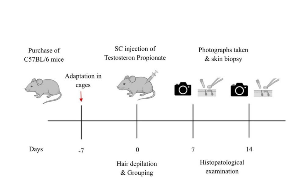
Figure 1 Illustration of the research procedure. Mice were acclimatized for one week, shaved on day 0 to synchronize follicles into telogen, then injected with TP for 7 or 14 days, followed by clinical and histopathological evaluation on day 7 (P1 - P3) and day 14 (P4 - P6).
Clinical evaluation
Clinical assessment of hair regrowth was performed through standardized digital photography on days 0, 7, and 14. On day 0, the dorsal hair of each mouse was carefully shaved to synchronize follicles into the telogen phase, marking the start of treatment. Mice received subcutaneous injections of testosterone propionate (TP) at specified doses (0.5, 1.0, or 1.5 mg/day) for 7 or 14 consecutive days, as assigned to their respective groups.
Photographs were taken under uniform lighting conditions and fixed camera settings to ensure consistency across time points. Each image captured the entire dorsal area, enabling visual comparison of hair regrowth patterns among treatment groups. Hair regrowth was qualitatively evaluated based on the degree of pigmentation and hair coverage, which correlate with the transition from telogen to anagen phase. A darker dorsal surface indicated active anagen induction, whereas pale or hairless regions reflected delayed or suppressed follicular cycling.
Clinical observations were documented throughout the experimental period to monitor behavioral changes, general health status, and local skin reactions to TP injection. All evaluations were performed by 2 independent observers blinded to the treatment groups to minimize bias.
Histopathological analysis
Skin biopsies were obtained from the dorsal region on days 7 and 14 for histopathological examination. Tissues were fixed in 10% neutral-buffered formalin, embedded in paraffin, sectioned at 5 µm, and stained with hematoxylin-eosin (H&E) for microscopic evaluation using a light microscope at 100x and 400x magnification. Histopathological assessment focused on dermal thickness (DT) and hair follicle density (HFD) as primary indicators of AGA-associated structural changes. Dermal thickness was measured vertically from the epidermal-dermal junction to the dermal-subcutaneous interface using calibrated ImageJ software (NIH, USA), following a previously established method [12]. Hair follicle density was quantified as the number of follicles per mm2 of dermal area, where the significant reduction reflects follicular miniaturization and successful AGA induction [16].
Immunohistochemical analysis
Immunohistochemical staining was performed to evaluate β-catenin expression and localization within hair follicles as a marker of canonical Wnt pathway activity. Paraffin sections were deparaffinized, rehydrated subjected to antigen retrieval, and incubated with primary anti- β-catenin antibody, followed by HRP-conjugated secondary antibody and DAB chromogen visualization. Counterstaining was performed using hematoxylin.
β-catenin expression was semi-quantitively assessed based on staining intensity and percentage of percentage of positive follicular cells. A reduction in β-catenin immunoreactivity, often associated with enhances glycogen synthase kinase-3β (GSK3β) activity, indicates inhibition of the Wnt/β-catenin signaling pathway —recognized as a key molecular hallmark of AGA [17].
Statistical analysis
All data were initially tested for normality using the Shapiro-Wilk test. Normally distributed variables, such as dermal thickness (DT), were analyzed using one-way analysis of variance (ANOVA), followed by the Least Significant Difference (LSD) post hoc test for pairwise comparisons. Non-normally distributed variables, including hair follicle density (HFD) and β-catenin expression scores, were evaluated using the Kruskal-Wallis test, followed by Dunn’s post hoc analysis.
Results were expressed as mean ± standard deviation (SD) for parametric data and as median (interquartile range) for non-parametric data. A p - value of < 0.05 was considered statistically significant. All analyses were performed using IBM SPSS Statistics version 25.0 (IBM Corp., Armonk, NY, USA).
Results and discussion
Clinical findings
This study aimed to establish a murine model of androgenetic alopecia (AGA) and determine the optimal testosterone propionate (TP) dose for consistent AGA induction. To identify the most effective regimen, clinical, histopathological, and immunohistochemical parameters were comparatively evaluated. Clinical observations were based on serial photographic documentation of the dorsal region on days 0, 7, and 14 (Figure 2).
The experimental design comprised seven groups: P1 (0.5 mg/day for 7 days), P2 (1.0 mg/day for 7 days), P3 (1.5 mg/day for 7 days), P4 (0.5 mg/day for 14 days), P5 (1.0 mg/day for 14 days), P6 (1.5 mg/day for 14 days), and a normal control group (N) without TP administration.
Among these, group P6 (1.5 mg/day, equivalent to ~50 mg/kg/day for 14 days, body weight ≈ 30 g) exhibited the most prominent AGA-like phenotype, characterized by diffuse hair loss over the dorsal surface and markedly delayed regrowth of new hair. This group demonstrated the most consistent and pronounced androgen-dependent alopecia compared to all others.
Interestingly, the short-term high-dose group (P3: 1.5 mg/day for 7 days) also showed early clinical signs of AGA, including focal alopecia and delayed anagen initiation, although the extent of hair loss was less severe than that observed in P6. Groups receiving lower doses (P1 - P2 for 7 days and P4 - P5 for 14 days) exhibited only mild to moderate thinning, with partial hair regrowth visible by day 14. The normal control group maintained complete hair coverage and normal pigmentation throughout the study period.
Histopathological findings
Histopathological evaluation revealed significant structural alterations in the skin following testosterone propionate (TP) administration, as summarized in Table 1. Compared with the normal control group, all TP-treated groups exhibited distinct histological changes consistent with androgenetic alopecia (AGA).
Dermal thickness (DT) analysis showed a significant reduction across all treatment groups (one-way ANOVA, p ≤ 0.001). As illustrated in Figure 3, the dermis appeared progressively thinner in response to increasing dose and duration of TP exposure. The most pronounced thinning was observed in group P6 (1.5 mg/day for 14 days), which demonstrated a mean DT of 367.87 ± 45.51 µm. Moderate reduction was also evident in P3 (1.5 mg/day for 7 days), confirming that high-dose TP could induce notable dermal atrophy even after short-term administration.
Similarly, hair follicle density (HFD) differed significantly among groups (Kruskal-Wallis, p = 0.028). All TP-treated groups displayed decreased follicular counts compared with controls, indicating follicular miniaturization and early catagen transition. The lowest HFD value was recorded in group P6 (31.00 ± 6.57 follicles/mm²), as shown in Figure 4, followed by P5 and P3, which also demonstrated reduced follicle numbers relative to the normal group.
These histological findings confirm a dose- and duration-dependent effect of testosterone propionate on dermal and follicular structures, supporting the establishment of a reproducible murine model of androgenetic alopecia.
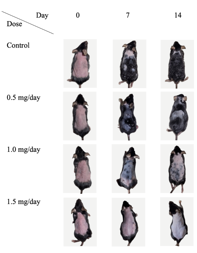
Figure 2 Clinical observations of the murine AGA model. Mice from each group were observed and photographed on days 0, 7, and 14.
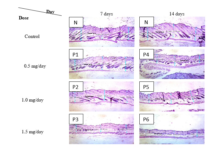
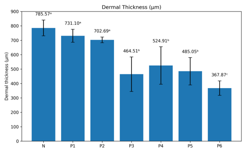
Figure 3 Dermal thickness measurement using hematoxylin and eosin staining (H&E,×10). (A) Representative histopathological images of dorsal skin sections from control and treatment groups at 7 and 14 days, showing dermal thickness measurements (blue lines). (B) Quantitative analysis of dermal thickness (µm) in control and treatment groups. Data are presented as mean ± SD. Bars with different superscript letters indicate statistically significant differences between groups (p < 0.05).
Table 1 Dermal thickness, hair follicle density, and β-catenin expression in C57BL/6 mice after testosterone propionate induction.

Notes: Values are presented as mean ± SD (n = 3). Dermal thickness was analyzed using one-way ANOVA (p < 0.001). Hair follicle density was analyzed using Kruskal-Wallis test (p = 0.028). β-catenin expression was analyzed using Kruskal-Wallis test (p = 0.017). Post hoc analyses were performed using LSD or Dunn’s test where appropriate. *p < 0.05 considered statistically significant.
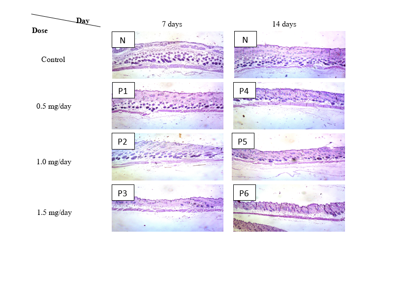
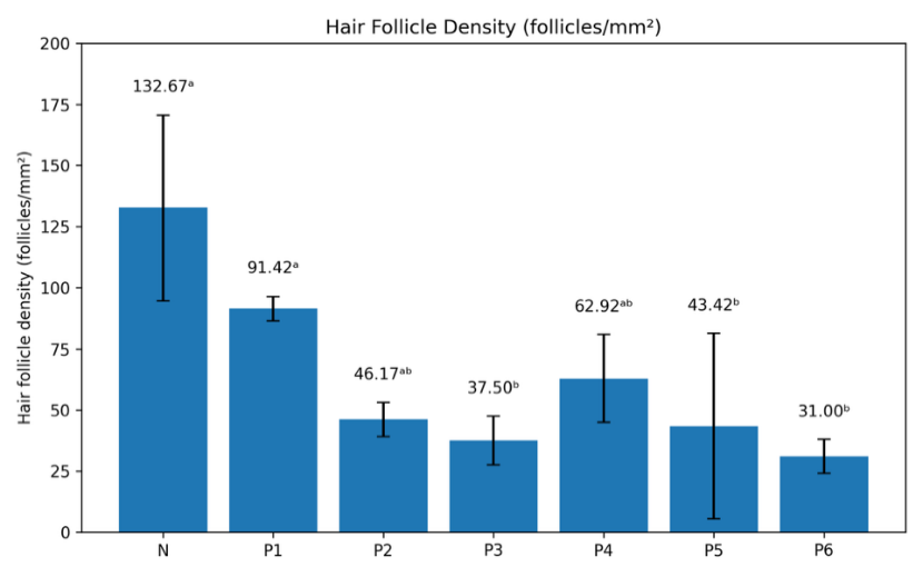
Figure 4 Hair follicle density assessment using hematoxylin and eosin staining (H&E,×10). (A) Representative histopathological images of dorsal skin sections from control and treatment groups at 7 and 14 days, showing the distribution and density of hair follicles. (B) Quantitative analysis of hair follicle density expressed as follicles/mm² in control and treatment groups. Data are presented as mean ± SD. Bars with different superscript letters indicate statistically significant differences between groups (p < 0.05).
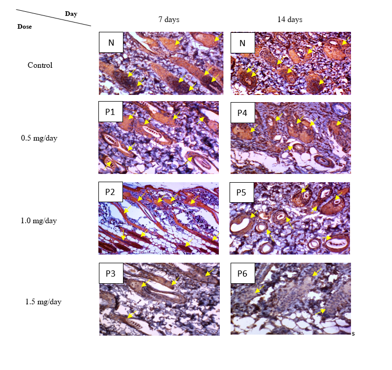
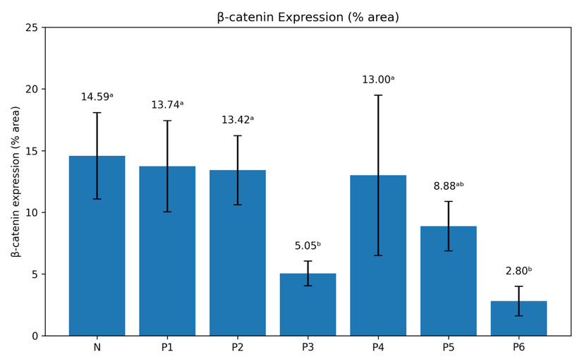
Figure 5 β-catenin expression in skin tissue assessed by immunohistochemistry. (A) Representative immunohistochemical images of dorsal skin sections from control and treatment groups at 7 and 14 days. Brown staining indicates β-catenin-positive expression in hair follicle structures (yellow arrows). (B) Quantitative analysis of β-catenin expression expressed as percentage of positive area (% area) in control and treatment groups. Data are presented as mean ± SD. Bars with different superscript letters indicate statistically significant differences between groups (p < 0.05).
Immunohistochemistry (β-catenin expression)
Based on Table 1, β-catenin expression in hair follicles was significantly reduced following testosterone propionate induction (Kruskal-Wallis, p = 0.017). The lowest expression was observed in P6, with a mean value of 2.80 ± 1.40 (Figure 5).
Taken together, these findings demonstrate dose- and duration-dependent effects of testosterone propionate on dermal thinning, decreased follicle density, and suppression of β-catenin expression. Notably, P6 was the most consistent and effective treatment group in reducing dermal thickness, follicle density, and β-catenin expression, thereby reinforcing the validity of the developed androgenetic alopecia model.
This study successfully established a testosterone-induced murine model of AGA using C57BL/6 mice. The findings demonstrated dose-dependent inhibition of hair growth, dermal thinning, follicular miniaturization, and reduced β-catenin expression, closely mirroring the pathology of human AGA. Clinically, observations indicated that the treatment group receiving 1.5 mg/day (equivalent to 50 mg/kg/day or 0.15 mL/mouse) for 14 days (P6) exhibited the most pronounced hair loss phenotype, characterized by extensive alopecic areas and delayed regrowth of new hair. This outcome is consistent with the phenotypic features of human AGA, which include progressive hair loss, follicular miniaturization, and delayed re-entry of hair follicles into the anagen phase [13,18]. Previous studies have similarly reported that testosterone administration accelerates the transition from anagen to telogen, thereby inducing hair loss patterns resembling AGA [14].
Low-dose groups (0.5 - 1 mg/day) or shorter treatment duration (7 days) exhibited only minimal changes, whereas the control group maintained normal hair growth. These findings are in agreement with Kusumawardani et al. [15], who demonstrated that sustained subcutaneous testosterone administration at 0.075 mL/day for 10 days effectively induced an androgenetic alopecia phenotype resembling human AGA. Macroscopically, the dorsal areas of mice receiving testosterone propionate displayed progressive hair thinning and the formation of bald patches [19]. According to previous reports, this thinning may begin as diffuse hair loss and subsequently progress into more clearly demarcated bald regions over time [9]. These clinical changes closely reflect the manifestation of human AGA, where progressive hair thinning is the defining characteristic [8].
Histopathological analysis with H&E staining reinforced the clinical findings. Dermal thickness (DT) in the high-dose treatment group (1.5 mg/day, 14 days) showed the most significant reduction compared with both the control and the lower-dose groups. Dermal thinning was associated with a decrease in the dermal matrix and impaired dermal papilla function, the regulatory center of the hair cycle. These findings are consistent with recent work by Park et al. [20], which demonstrated that androgens alter dermal morphology through fibroblast apoptosis and reduced extracellular matrix production. Similarly, Hong et al. [18] reported that testosterone exposure in a murine AGA model resulted in histopathological changes, including dermal thinning and hair follicle degeneration, which could be ameliorated with specific herbal therapies. Fu et al. [13] also showed that subcutaneous testosterone administration caused a significant reduction in dermal thickness, while pharmacological interventions were able to restore dermal structure.
Other histological findings also revealed hair follicle miniaturization, a hallmark of AGA. These results are consistent with the reports of Woo et al. [21] and Kusumawardani et al. [15], both of which showed that hair follicle density decreased significantly following testosterone induction, with the high-dose groups displaying the most consistent AGA-like phenotype. Follicular miniaturization and the reduction of active follicles in the anagen phase observed in P6 mirrored the defining characteristics of AGA. Woo et al. [21] also described in C57BL/6 mice that testosterone exposure slowed hair growth and reduced follicle density in a pattern resembling human androgenetic alopecia. Furthermore, Hong et al. [18] and Fu et al. [13] confirmed that testosterone propionate induces significant follicular miniaturization and decreased HFD, both of which could be reversed by natural extract-based or pharmacological interventions. More recently, Kusumawardani et al. [15] corroborated these findings, showing that the most profound decrease in hair follicle density occurred in the highest-dose testosterone group, which was used as a primary parameter for successful induction of the AGA model. This explains why the lower-dose groups still displayed relatively preserved follicle density, whereas the high-dose groups showed diffuse hair loss accompanied by markedly reduced follicle numbers.
Immunohistochemical analysis of β-catenin expression provided mechanistic insights. β-catenin is a key component of the Wnt/β-catenin signaling pathway, which plays a crucial role in maintaining the anagen phase and supporting hair follicle regeneration. The most pronounced decrease in β-catenin expression was observed in the high-dose group (1.5 mg/day, 14 days), indicating that testosterone induction significantly suppressed the Wnt/β-catenin pathway. These findings are consistent with recent studies by Hong et al. [18] and Woo et al. [21], which reported that testosterone-induced murine AGA models exhibited reduced β-catenin expression accompanied by more evident hair loss. Furthermore, Kim et al. [6] demonstrated that enhancing Wnt/β-catenin pathway activity through pharmacological modulation restored hair follicle function and promoted dermal papilla cell proliferation. Similarly, Mehta et al. [22] confirmed that novel therapeutic strategies targeting this pathway hold great potential for mitigating androgen-induced hair loss.
Taken together, the suppression of β-catenin expression observed in this study supports its utility as a molecular indicator for evaluating successful AGA induction in experimental models. This feature may be particularly valuable for future preclinical studies assessing interventions aimed at restoring Wnt/β-catenin signaling and normal hair follicle cycling [9,18].
Although higher androgen doses or longer exposure durations could theoretically induce more severe alopecic changes, such conditions may increase the risk of systemic androgenic effects and reduce translational relevance. Excessive supraphysiological androgen exposure may also confound hair-specific outcomes by inducing generalized skin atrophy or systemic toxicity. Therefore, the selected regimen of 1.5 mg/day for 14 days represents an optimal balance, reliably inducing stable AGA-like features while maintaining animal welfare, experimental reproducibility, and relevance to human disease.
Limitation
This study has limitations inherent to its exploratory, model-development design. The observation period was limited to 7 - 14 days to capture early androgen-induced follicular changes while avoiding prolonged supraphysiological exposure; therefore, long-term follicular dynamics characteristic of chronic human androgenetic alopecia were not assessed. In addition, outcome measures were focused on key clinical, histopathological, and molecular indicators, while other parameters such as the anagen-to-telogen ratio, hair follicle length, hair shaft diameter, and androgen receptor expression were not evaluated and warrant investigation in future studies. Despite these limitations, the study establishes a standardized and reproducible murine AGA model with clear dose- and duration-dependent features, suitable for subsequent mechanistic and preclinical investigations.
Conclusions
This study demonstrates that sustained subcutaneous administration of testosterone propionate reliably induces androgenetic alopecia (AGA)-like features in C57BL/6 mice. The validity of this model is supported by consistent macroscopic findings of inhibited hair regrowth, histopathological evidence of dermal thinning and reduced hair follicle density, and immunohistochemical suppression of β-catenin expression.
By systematically comparing different androgen exposure regimens, this study underscores the critical role of both dose and treatment duration in stabilizing androgen-dependent alopecia phenotypes. The established murine model closely reproduces key clinical, structural, and molecular characteristics of human AGA and provides a robust and reproducible experimental platform for future mechanistic studies and preclinical evaluation of therapeutic strategies targeting androgen signaling and the Wnt/β-catenin pathway.
Acknowledgements
We would like to thank all staff of the Department of Dermatology and Venerology of Dr. Moewardi Hospital, all staff of the Department of Anatomical Pathology, Faculty of Medicine, Sebelas Maret University, Jawa Tengah 57126, Indonesia and also all staff of the Food and Nutrition Study Laboratory Center of Gajah Mada University, Daerah Istimewa Yogyakarta 55281, Indonesia for providing technical assistance and laboratory facilities essential to this study.
Declaration of Generative AI in Scientific Writing
The authors acknowledge the use of generative AI tools (ChatGPT by OpenAI) in the preparation of this manuscript, specifically for language editing and grammar correction. No content generation or data interpretation was performed by AI. The authors take full responsibility for the content and conclusion of this work.
CRediT author statement
Adniana Nareswari: Conceptualization; Methodology; Formal analysis; Investigation; Writing – Original draft. Didik Gunawan Tamtomo: Methodology; Investigation; Data curation. Harijono Kario Sentono: Validation; Supervision; Visualization. Ratih Puspita Febrinasari: Conceptualization; Resources; Investigation; Formal analysis. Muhammad Eko Irawanto: Methodology; Data curation; Validation; Data interpretation. Brian Wasita: Validation; Writing – Review & Editing; Data interpretation. Novan Adi Setyawan: Data interpretation; Writing – Review & Editing. Paramasari Dirgahayu: Conceptualization; Supervision; Data interpretation.
References
[1] OAA Ahmed and WY Rizg. Finasteride nano-transferosomal gel formula for management of androgenetic alopecia: Ex vivo investigational approach. Drug Design, Development and Therapy 2018; 12, 2259-2265.
[2] EL Aukerman and M Jafferany. The psychological consequences of androgenetic alopecia: A systematic review. Journal of Cosmetic Dermatology 2023; 22(1), 89-95.
[3] S Ntshingila, NP Khumalo, M Engel and AT Arowolo. An appraisal of laboratory models of androgenetic alopecia: A systematic review. Skin Health and Disease 2021; 1(2), 15.
[4] N Choi, S Shin, SU Song and JH Sung. Minoxidil promotes hair growth through stimulation of growth factor release from adipose-derived stem cells. International Journal of Molecular Sciences 2018; 19(3), 691.
[5] AJ Smith, RE Clutton, E Lilley, KEA Hansen and T Brattelid. PREPARE: Guidelines for planning animal research and testing. Laboratory Animals 2018; 52(2), 135-141.
[6] HR Kim, JU Park, SH Lee, JY Park, W Lee, KM Choi, SY Kim and MH Park. Hair growth effect and the mechanisms of Rosa rugosa extract in DHT-induced alopecia mice model. International Journal of Molecular Sciences 2024; 25(21), 11362.
[7] D Bellani, R Patil, A Prabhughate, R Shahare, M Gold, R Kapoor and D Shome. Pathophysiological mechanisms of hair follicle regeneration and potential therapeutic strategies. Stem Cell Research & Therapy 2025; 16, 302.
[8] CC Akoh and K Sukhdeo. Finasteride counseling for male androgenetic alopecia should reflect updated findings on prostate cancer risk. Skin Appendage Disorders 2020; 6, 130-131.
[9] Y Li, T Dong, S Wan, R Xiong, S Jin, Y Dai and C Guan. Application of multi-omics techniques to androgenetic alopecia: Current status and perspectives. Computational and Structural Biotechnology Journal 2024; 23, 2623-2636.
[10] ND Sert, V Hurst, A Ahluwalia, S Alam, MT Avey, M Baker, WJ Browne, A Clark, IC Cuthill, U Dirnagl, M Emerson, P Garner, ST Holgate, DW Howells, NA Karp, SE Lazic, K Lidster, CJ MacCallum, M Macleod, EJ Pearl, ..., H Würbel. The ARRIVE guidelines 2.0: Updated guidelines for reporting animal research. PLoS Biology 2020; 18(7), 3000410.
[11] MFW Festing and DG Altman. Guidelines for the design and statistical analysis of experiments using laboratory animals. ILAR Journal 2002; 43(4), 244-258.
[12] GJ Leirós, AI Attorresi and ME Balañá. Hair follicle stem cell differentiation is inhibited through cross-talk between Wnt/β-catenin and androgen signalling in dermal papilla cells from patients with androgenetic alopecia. British Journal of Dermatology 2012; 166(5), 1035-1042.
[13] D Fu, J Huang, K Li, Y Chen, Y He, Y Sun, Y Guo, L Du, Q Qu, Y Miao and Z Hu. Dihydrotestosterone-induced hair regrowth inhibition by activating androgen receptor in C57BL/6 mice simulates androgenetic alopecia. Biomedicine & Pharmacotherapy 2021; 137, 111247.
[14] X Yan, M Cheng, X Shen, S Zhao, F Tang, J Tang, C Ao, L Xue, S Zhu, F Wang and WQ Yang. Development and validation of androgen-induced hair loss and anagen induction mouse models for pharmacological evaluation of anti-androgenic agents. Journal of Pharmacology and Pharmacological Sciences 2024; 6, 192.
[15] A Kusumawardani, N Mulianto, A Nanareswari, P Monica and T Oktaviani. Testosterone-induced androgenetic alopecia mice model: A preliminary study. International Journal of Applied Pharmaceutics 2025; 17(2), 78-82.
[16] F Kaliyadan, A Nambiar and S Vijayaraghavan. Androgenetic alopecia: An update. Indian Journal of Dermatology, Venereology and Leprology 2013; 79(5), 613-625.
[17] CH Ho, T Sood and PM Zito. Androgenetic alopecia. StatPearls Publishing, American, 2025.
[18] GL Hong, HJ Lee, YJ Kim, KH Kim and JY Jung. Stauntonia hexaphylla extract ameliorates androgenic alopecia by inhibiting androgen signaling in testosterone-induced alopecia mice. Iranian Journal of Pharmaceutical Research 2023; 21(1), 133333.
[19] KB Chung, YI Lee, YJ Kim, HA Do, J Suk, I Jung, DY Kim and JH Lee. Quantitative analysis of hair luster in a novel ultraviolet-irradiated mouse model. International Journal of Molecular Sciences 2024; 25(3), 1885.
[20] SH Park, E Lee, D Kim, JW Yoon, SM Jeon, YH Song, J Bang and CH Jung. 2′-Fucosyllactose alleviates hair loss in testosterone-induced androgenic alopecia mice. Journal of Food Biochemistry 2025; 2025, 5625208.
[21] MJ Woo, HY Kang, SJ Paik, HJ Choi, S Uddin, S Lee, SY Kim, S Choi and SK Jung. The in vivo and in vitro effects of Terminalia bellirica (Gaertn.) Roxb. fruit extract on testosterone-induced hair loss. Journal of Microbiology and Biotechnology 2023; 33(11), 1467-1474.
[22] A Mehta, M Motavaf, D Raza, AJ McLure, KD Osei-Opare, LA Bordone and AA Gru. Revolutionary approaches to hair regrowth: Follicle neogenesis, Wnt/β-catenin signaling, and emerging therapies. Cells 2025; 14, 779.