Trends
Sci.
2026;
23(8):
12697
Phytochemical Constituents and In Vitro Antioxidant and Anticancer Activities of Hornstedtia conoidea Ridl. (Zingiberaceae) from the Simeulue Islands
Zumaidar1,*, Nuzul Asmilia2, Saudah3, Indriaty4 and Muhammad Alfarabi5
1Department of Biology, Faculty of Mathematics and Natural Sciences, Universitas Syiah Kuala,
Banda Aceh 23111, Indonesia
2Clinical Laboratory, Faculty of Veterinary Medicine, Universitas Syiah Kuala, Banda Aceh 23111, Indonesia
3Biology Education, Graduate School, Universitas Serambi Mekkah, Banda Aceh 23245, Indonesia
4Department of Biology Education, Faculty of Teacher Training and Education, Universitas Samudra,
Langsa 24354, Indonesia
5Department of Biochemistry, Faculty of Medicine, Universitas Kristen Indonesia, Jakarta 13630, Indonesia
(*Corresponding author: [email protected])
Received: 18 November 2025, Revised: 22 December 2025, Accepted: 25 January 2026, Published: 25 March 2026
Abstract
Hornstedtia conoidea (Zingiberaceae) is a traditional medicinal plant in the Simeulue Islands. Previous studies on the Hornstedtia genus have reported various pharmacological activities, including antioxidant, toxicity, antimicrobial, and glucosidase inhibitory effects; however, its anticancer activity, particularly against breast and cervical cancer, has not yet been scientifically validated. This study aimed to examine the antioxidant and anticancer activities of H. conoidea leaf extracts. The extraction process used solvents with different polarities (methanol, ethyl acetate, and n-hexane). Phytochemical screening and measurements of total phenolic and flavonoid contents were conducted for each extract. Antioxidant activity was evaluated using the 2,2-diphenyl-1-picrylhydrazyl (DPPH) assay, and antiproliferative activity against breast (MCF7) and cervical (HeLa) cancer cells was assessed using the 3-(4,5-dimethylthiazol-2-yl)-2,5-diphenyltetrazolium bromide (MTT) assay. The results showed that the methanol leaf extract of H. conoidea contained alkaloids, phenols, flavonoids, steroids, and terpenoids, with the highest total phenolic (314.69 ± 0.65 mg/g) and flavonoid (43.62 ± 1.20 mg/g) contents among the tested extracts. This extract also exhibited the strongest antioxidant activity, with an IC₅₀ of 14.21± 0.100 µg/mL against DPPH. In addition, the methanol extract demonstrated higher antiproliferative activity against cancer cells compared to the other extracts, with IC₅₀ values of 287.09 ± 3.33 µg/mL for MCF7 cells and 384.78 ± 4.21µg/mL for HeLa cells; however, this activity is classified as weak. The methanolic and ethyl acetate leaf extracts of H. conoidea exhibited strong antioxidant activity, indicating their potential for further development as natural antioxidant agents. In contrast, the in vitro antiproliferative effects against breast and cervical cancer cells remain limited and require further investigation.
Keywords: Aceh, Antiproliferation, Breast cancer, Cervical cancer, Flavonoid content, Phenolic content
Introduction
Oxidative stress-induced DNA damage is a key factor in the pathogenesis of cancer, which accounted for a substantial proportion of global mortality in 2020, especially in Asia [1,2]. In 2022, 9.8 million new cases of cancer in Asia, with 5.4 million deaths. Lung cancer is most common in male and breast cancer in female patients [3]. According to the epidemiological data,
China has the highest rate of cancer in the Asian region with lung cancer was the most prevalent type of cancer diagnosis in male and breast cancer in female patients [2]. In Indonesia, a majority of cancer patients registered at hospitals in several regions were women aged over 39 years with a diagnosis of breast cancer and cervical cancer [4].
The presence of dysregulation in redox signals, apoptosis, and proliferation processes has been reported in cancer cells, a phenomenon attributable to oxidative stress triggered by elevated concentrations of cellular free radicals [5]. Antioxidants derived from dietary and medicinal plants can mitigate cellular oxidative damage, and traditional medicine offers a cost-effective source of natural antioxidants with fewer side effects. The Zingiberaceae family comprises approximately 1,300 aromatic herb species mainly distributed in tropical Asia, including Hornstedtia conoidea, which exhibits antioxidant and anticancer potential but remains pharmacologically underexplored [6-8]. The genus Hornstedtia, which includes around 60 species, is traditionally used for culinary and medicinal purposes, with seeds, rhizomes, and leaves utilized to treat digestive disorders and as spices [9-12]. Previous pharmacological studies have reported antioxidant, antimicrobial, glucosidase-inhibitory, and toxicity-modulating activities in Hornstedtia species, indicating their considerable therapeutic potential [10,13].
The anticancer potential of H. conoidea has not been widely reported scientifically in published research. Previous studies employing simple maceration techniques using methanol and ethanol as solvents have shown that this plant extract has antioxidant and cytotoxic activities [12,13]. However, the phytochemical composition of the leaves of this plant has not been extensively explored. Therefore, it is necessary to profile the compounds contained in the leaves using various solvents with different polarities to demonstrate their various pharmacological effects [14,15]. Additionally, research on H. conoidea, especially the species native to Aceh, Indonesia, is limited.
Simeulue Island, located in the Indian Ocean northwest of Sumatra and part of Aceh Province, is a habitat for H. conoidea, which grows naturally in the Mount Sibao region [16]. Locally, this plant is known as tagbak or gensong [17]. This plant has not been cultivated unlike other species of the Zingiberaceae family, and there is no detailed information regarding the pharmacological properties of H. conoidea from the Simeulue Islands, particularly its antioxidant and anticancer activities. This study aimed to evaluate the antioxidant and antiproliferative activities against cancer cells of H. conoidea leaf extracts from the Simeulue Island region of Aceh, Indonesia. The phytochemical, total phenolic, and total flavonoid contents of the leaf extracts were analyzed. This study is important for assessing the pharmacological potential of H. conoidea and for increasing its economic value. Furthermore, this scientific information can increase awareness among local communities about cultivating this plant and maintaining its sustainability.
Materials and methods
Sample collection and extraction
Hornstedtia conoidea was obtained from Mount Sibao, Simeulue Island, Aceh, Indonesia, at coordinates 2°34'36.8'' N and 96°13'30.4'' E (Figure 1). Taxonomic identification of this plant was performed at the Herbarium Acehense, Department of Biology, Faculty of Mathematics and Natural Sciences, Universitas Syiah Kuala, Banda Aceh, Indonesia. Fresh leaf of H. conoidea (2 kg) were washed with distilled water, air-dried without sunlight at room temperature for seven days, and ground into a coarse powder. Extraction was performed by maceration using 96% methanol (Merck), pure (100%, v/v) ethyl acetate (Merck), and pure (100%, v/v) n-hexane (Merck). Briefly, 100 g of the powdered sample was subjected to maceration using 500 mL of each solvent at a solid-to-solvent ratio of 1:5 (w/v) for 24 h at room temperature. After filtration, the residue was re-extracted under the same conditions, and this procedure was repeated until the extraction solvent became colorless, indicating exhaustive extraction. The resulting filtrates were concentrated through reduced pressure using a vacuum rotary evaporator (Büchi Rotavapor R-300) at 40 - 50 °C, and the crude extract from each solvent was stored at 20 °C in an amber bottle for further analyses. This extract is hereafter referred to as Hornstedtia conoidea of methanol (HC-M), Hornstedtia conoidea of ethyl acetate (HC-EA), and Hornstedtia conoidea of n-hexane (HC-H).
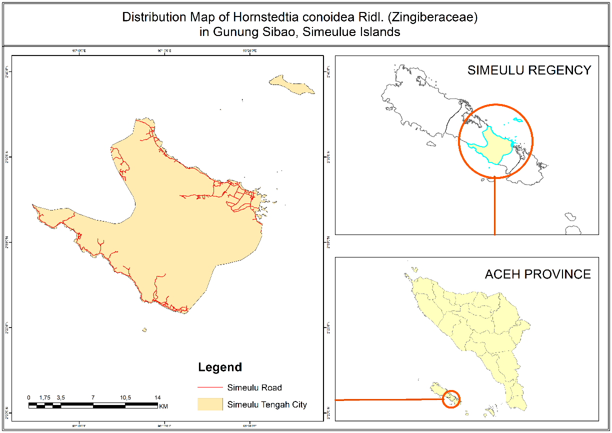
Figure 1 Area collection of Hornstedtia conoidea on Mount Sibao, Simeulue Island, Aceh, Indonesia.
Phytochemical identification
Qualitative phytochemical screening tests were conducted to detect the presence of alkaloids, phenols, flavonoids, terpenoids, and steroids in the extracts. Dragendorff's reagent was used to detect alkaloids, with a positive result indicated by the formation of an orange color. FeCl3 reagent was used to detect phenols, with a positive result indicated by the formation of a black color. AlCl3 reagent was used to detect flavonoids, with a positive result indicated by the formation of a yellowish-green color. Vanillin sulphate reagent was used to detect terpenoids, with a positive result indicated by the formation of a violet color. Cerium sulphate reagent was used to detect steroids, with a positive result indicated by the formation of a faded brown color [18,19].
Total phenolic content (TPC)
The Folin-Ciocalteu method was used to determine the total phenolic content of the extract, and gallic acid (Sigma-Aldrich) was used as the standard compound [20]. Standard gallic acid solutions were prepared in methanol (Merck) at different concentrations (5, 10, 15, 20, 25, and 30 µg/mL). Extract solutions (1 mL) with various concentrations (100 - 500 µg/mL) were mixed with 10% Folin-Ciocalteu reagent and stirred for 1 min. After incubation (room temperature) for 5 min, the solution was mixed with 2 mL of 1 M Na2CO3 (Merck) solution and incubated for 10 min at 50 °C. The solution was cooled for 30 min at room temperature and its absorbance was measured at 746 nm. The total phenol content was determined using a standard curve of absorbance values correlated with the standard concentration of gallic acid, and the results were expressed as milligrams of gallic acid equivalents per gram of fresh weight (mg/g).
Total flavonoid content (TFC)
Total flavonoid content was determined using AlCl3 (Sigma-Aldrich) reagent and quercetin was used as the standard solution [21]. Standard quercetin solutions in methanol were prepared at different concentrations (2, 4, 6, 8, and 10 µg/mL). Samples prepared at various concentrations (5 - 200 µg/mL) were mixed with 2% AlCl3 and 5% sodium acetate (Merck). After incubation for 2.5 h at room temperature, absorbance was measured at 440 nm. The data were expressed as mg/g of quercetin equivalents in milligrams per gram (mg/g) of dry extract.
Antioxidant activity
The DPPH method was used to measure antioxidant activity by measuring free radical scavenging [22]. A total of 2 mL of extract solution (1.56, 3.125, 6.25, 12.5, 25 µg/mL) was dissolved in methanol (Merck), followed by 2 mL of DPPH (Sigma-Aldrich) solution (0.1 mM). The mixture was incubated for 30 min, and the absorbance was measured at 517 nm. DPPH solution without the sample was used as a negative control, and ascorbic acid (Sigma-Aldrich) solution (1, 3, 6, 9 and 12 µg/mL) was used as the positive control. All measurements were performed in triplicate, and the results were expressed as mean ± standard deviation (SD). The free radical scavenging activity of the extracts and the positive control was expressed as the percentage of inhibition, calculated based on the difference between the absorbance of the negative control and that of the sample relative to the absorbance of the negative control. A lower absorbance value indicated a higher free radical scavenging activity. The IC₅₀ value was determined from the dose-response curve of percentage inhibition versus concentration and calculated based on triplicate measurements. The IC50 of antioxidant activity was determined using a linear regression between concentration (X-axis) and percent inhibition (Y-axis) through the probit analysis of concentration log data with the probit free radical scavenger (DPPH) percentage.
Antiproliferative cancer cell assay
The MTT (3-(4,5-dimethylthiazol-2-yl)-2,5-diphenyltetrazolium bromide) assay was used to determine the antiproliferative activity against cancer cells [22]. MCF7 breast cancer cells and HeLa cervical cancer cells used in this assay were obtained from the Cell Culture Laboratory, Research Center of Raw Materials for Drugs and Traditional Medicine-LAPTIAB, National Research and Innovation Agency (BRIN), Puspiptek Serpong in South Tangerang, Banten, Indonesia. Cells were grown in high-glucose Dulbeccoʼs minimal Eagleʼs medium (Gibco, Thermo Fisher Scientific) containing 10% foetal bovine serum (Gibco, Thermo Fisher Scientific), 1% penicillin-streptomycin (Gibco, Thermo Fisher Scientific), and 0.5% amphotericin (Gibco, Thermo Fisher Scientific) until they reached 80% - 90% confluence. These cells were seeded in 96-well plates at a density of 10,000 cells per well in 100 μL of medium and incubated for 24 h (5% CO2; 37 °C). A 96-well plate containing control medium (without cells and samples), negative controls, samples, and positive controls was used for analysis. Samples were dissolved in dimethyl sulfoxide (Sigma-Aldrich) at serial concentrations (62.5, 125, 250, 500, 750, and 1000 μg/mL). Approximately 100 µL of this was mixed with the cells on the plate and incubated for 24 h (5% CO2; 37 °C), and each well was washed with phosphate buffered saline (Sigma-Aldrich) before adding 0.5 mg/mL MTT (Invitrogen, Thermo Fisher Scientific). After 4 h of incubation, purple formazan crystals were formed from the reaction of MTT with the mitochondria of the surviving cells, and the reaction was stopped by adding 10% sodium dodecyl sulfate (Sigma-Aldrich). The absorbance of the purple colour was measured at 570 nm after overnight incubation. The negative control was a cell culture without the sample. Doxorubicin (CKD OTTO Pharmaceuticals) served as a positive control (4, 6, 8, 10, and 12 µg/mL), and the blank solution was the control medium. All experiments were conducted in triplicate, and the results were expressed as mean ± standard deviation (SD). Cell viability was expressed as a percentage and calculated based on the ratio of the absorbance difference between the sample and blank solution to the absorbance difference between the negative control and blank solution. The higher the absorbance, the higher the percentage of viable cells. The percentage inhibition of proliferation was estimated as the difference in cell viability between the sample and the maximum cell viability (100%). The IC₅₀ values in the MTT assay were determined using linear regression analysis of the relationship between extract concentration (X-axis) and percentage of cell growth inhibition (Y-axis).
Results and discussion
Phytochemical profile
The analysis revealed that the secondary metabolites in HC-M and HC-EA included alkaloids, phenols, flavonoids, terpenoids, and steroids. HC-H contained only terpenoids and steroids (Table 1). This indicates that the type and concentration of the solvent can affect the extraction of bioactive compounds from natural products [23]. The polarity of the solvent also affects the extraction process [24]. Therefore, in samples extracted using n-hexane, a solvent with a low polarity, only groups of metabolites with highly hydrophobic chemical structures, such as terpenoids, were detected [25]. In addition, the results of this study are consistent with previous research showing that medicinal plants contain alkaloids, flavonoids, and terpenoids [26]. Metabolite profiling of Hornstedtia leaves showed that they contain essential oils in the form of α-pinene, camphene, and camphor, in addition to secondary metabolites such as flavonoids and terpenoids [27].
Table 1 Phytochemical profile of Hornstedtia conoidea leaf extracts.
Sample |
Compound |
||||
Alkaloid |
Phenol |
Flavonoid |
Terpenoid |
Steroid |
|
HC-M |
+ |
+ |
+ |
+ |
+ |
HC-EA |
+ |
+ |
+ |
+ |
+ |
HC-H |
- |
- |
- |
+ |
+ |
HC-M (methanol extract), HC-EA (ethyl acetate extract), HC-H (n-hexane extract), + (detected), - (not detected)
Total phenolic and flavonoid content
HC-M had the highest phenol and flavonoid content compared to the other extracts. The total phenol content in HC-M was 314.69 ± 0.65 mg GAE/g, and the total flavonoid content in HC-M was 43.62 ± 1.20 mg QE/g (Table 2). The total phenol content of HC-M was three-fold higher than that of HC-EA and four-fold higher than that of HC-H. The total amount of flavonoids in HC-M was one and a half-fold greater than that in HC-EA and five-fold greater than that in HC-H. Methanol had the highest polarity in this study, making it capable of effectively extracting metabolites with hydrophilic chemical structures, such as phenols and flavonoids [28]. The total phenolic and flavonoid content can vary because the metabolites contained in the extract can be affected by the extraction process [23]. Furthermore, the location where the plant grows also affects the metabolite levels contained in an extract. For example, H. conoidea grown in Kibawe, Bukidnon, Philippines, has a total phenol content of 1.67 mg GAE/g [12]. Another report showed that the leaves of this plant, which grows in Mindanao, Philippines, contain 6.87 mg QE/g of flavonoids, which is different from the total flavonoid content in HC-M, HC-EA, and HC-H [13].
Table 2 Total phenolic and flavonoid content of Hornstedtia conoidea leaf extracts.
Sample |
Total phenolics (mg /g) |
Total flavonoids (mg /g) |
HC-M |
||
HC-EA |
||
HC-H |
HC-M (methanol extract), HC-EA (ethyl acetate extract), HC-H (n-hexane extract)
Antioxidant activity
Each extract demonstrated its ability to scavenge free radicals (DPPH), with activity increasing as the extract concentration increased. At the highest extract concentration (25 µg/mL), HC-M inhibited free radicals by 76.93%, HC-EA by 72.75%, and HC-H by 12.99%. The IC50 values of HC-M, HC-EA were 14.21± 0.100 µg/mL and 39.01± 0.160 µg/mL, respectively (Table 3). According to the results of this analysis, HC-M exhibited the strongest antioxidant activity among the extracts.
Table 3 Antioxidant activity of Hornstedtia conoidea leaf extracts.
Samples |
Concentration (μg/mL) |
Free Radical Inhibition (%) |
IC50 (µg/mL) |
HC-M |
1.56 |
8.27 ± 0.001 |
14.21 ± 0.100 |
3.12 |
16.86 ± 0.004 |
||
6.25 |
27.63 ± 0.001 |
||
12.5 |
53.21 ± 0.005 |
||
25 |
76.93 ± 0.002 |
||
HC-EA |
1.56 |
10.52 ± 0.008 |
39.01 ± 0.160 |
3.12 |
16.18 ± 0.002 |
||
6.25 |
37.39 ± 0.007 |
||
12.5 |
57.05 ± 0.009 |
||
25 |
72.75 ± 0.009 |
||
HC-H |
1.56 |
2.75 ± 0.010 |
> 1000 |
3.12 |
4.94 ± 0.004 |
||
6.25 |
7.48 ± 0.002 |
||
12.5 |
10.45 ± 0.007 |
||
25 |
12.99 ± 0.008 |
||
Ascorbic acid |
1.0 |
63.87 ± 0.001 |
4.47 ± 0.025 |
3.0 |
66.26 ± 0.003 |
||
6.0 |
68.11 ± 0.001 |
||
9.0 |
79.32 ± 0.002 |
||
12 |
89.55 ± 0.002 |
HC-M (methanol extract), HC-EA (ethyl acetate extract), HC-H (n-hexane extract).
The results of triplicate experiments are presented with standard deviations.
The IC50 value in this test indicates the concentration of a compound or extract that can inhibit free radicals by 50%. Therefore, the lower the IC50 value measured for a compound or extract to inhibit free radicals, the higher the antioxidant activity of that compound or extract [22]. The results showed that HC-M possessed the highest antioxidant activity compared to the other extracts. This is possible based on previous analysis results that HC-M contains the highest phenols and flavonoids compared to other extracts. The Zingiberaceae family was recognized for its content of flavonoids and phenolic compounds, with the concentration of these compounds being influenced by the geographical location of the plantʼs cultivation. These compounds exhibit antioxidant properties by scavenging free radicals [29,30]. At the molecular level, higher phenolic and flavonoid contents are associated with enhanced antioxidant capacity due to their ability to donate hydrogen atoms or electrons, chelate pro-oxidant metal ions, and stabilize radical intermediates. The presence of hydroxyl groups in flavonoid structures plays a crucial role in these mechanisms, facilitating effective interactions with reactive oxygen species and contributing to the overall antioxidant potential of the extracts [31,32]. However, the antioxidant activity of HC-M was weaker than that of ascorbic acid. As a single compound, ascorbic acid can be sourced from nature or produced synthetically and can act as an antioxidant by acting as a direct electron donor to free radicals. This mechanism can increase the antioxidant activity of ascorbic acid compared to extracts from natural products containing many compounds [33]. The IC50 range value of ascorbic acid in antioxidant assays was 0.2 - 22.5 μg/mL [34].
Antiproliferative activity against cancer cells
The analysis results showed that each H. conoidea leaf extract inhibited the proliferation of MCF7 and HeLa cancer cells (Table 4). This inhibition increased with increasing extract concentration. Except for HC-M, at the lowest test concentration (62.5 μg/mL), the extracts did not inhibit the proliferation of MCF7 and HeLa cells. The highest inhibition of cancer cell proliferation was observed at the highest tested concentration (1,000 μg/mL) in both MCF-7 and HeLa cell lines. At this concentration, the methanolic extract (HC-M) exhibited the highest inhibitory activity against MCF-7 cells (91.96%), followed by the ethyl acetate extract (HC-EA; 82.97%) and the hexane extract (HC-H; 42.99%). Similarly, in HeLa cells, HC-M showed the highest inhibition (89.16%), followed by HC-H (50.37%) and HC-EA (26.34%). The methanolic extract (HC-M) exhibited the highest cytotoxic potency among all tested extracts, as indicated by its lowest IC₅₀ value against MCF-7 cells (287.09 ± 3.33 µg/mL). This result suggests a stronger inhibitory effect of HC-M on MCF-7 cell proliferation compared to the other extracts. A similar trend was observed in HeLa cells, where HC-M also demonstrated superior activity, with an IC₅₀ value of 384.78 ± 4.21 µg/mL, indicating greater effectiveness relative to the remaining extracts. Notably, the lower IC₅₀ value obtained for MCF-7 cells compared to HeLa cells implies that HC-M is more effective in inhibiting the proliferation of MCF-7 cells, suggesting a higher sensitivity of MCF-7 cells to the methanolic extract. However, in this analysis, the inhibition of all extracts was weaker than that of doxorubicin, which was used as a positive control. The IC50 of doxorubicin was 2.08 µg/mL for MCF7 cells and 1.82 µg/mL for HeLa cells (Table 4). Doxorubicin is a chemotherapeutic agent used for the treatment of various types of tumours, such as breast, gastric, lung, ovarian, thyroid, and soft tissue sarcoma, by inhibiting cellular macromolecular biosynthesis, inhibiting topoisomerase II, and relaxing DNA supercoils, thereby preventing DNA replication [35].
According to the test results, HC-M is an extract with good potential to inhibit the proliferation of MCF7 and HeLa cancer cells. This can be observed from the inhibition values at each test concentration and the IC50 values, indicating that the antiproliferative activity of HC-M cancer cells is higher than that of the other extracts. The lower the IC50 value of an extract for inhibiting cancer cell proliferation, the higher its antiproliferative activity against cancer cells [22]. This was evidenced by its higher inhibition percentages at the tested concentrations and its comparatively lower IC₅₀ values, indicating greater antiproliferative effectiveness relative to the other extracts. However, based on commonly used classification criteria, the IC₅₀ values of HC-M (> 200 µg/mL) indicate weak cytotoxic activity. Overall, these findings suggest that the antiproliferative effect of HC-M remains preliminary and limited, warranting further fractionation, chemical characterization, and mechanistic studies to identify the active constituents and enhance efficacy. In contrast, the antioxidant activity of HC-M can be classified as very strong, as indicated by its low IC₅₀ value (< 50 µg/mL), suggesting the presence of potent bioactive compounds that may contribute to its overall biological profile. Members of the Zingiberaceae family are rich in bioactive phytochemicals, particularly flavonoids, terpenoids, and essential oils, which are known to exhibit cytotoxic and antiproliferative activities through the modulation of cancer-related signaling pathways while maintaining relatively low toxicity toward normal cells [36]. In line with this, the ethanolic leaf extract of H. conoidea has demonstrated significant in vitro cytotoxic activity, with an LC₅₀ value of 24.61 mg/L [13]. Flavonoids are considered key contributors to these effects due to their ability to regulate critical intracellular pathways such as PI3K/Akt/mTOR, which govern cell growth, survival, and cell cycle progression [37]. Furthermore, synergistic interactions among phenolic and flavonoid compounds within plant extracts may enhance anticancer efficacy through multiple mechanisms, including apoptosis induction, oxidative stress modulation, MAPK signaling regulation, and PI3K/Akt inhibition. Similar observations in other Zingiberaceae species, such as Etlingera elatior, indicate that higher phenolic and flavonoid contents—often influenced by environmental factors like cultivation altitude—are associated with stronger cytotoxic activity. Overall, these findings underscore the combined influence of phytochemical composition and environmental conditions on the antiproliferative potential of Zingiberaceae-derived extracts [30].
Table 4 Antiproliferative activity of Hornstedtia conoidea leaf extracts.
Samples |
Concentration (μg/mL) |
MCF7 Cell Inhibition (%) |
HeLa Cell Inhibition (%) |
IC50 (µg/mL) for MCF7 Cell |
IC50 (µg/mL) for HeLa Cell |
HC-M |
62.5 |
‒1.50 ± 0.90 |
‒29.54 ± 9.20 |
||
125 |
8.68 ± 5.70 |
10.79 ± 5.70 |
|||
250 |
40.86 ± 5.10 |
35.75 ± 2.60 |
|||
500 |
83.42 ± 2.90 |
63.09 ± 1.40 |
|||
750 |
86.75 ± 1.70 |
70.77 ± 5.30 |
|||
1000 |
91.96 ± 0.60 |
89.16 ± 0.70 |
|||
HC-EA |
62.5 |
3,09 ± 9.00 |
2.56 ± 7.20 |
648.18 ± 5.46 |
709.36 ± 9.09 |
125 |
19,72 ± 5.30 |
8.34 ± 0.10 |
|||
250 |
27,61 ± 0.40 |
5.06 ± 2.70 |
|||
500 |
31,51 ± 9.10 |
6.89 ± 1.40 |
|||
750 |
81,39 ± 2.00 |
22.57 ± 3.70 |
|||
1000 |
82,97 ± 1.90 |
26.34 ± 4.00 |
|||
HC-H |
62.5 |
1.95 ± 4.90 |
21.68 ± 7.30 |
> 1000 |
> 1000 |
125 |
5.44 ± 4.50 |
28.56 ± 12.30 |
|||
250 |
7.74 ± 5.40 |
31.26 ± 4.50 |
|||
500 |
8.53 ± 4.60 |
31.3 ± 4.90 |
|||
750 |
26.81 ± 2.50 |
54.65 ± 9.90 |
|||
1000 |
42.99 ± 5.70 |
50.37 ± 5.20 |
|||
Doxorubicin |
4.0 |
52.37 ± 2.39 |
75.99 ± 1.59 |
2.08 ± 3.28 |
1.82 ± 2.45 |
6.0 |
58.22 ± 9.85 |
80.72 ± 3.33 |
|||
8.0 |
64.22 ± 4.71 |
80.25 ± 0.70 |
|||
10 |
64.92 ± 2.16 |
87.48 ± 3.49 |
|||
12 |
63.84 ± 6.42 |
81.78 ± 1.34 |
HC-M (methanol extract), HC-EA (ethyl acetate extract), HC-H (n-hexane extract).
The results of triplicate experiments have been presented with standard deviation.
Conclusions
Hornstedtia conoidea leaf extracts contain various bioactive secondary metabolites, with methanol identified as the most effective solvent for extracting phenolic and flavonoid compounds. The methanol and ethyl acetate extracts demonstrated notable antioxidant activity, highlighting their potential as natural antioxidant sources. In contrast, their in vitro antiproliferative effects against breast (MCF-7) and cervical (HeLa) cancer cell lines were relatively weak, as indicated by high IC₅₀ values. These findings suggest that, while the extracts possess promising antioxidant properties, their anticancer potential remains limited at the crude extract level. Therefore, further studies involving bioactivity-guided fractionation, comprehensive phytochemical characterization, and in vivo evaluation are necessary to identify the responsible bioactive compounds and to elucidate the mechanisms underlying the observed biological activities.
Acknowledgements
The authors are grateful to the Directorate of Research, Technology, and Community Service (DRTPM), Directorate General of Higher Education, Research, and Technology of the Ministry of Education, Culture, Research, and Technology, Indonesia for funding our work through Regular Fundamental Research (PFR) (Contract Number: 168/E5/PG.02.00.PL/2023), Research Program for the 2023.
Declaration of generative AI in scientific writing
The authors take full responsibility for the content and conclusions of this work. The authors acknowledge the use of a generative AI tool in the preparation of this manuscript, particularly for language editing and grammar correction. No content generation or data interpretation was performed by AI.
CRediT author statement
Zumaidar: Conceptualization; Validation; Supervision. Nuzul Asmilia: Methodology; Investigation. Saudah: Software; Visualization; Resources. Indriaty: Data Curation; Writing - Original Draft. Muhammad Alfarabi: Formal Analysis; Writing - Review & Editing.
References
[1] G Pizzino, N Irrera, M Cucinotta, G Pallio, F Mannino, V Arcoraci, F Squadrito, D Altavilla and A Bitto. Oxidative stress: Harms and benefits for human health. Oxidative Medicine and Cellular Longevity 2017; 2017(1), 8416763.
[2] H Sung, J Ferlay, RL Siegel, M Laversanne, I Soerjomataram, A Jemal and F Bray. Global cancer statistics 2020: GLOBOCAN estimates of incidence and mortality worldwide for 36 cancers in 185 countries. CA: A Cancer Journal for Clinicians 2021; 71(3), 209-249.
[3] The Global Cancer Observatory, Available at: https://gco.iarc.who.int/media/globocan/factsheets/populations/935-asia-fact-sheet.pdf, accessed December 2025.
[4] S Gondhowiardjo, N Christina, NPD Ganapati, S Hawariy, F Radityamurti, VF Jayalie, S Octavianus, AP Putra, SM Sekarutami, GB Prajogi, A Giselvania, M Adham, ARAH Hamid, E Widyastuti, Y Prabowo, T Aninditha, G Purwoto, RA Aman, TP Siregar, AD Siswoyo, ..., Priyambodho. Five-year cancer epidemiology at the national referral hospital: Hospital-based cancer registry data in Indonesia. JCO Global Oncology 2021; 7, 190-203.
[5] F Xing, Q Hu, Y Qin, J Xu, B Zhang, X Yu and W Wang. The relationship of redox with hallmarks of cancer: The importance of homeostasis and context. Frontiers in Oncology 2022; 12, 862743
[6] S Skalli, R Hassikou and M Arahou. An ethnobotanical survey of medicinal plants used for diabetes treatment in Rabat, Morocco. Heliyon 2019; 5(3), 01421.
[7] RN Alolga, F Wang, X Zhang, J Li, LSP Tran and Y Yin. Bioactive compounds from the Zingiberaceae family with known antioxidant activities for possible therapeutic uses. Antioxidants 2022; 11(7), 1281.
[8] YQ Zhou, H Liu, MX He, R Wang, QQ Zeng, Y Wang, WC Ye and QW Zhang. A review of the botany, phytochemical, and pharmacological properties of Galangal. Academic Press, London, 2018, p. 351-396.
[9] Saudah, Zumaidar, Darusman, Fitmawati, DI Roslim and Ernilasari. Ethnobotanical knowledge of Etlingera elatior for medicinal and food uses among ethnic groups in Aceh Province, Indonesia. Biodiversitas 2022; 23(8), 4361-4370.
[10] SE Hashim and HM Sirat. Chemical composition, antioxidant, antimicrobial, and α-glucosidase activities of essential oils of Hornstedtia scyphifera (Zingiberaceae). Natural Product Communications 2018; 13(2), 229-232.
[11] NP Menzed and FM Acma. In vitro studies on pollen viability, pollen germination and pollen tube growth of Hornstedtia conoidea Ridl. - A Philippine endemic ginger species. Journal of Tropical Life Science 2018; 8(3), 303-310.
[12] GB Barbosa and MCY Nueva. Antioxidant activity and phenolic content of Hornstedtia conoidea (Zingiberaceae). Asian Journal of Biological and Life Sciences 2019; 8(1), 1-7.
[13] GB Barbosa, CV Alinapon, MM Enot, GJD Baltazar, EO Buncales, AG Gultiano, and AMG Ang. Cytotoxicity, antioxidant activity, and total phenolic content of the ethanolic leaf extract of Hornstedtia conoidea Ridl. Asian Journal of Biological and Life Sciences 2022; 11(1), 166-172.
[14] B Ginting, Mustanir, Nurdin, Maulidna, Murniana, and Safrina. Evaluation of antioxidant and anticancer activity of Myristica fragrans Houtt. Bark. Pharmacognosy Journal 2021; 13(3), 780-786.
[15] Masyudi, M Hanafiah, Rinidar, S Usman and Marlina. Phytochemical screening and GC-MS analysis of bioactive compounds of Blumea balsamifera leaf extracts from South Aceh, Indonesia. Biodiversitas 2022; 23(3), 1344-1352.
[16] AP Keim, A Agusta, MF Royyani, O Efendy and BA Qarim. Botani pulau simeulue (spermatophyta). Unimal Press, Lhokseumawe, Indonesia, 2019.
[17] GA Otunola and AJ Afolayan. A review of the antidiabetic activities of ginger. IntechOpen 2020. https://doi.org/10.5772/intechopen.88899
[18] JR Shaikh and MK Patil. Qualitative tests for preliminary phytochemical screening: An overview. International Journal of Chemical Studies 2020; 8(2), 603-608.
[19] S Dubale, D Kebebe, A Zeynudin, N Abdissa and S Suleman. Phytochemical screening and antimicrobial activity evaluation of selected medicinal plants in Ethiopia. Journal of Experimental Pharmacology 2023; 15, 51-62.
[20] YH Lee, C Choo, MI Watawana, N Jayawerdana and VY Waisundara. An appraisal of eighteen commonly consumed edible plants as functional food based on their antioxidant and starch hydrolase inhibitory activities. Journal of the Science of Food and Agriculture 2015; 95(14), 2956-2964.
[21] S Aryal, MK Baniya, K Danekhu, P Kunwar, R Gurung and N Koirala. Total phenolic content, flavonoid content, and antioxidant potential of wild vegetables from western Nepal. Plants 2019; 8(4), 96.
[22] M Alfarabi, M Safithri, JM Cing, ES Arodes, D Andrianto, R Kurniasih and HEC Hutapea. Antioxidant and antiproliferative activity of black rice extract against colon cancer cells in-vitro and in-silico. Trends in Sciences 2025; 22(10), 10397.
[23] IE Mannoubi. Impact of different solvents on extraction yield, phenolic composition, in vitro antioxidant and antibacterial activities of deseeded Opuntia stricta fruit. Journal of Umm Al-Qura University for Applied Sciences 2023; 9, 176-184.
[24] A Akbar, NH Soekamto, Firdaus and Bahrun. Antioxidant of n-hexane, ethyl acetate and methanol extracts of Padina sp with DPPH method. IOP Conference Series: Earth and Environmental Science 2021; 800, 012019.
[25] MS Sankhla, K Parihar, R Kumar, DS Bhagat, SS Sonone, GK Singh, V Nagar, G Awasthi and CS Yadav. Ecofriendly approach for steroids, terpenes, and alkaloids-based biosurfactant. Biointerface Research in Applied Chemistry 2023; 13(2), 114.
[26] WB Adeosun and DT Loots. Medicinal plants against viral infections: A review of metabolomics evidence for the antiviral properties and potentials in plant sources. Viruses 2024; 16(2), 218.
[27] J Kappen, A David, K Pieplow, A Wujtschik, I Ware, D Dhar, C Wagner, MD Davari, K Franke and LA Wessjohann. Exploring Hornstedtia scyphifera: An extensive multimethod phytochemical investigation reveals the chemical composition and bioactive potential. Discover Plants 2025; 2, 6.
[28] ZX Ng, SN Samsuri and PH Yong. The antioxidant index and chemometric analysis of tannin, flavonoid, and total phenolic extracted from medicinal plant foods with the solvents of different polarities. Journal of Food Processing and Preservation 2020; 44(9), 14680.
[29] AA Belew and SH Gebre. Comparative assessment of phenolic and flavonoid contents and antioxidant activities in methanol extracts of spices from Jigjiga market, Ethiopia. Pharmacological Research - Natural Products 2025; 6, 100168.
[30] N Asmilia, W Sari, R Nursanty, Saudah and Zumaidar. Phytochemical diversity and anticancer potential of Etlingera elatior fruits across ecological regions in Aceh, Indonesia. Biodiversitas 2025; 26(11), 5423-5429.
[31] SA de Souza Farias, KS da Costa and JBL Martins. Analysis of conformational, structural, magnetic, and electronic properties related to antioxidant activity: Revisiting flavan, anthocyanidin, flavanone, flavonol, isoflavone, flavone, and flavan-3-ol. ACS Omega 2021; 6(13), 8908-8918.
[32] VR Puspa, Zumaidar, Nurdin and Fitmawati. Phytochemicals and bioactivities of Erigeron sumatrensis Retz. from Gayo Highlands: Antioxidant, antidiabetic, anticancer. Journal of Human, Earth, and Future 2025; 6(1), 41-66.
[33] KLB Santos, VAN Bragança, LV Pacheco, SSB Ota, CPO Aguiar and RS Borges. Essential features for antioxidant capacity of ascorbic acid (vitamin C). Journal of Molecular Modeling 2022; 28, 1-9.
[34] F Martino-Morales, AJ Alonso-Castro, JR Zapata-Morales, C Carranza-Álvarez and OH Aragon-Martinez. Use of standardized units for a correct interpretation of IC50 values obtained from the inhibition of the DPPH radical by natural antioxidants. Chemical Papers 2020; 74, 3325-3334.
[35] R Sangeeta. An overview of doxorubicin formulations in cancer therapy. Journal of Cancer Research and Therapeutics 2014; 10(4), 853-858.
[36] T Soumya, PR Jayasree and PRM Kumar. Zingiberaceae plants: A cornucopia of promising chemotherapeuticals for cancer cure. Springer, Cham, Switzerland, 2023, p. 427-462.
[37] TA Zughaibi, M Suhail, M Tarique and S Tabrez. Targeting PI3K/Akt/mTOR pathway by different flavonoids: A cancer chemopreventive approach. International Journal of Molecular Sciences 2021; 22(22), 12455.