Trends
Sci.
2026;
23(8):
13308
UHPLC Analysis of Phenolic Compounds from Phanera strychnifolia Leaf Extracts, and Their Alpha-glucosidase and Alpha-Amylase Inhibitory Activities Supported by Molecular Docking
Sumet
Kongkiatpaiboon1,2,*,
Ngampuk Tayana1,2,
![]() ,
Boonwiset Seaho2,
,
Boonwiset Seaho2,
![]() ,
,
Johann
Schinnerl3,
![]() ,
Nongnaphat Duangdee1,2,
,
Nongnaphat Duangdee1,2,
![]() ,
Wichayasith Inthakusol1,2,
,
Wichayasith Inthakusol1,2,
![]() ,
,
Chawannuch
Mudjupa4,
![]() and Savita Chewchinda5,
and Savita Chewchinda5,
![]()
1Drug Discovery and Development Center, Office of Advanced Science and Technology,
Thammasat University, Rangsit Campus, Pathum Thani 12121, Thailand
2Thammasat University Research Unit in Cannabis and Herbal Product Innovation,
Thammasat University, Rangsit Campus, Pathum Thani 12121, Thailand
3Department of Botany and Biodiversity Research, University of Vienna, Rennweg 14, A-1030-Vienna, Austria
4Pharmaceutical Chemistry and Natural Products Research Unit, Faculty of Pharmacy,
Mahasarakham University, Maha Sarakham 44150, Thailand
5Department of Food Chemistry, Faculty of Pharmacy, Mahidol University, Bangkok 10400, Thailand
(*Corresponding author’s e-mail: [email protected], [email protected])
Received: 9 January 2026, Revised: 11 February 2026, Accepted: 18 February 2026, Published: 25 March 2026
Abstract
Phanera strychnifolia is a traditional medicinal plant showing remarkable antihyperglycemic activity. To develop as a phytopharmaceutical product for Diabetes, an in-depth investigation of the α-glucosidase and α-amylase inhibitory activities of the main constituents was conducted. The developed UHPLC method was validated for qualitative and quantitative analyses of the major constituents gallic acid (1), trilobatin (2), yanangdaengin (3), and phloretin (4). The performed stress tests provide information about storage conditions and about the stabilities of these compounds under various conditions. The optimized extraction with ethanol/water (1:1, v/v) gave the highest yield of gallic acid (1), and the dihydrochalcones trilobatin (2), yanangdaengin (3), and phloretin (4). Yanangdaengin (3) exhibited potent α-glucosidase inhibitory activity (IC50 of 10.42 µM), which was about 30 times higher active than the commercial drug acarbose (IC50 of 329.09 µM). This result was also confirmed by computerized molecular docking. Our work confirms the suitability of hydrophilic leaf extracts from P. strychnifolia as a potential herbal medicine for the treatment of postprandial hyperglycemia and provides valuable information for the phytopharmaceutical development of this plant.
Keywords: Bauhinia strychnifolia, Diabetes mellitus, Extraction, Lysiphyllum strychnifolium, α-Glucosidase inhibitory activity, Molecular docking, Quantitative analysis
Introduction
Diabetes mellitus (DM) is a chronic, metabolic disorder characterized by high levels of blood glucose. It is caused by either a lack of insulin secretion or impaired insulin action, or both [1]. Most Diabetes patients are type 2 where the pathogenesis is caused by two main factors: Defective insulin secretion by pancreatic β-cells and the inability of insulin-sensitive tissues to respond to insulin [2]. In 2021, approximately 529 million people suffered from Diabetes mellitus, and forecasts predict that this number will rise to 1.31 billion by 2050 [3]. Chronic hyperglycemia can cause several functional and structural abnormalities due to increased oxidative stress, inflammation, protein glycation, and formation of advanced glycation end products (AGEs) [4]. Currently, Diabetes mellitus is treated with antihyperglycemic drugs and insulin therapy. However, there are some adverse effects such as vascular complications, hypoglycemia, flatulence, diarrhea, weight gain, weakness, fatigue, lactic acidosis, abdominal pain, and hepatotoxicity known from the used drugs [4]. Such adverse effects might be reduced by application of natural products because some of them are able to reduce the blood glucose levels in patients suffering from Diabetes mellitus [5].
Phanera strychnifolia (Craib) Jiang et al. [6] (Fabaceae) or “Kha-Yan” or “Ya-Nang-Daeng” in Thai [7] is native to Thailand and Peninsula Malaysia and used in the folk medicine for detoxification [8]. Its water extract showed remarkable antihyperglycemic activity in mice by inhibition of -glucosidase in a concentration-dependent manner and also inhibited the glucose uptake. This aqueous extract reduced fasting blood glucose levels in both the OGTT and 28-day consecutive administration models in streptozotocin induced diabetic mice. Further, it downregulated SGLT1 and GLUT2 mRNA expression in Caco-2 cells [9]. Moreover, Sato et al. [10] reported anti-inflammatory and anti-hyperuricemic effects of this plant extract.
Phytochemical studies of the leaves revealed the presence of mainly dihydrochalcones such as trilobatin, phloretin and its glycoside yanangdaengin (phloretin 4'-O-(6''-O-galloyl)-β-d-glucoside) as well as the polyol d-pinitol [11,12] in the leaf extracts. Yanangdaengin showed significant α-glucosidase inhibitory activity [12]. Due to the potential of P. strychnifolia as a source of potent plant-derived α-glucosidase inhibitors, we conducted an in-depth investigation of the main constituents of this plant species, including quantification of the main constituents by UHPLC, analyzing their stability under various harsh conditions. Further, we assessed the α-glucosidase and α-amylase activity inhibition and performed molecular docking to confirm and elucidate binding interactions with α-glucosidase. The present study provides useful information for drug discovery and development based on this plant from natural sources.
Materials and methods
Plant materials
P. strychnifolia leaves were purchased from a local supplier at Nongkhai province. The sample was identified by comparison with an authentic sample by the authors. The voucher specimen (SKPS001) was deposited at the Drug Discovery and Development Center, Office of Advanced Science and Technology, Thammasat University, Thailand.
Chemicals
HPLC grade methanol was purchased from Fisher Scientific, UK. Deionized water was purified by Ultra Clear, Siemen Technologies Corp. (Germany). Gallic acid, phloretin, 3,5-dinitrosalicylic acid (DNSA), acarbose hydrate, 4-nitrophenyl α-d-glucopyranoside (p-NPG), p-nitrophenol, potassium sodium l-(+)-tartrate tetrahydrate were purchased from TCI, Japan. α-amylase from porcin pancreas, and α-glucosidase from Saccharomyces cerevisiae were purchased from Sigma-Aldrich, USA. Starch was purchased from Merck, Germany. Sodium phosphate dibasic and sodium dihydrogen orthophosphate were purchased from Loba Chemie, India. Sodium hydroxide was purchased from Fisher Scientific, UK. All reagents were of analytical grade if not stated otherwise.
Preparation of crude extracts
Dried and ground leaves of P. strychnifolia were extracted exhaustively with n-hexane, ethyl acetate, ethanol, water, and ormixture of ethanol-water (1:1, v/v) using ultrasonication at room temperature or using heating reflux with boiling water using water bath for 30 min and filtered through a Whatman filter paper No.1. The extracts obtained were combined, evaporated using a rotary evaporator, dried and the yield determined.
UHPLC Analysis
Apparatus
The analysis was performed on a Vanquish UHPLC system (Thermo Fisher Scientific Inc.) equipped with a Thermo Scientific Vanquish-Binary Pump F, a Thermo Scientific Vanquish-Split sampler FT, a Thermo Scientific Vanquish-column compartment H, and a Thermo Scientific Vanquish-diode array detector FG. The separation was done on a Hypersil BDS C18 column (50×2.1 mm2 i.d., 2.4 m grain size). The mobile phases were (A) 0.1% formic acid in water and (B) methanol. A mobile phase time program was set up at a constant flow rate of 0.5 mL/min with linear gradient elution from 100% A for 2 min, 0% B to 40% B in 1 min, 40% to 55% B in 4 min, and 100% B for 2 min. Prior to each injection, the column was equilibrated with 100% A for 3 min. The column temperature was set at 25 °C and the diode array detection was set at a wavelength of 280 nm. The injection volume was 2 L for all samples and standards.
Stock and working solution of standard compounds
Standard stock solutions of gallic acid (1), trilobatin (2), and yanangdaengin (3) were prepared in methanol-water (1:1, v/v) to obtain a final concentration of 2.0 mg/mL. Phloretin (4) was prepared in methanol at a final concentration of 2.0 mg/mL Working standard solutions were obtained by appropriate dilution of the stock solution with methanol-water (1:1, v/v) to obtain the desired concentration.
Sample preparation
Each sample was accurately weighed and extracted with methanol-water (1:1, v/v) at concentrations of 2–10 mg/mL in an ultrasonic bath for 30 min. Each solution was passed through a 0.22 m nylon membrane filter before injection.
Method validation
The method was validated according to the ICH guidelines [13]. The validation parameters were linearity, precision, accuracy, limit of detection (LOD), and limit of quantitation (LOQ).
Linearity
Linearity relationship between each analyte concentration and the response was evaluated across nine known concentrations in the range of 3.9–1,000 g/mL. The calibration curves were plotted using the peak area versus the concentration of the standard. Regression lines were calculated using the least square method.
Precision
Intra- and inter-day precision were measured by analyzing 100 g/mL standard solution. The intra-day precision (representing repeatability of the method) was determined seven times within one day, while the inter-day precision (representing intermediate precision) was determined for three different days. The percentage relative standard deviation (%RSD) was then calculated and used to indicate the precision.
Accuracy
Spiking study was conducted to evaluate the accuracy of the method. Three different levels of standard mixtures of pre-analyzed standard solution were spiked to the known concentration sample extract. Spiked samples were prepared in triplicate for each concentration level. The results from measurements were calculated. The recovery was calculated as:
recovery (%)= 100×(amount found – original amount)/amount spiked.
Limit of detection (LOD) and limit of quantitation (LOQ)
Determination of the signal-to-noise ratio (S/N) was performed by comparing signals from samples with known low concentrations of analyte with noise situated around the peak of interest under the proposed chromatographic condition. A minimum concentration that produced a S/N of 3:1 was considered for the LOD and of 10:1 for the LOQ
Stress testing
The stress conditions included base hydrolysis, acid hydrolysis, oxidation, and photolytic conditions were done following our previous method [14]. It was done by adding 50 µL of reagent to 1 mL of extract sample. Concentrated hydrochloric acid (36% w/w), 5N sodium hydroxide, and hydrogen peroxide (30% w/w) were used as reagents for acid hydrolysis, base hydrolysis, and oxidative stress, respectively. Deionized water was used as solvent. The spiked solutions were incubated at 60 °C for 60 min. The photolytic stress was done by spiking the deionized water and exposing it to light (4500 Lux) for 72 h. Each sample was then analyzed with the developed UHPLC method. The peak purity of stressed samples was monitored by the diode array detector in the wavelength range of 200–800 nm.
Total phenolic contents
Determination of total phenolic contents was conducted using Folin-Ciocalteu method [15]. Each sample (20 µL) was mixed with 50 µL of Folin-Ciocalteu’s solution (diluted 1:10 with deionized water) in a 96-well plate. Then 80 µL of 7.5% (w/v) sodium bicarbonate was added and incubated for 30 min in the dark at room temperature. The absorbance was measured at 765 nm using a microplate reader (SPECTROstar Nano, BMG Labtech, Germany). The standard curve was established using gallic acid (1). Total phenolic content in the extracts was expressed as gallic acid equivalent per gram of extract (mg GAE/g of extract).
-Amylase and -glucosidase inhibitory activity
The -amylase enzyme was prepared in 0.02 M sodium phosphate buffer (pH 6.9) with 0.006 M sodium chloride. Acarbose (positive control) was prepared in deionized water. n-Hexane and ethyl acetate extracts of P. strychnifolia leaves were prepared in methanol while ethanolic, 50% ethanol in water, and water extracts were prepared in methanol-water (1:1, v/v). Gallic acid (1), trilobatin (2), yanangdaengin (3) were prepared in methanol-water (1:1, v/v) while phloretin (4) was prepared in methanol. The final concentration of solvents used was 10% and did not affect the activity of the enzyme. The starch solution was prepared at concentration of 1% (w/v) in boiling water. The DNSA reagent was prepared as follows: 3,5-Dinitrosalicylic acid (1 g) was dissolved in 50 mL water, 30 g sodium potassium tartrate added, after mixed well then added 20 mL 2N sodium hydroxide, then adjusted the volume to 100 mL with deionized water.
The -amylase inhibitory activity was determined by the release of glucose using the 3,5-dinitrosalicylic acid (DNSA) method using 96-well plate modified from [16]. Briefly, 20 L of sample or standard solution, 80 µL of sodium phosphate buffer, 50 L of -amylase solution (2 U/mL), were mixed and incubated at 37 C for 10 min, then 50 L of 1% (w/v) starch solution were added and incubated at 37 °C for 3 min. The reaction blank was done using phosphate buffer instead of -amylase while corresponding solvent, i.e., water, methanol-water (1:1, v/v), or methanol, were used as sample blanks. The reaction was then terminated by adding 50 L of DNSA solution and boiling at 100 °C for 15 min. After cooling to room temperature for 15 min, the absorbance was measured at 540 nm using a microplate reader (SPECTROstar Nano, BMG Labtech, Germany). The enzymatic activity was calculated compared with that of the assay using corresponding solvent without any inhibitor.
For the -glucosidase assay 4-nitrophenyl α-d-glucopyranoside (p-NPG) was prepared in 0.1 M sodium phosphate buffer (pH 6.8). Acarbose (positive control) and d-pinitol were prepared in deionized water. The n-hexane and ethyl acetate extracts were prepared in methanol while ethanolic, 50% ethanol in water, and water extracts were prepared in methanol-water (1:1, v/v). Gallic acid (1), trilobatin (2), yanangdaengin (3) were prepared in methanol-water (1:1, v/v) while phloretin (4) was prepared in methanol. The final concentration of solvent (10%) did not affect the enzymatic activity. Sodium bicarbonate was prepared in water at concentration of 1 mM.
The -glucosidase inhibitory activity was carried out according to a previously published method [16] with slight modifications using p-NPG as substrate. Briefly, 10 L of sample or standard solution was mixed with 40 L of 20 mM p-NPG in sodium phosphate buffer (pH 6.9), and 40 L of 0.1 U/mL -glucosidase in sodium phosphate buffer (pH 6.9). The reaction blank was done using phosphate buffer instead of -glucosidase while corresponding solvent, i.e., water, methanol-water (1:1, v/v), or methanol, were used as sample blanks. The reaction was incubated at 37 °C in microplate reader. After 20 min, 100 µL of 1 mM sodium bicarbonate was added. The absorbance at 405 nm was measured after 20 min using a microplate reader (SPECTROstar Nano, BMG Labtech, Germany). The activity of the enzyme was calculated compared to that of the assay using corresponding solvent without any inhibitor.
The most potent inhibitor of -glucosidase, yanangdaengin (3), was further used for the determination of the enzyme kinetics. Lineweaver-Burk and Dixon plots were performed based on previous studies. Briefly, various concentrations of yanangdaengin (3) (0–40 µg/mL) and substrate p-NPG (0.125–4 mM) were used in the reactions. The absorbance at 405 nm was measured every 2 min for 20 min at 37 C. Calibration curve of p-nitrophenol in phosphate buffer was done to calculate the amount p-nitrophenol. The rate of reaction (V) based on p-nitrophenol release rate was plotted as Lineweaver-Burk plot (1/V versus 1/[p-NPG]) and Dixon plot (slope versus [inhibitor]). The mode of enzyme inhibition was identified according to the Lineweaver-Burk plot between the reciprocal value (1/V and 1/[p-NPG]) where V was the reaction rate and [p-NPG] was the concentration of p-NPG. Kinetic parameters consist of the Michaelis-Menten constant (Km) and the maximum velocity of enzyme activity (Vmax) were calculated from the Lineweaver-Burk plot, while the inhibition constant (Ki) was calculated from the Dixon plot.
Computerized molecular docking
Molecular docking of acarbose, gallic acid (1), trilobatin (2), yanangdaengin (3), and phloretin (4) was performed according to the previously published method [17]. The 3D crystal structure of α-glucosidase complexed with α-d-glucopyranose [18] (PDB ID: 3A4A, resolution 1.60 Å) was obtained from RCSB (https://www.rcsb.org/; accessed on 10 Oct 2025). The 3D structures of gallic acid (1), trilobatin (2), phloretin (4), and acarbose were retrieved from PubChem (https://pubchem.ncbi.nlm.nih.gov/; accessed on 10 Oct 2025), while the structure of yanangdaengin (4) was drawn in ChemDraw15.1. These structures were subjected to geometry optimization and energy minimization using IQmol program (Version 2.15.0). Further preparation involved modifying the charges and torsion angles with AutoDockTools v1.5.7 and saving the structures in the PDBQT format. The α-glucosidase structure was prepared using AutoDockTools, by removing co-crystallized ligands and water molecules, adding polar hydrogens, and computing Gasteiger charges. A grid box encompassing the amino acid regions ASP69, HIS112, ARG213, ASP215, GLU277, HIS351, ASP352, and ARG442, was generated. The grid size was set as x= 45, y= 45, and z= 45. The grid center coordinate was set as x= 21.395, y= −7.515, and z= 23.75.
Molecular docking was performed using AutoDock Vina [19,20], with an exhaustiveness value of 64, CPU number of 8, energy range number of 3, and default parameters. The best docking pose was selected based on the lowest binding energy. The reliability of the docking protocol was validated by redocking the co-crystallized ligand, α-d-glucopyranose, into the active site. The root means square deviation (RMSD) between the redocked pose and the original crystallographic pose was then calculated. Finally, protein-ligand interactions were visualized using Discovery Studio Visualizer v.21.1.0.20298.
Results
The extraction with ethanol/water (1:1) under sonication at room temperature of dried and ground P. strychnifolia leaves exhibited the highest yield of 27.27% (w/w). The application of the other solvents water, ethanol, and ethyl acetate yielded lower amounts (Table 1). Extraction with ethanol/water (1:1) under reflux afforded a yield of 33.58% (w/w). The highest total phenolic content was determined in the ethanol (heating reflux) extract (382.04 mg GAE/g) followed by 50% ethanol (heating reflux), ethanol (sonication), ethanol (heating reflux), ethyl acetate, water (heating reflux), water (sonication), and n-hexane extract which exhibited 361.89, 327.37, 319.07, 216.33, 215.61, 180.24, and 30.35 mg GAE/g, respectively (Table 1).
Table 1 Extraction yield, gallic acid (1), trilobatin (2), yanangdaengin (3), and phloretin (4) concentration, total phenolic content, α-amylase and α-glucosidase inhibitory activity of leaf extracts from P. strychnifolia.
Extraction solvents |
Yield
of crude extract |
Active content in dried crude extract (% w/w) |
Total Phenolics (mg GAE/g) |
-amylase inhibition IC50 (g/mL) |
-glucosidase inhibition IC50 (g/mL) |
|||
gallic acid (1) |
trilobatin (2) |
yanangdaengin (3) |
phloretin (4) |
|||||
n-Hexane |
2.75 ± 0.19 |
- |
0.0040 ± 0.0011 |
0.0045 ± 0.0014 |
- |
30.35 ± 0.77 |
>1,000 |
>1,000 |
Ethyl acetate |
6.09 ± 0.14 |
0.09 ± 0.01 |
10.88 ± 1.68 |
3.52 ± 0.60 |
0.10 ± 0.02 |
216.33 ± 8.86 |
>1,000 |
356.25 ± 44.81 |
Ethanol (sonication) |
8.32 ± 0.50 |
0.22 ± 0.02 |
15.06 ± 0.15 |
3.87 ± 0.15 |
0.11 ± 0.04 |
327.37 ± 9.70 |
7.06 ± 2.47 |
5.37 ± 0.75 |
Ethanol (heating reflux) |
27.69 ± 0.94 |
0.17 ± 0.07 |
14.13 ± 1.16 |
3.19 ± 0.38 |
0.06 ± 0.01 |
382.04 ± 1.95 |
9.05 ± 1.49 |
5.08 ± 1.47 |
50% Ethanol (sonication) |
27.27 ± 2.62 |
0.45 ± 0.05 |
10.49 ± 1.04 |
2.22 ± 0.13 |
0.02 ± 0.01 |
319.07 ± 3.89 |
15.82 ± 4.09 |
3.70 ± 1.92 |
50% Ethanol (heating reflux) |
33.58 ± 2.13 |
0.46 ± 0.01 |
10.10 ± 0.40 |
2.06 ± 0.02 |
~0.01 |
361.89 ± 1.72 |
8.31 ± 3.13 |
3.76 ± 0.87 |
Water (sonication) |
17.36 ± 0.13 |
0.68 ± 0.05 |
4.24 ± 0.20 |
0.41 ± 0.04 |
- |
180.24 ± 8.05 |
>1,000 |
16.12 ± 3.62 |
Water (decoction) |
12.38 ± 1.39 |
0.69 ± 0.01 |
3.18 ± 0.52 |
0.30 ± 0.01 |
- |
215.61 ± 4.40 |
>1,000 |
22.62 ± 4.79 |
Acarbose
|
- |
- |
- |
- |
- |
|
6.65
± 0.09 |
212.46
± 31.74 |
Gallic acid (1) |
- |
- |
- |
- |
- |
|
N/A |
>1,000 |
Trilobatin (2) |
- |
- |
- |
- |
- |
|
>1,000 |
410.04
± 55.86 |
Yanangdaengin (3) |
- |
- |
- |
- |
- |
|
>1,000 |
6.13
± 1.61 |
Phloretin (4) |
- |
- |
- |
- |
- |
|
>1,000 |
41.35
± 20.84 |
d-Pinitol |
- |
- |
- |
- |
- |
|
>1,000 |
>1,000 |
aData expressed as mean ± SD (n= 3).
The developed UHPLC method allowed a quick and reliable qualitative and quantitative analyses of the extracts obtained from P. strychifolia leaves and its major contituents gallic acid (1), trilobatin (2), yanangdaengin (3), and phloretin (4) (Figure 1, Figure 2). The performed validation was conducted according to the ICH guidelines [13] and comprised of linearity, precision, accuracy, limit of detection, and limit of quantitation. The linearity was tested between 2–500 µg/mL of all analytes. Excellent linear relationships between peak areas versus concentrations were demonstrated by coefficients of determination (r2) (Table 2). The LOD and LOQ values were 0.02 and 0.06 µg/mL, respectively, indicating a high sensitivity of the method. The repeatability and intermediate precision based on analysis using the same analyte on three different days provided a percentage of relative standard deviation less than 1% (Table 3), showing good precision. The accuracy of the method was tested assessing the recovery. The results showed that the recovery of gallic acid (1), trilobatin (2), yanangdaengin (3), and phloretin (4) were in the range of 99.45%–101.45% (average 100.56 %), 100.47%–100.65% (average 100.54%), 101.25%–101.48% (average 101.34%), and 100.97%–106.18% (average 104.08%), respectively (Table 4). The amounts detected of gallic acid (1), trilobatin (2), yanangdaengin (3), and phloretin (4) in each dried crude extract were analyzed using the developed UHPLC method. The results are shown in Table 1. The highest content of trilobatin (2), yanangdaengin (3), and phloretin (4) was detected in the ethanol extract followed by ethyl acetate, 50% aqueous ethanol, and water extract while the highest content of gallic acid (1) was detected in water extract followed by 50% ethanol, ethanol, and ethyl acetate extract.
Table 2 Method validation parameters for quantitative analysis of gallic acid (1), trilobatin (2), yanangdaengin (3), and phloretin (4).
Parameter |
Results |
|||
Gallic acid (1) |
Trilobatin (2) |
Yanangdaengin (3) |
Phloretin (4) |
|
Linear range, |
2–500 |
2–500 |
2–500 |
2–500 |
Equationa |
y= 0.19x + 0.1443 |
y= 0.1655x + 0.3147 |
y= 0.1991x + 0.2684 |
y= 0.2793x + 0.3918 |
Coefficient of determination (r2) |
0.9999 |
0.9999 |
0.9999 |
0.9999 |
LOD |
0.02 |
0.02 |
0.02 |
0.02 |
LOQ |
0.06 |
0.06 |
0.06 |
0.06 |
aY is the peak area detected at 280 nm and X is the concentration of the analyte. The linear range, LOD and LOQ are given in µg/mL.
Table 3 Intraday and interday precision for quantitative analysis of gallic acid (1), trilobatin (2), yanangdaengin (3), and phloretin (4).
Compound |
Intraday, % RSD |
Interday, %RSD |
||
Day1 |
Day2 |
Day3 |
||
Gallic acid (1) |
0.19 |
0.13 |
0.07 |
0.23 |
Trilobatin (2) |
0.17 |
0.24 |
0.29 |
0.27 |
Yanangdaengin (3) |
0.20 |
0.43 |
0.26 |
0.17 |
Phloretin (4) |
0.17 |
0.12 |
0.19 |
0.95 |
Table 4 Recovery study of gallic acid (1), trilobatin (2), yanangdaengin (3), and phloretin (4).
Level |
Compound |
Theoreticala (µg/mL) |
Foundb (µg/mL) |
Recoveryb (%) |
1 |
Gallic acid (1) |
7.8712 |
7.9852 ± 0.0566 |
101.45 ± 0.72 |
Trilobatin (2) |
377.4901 |
379.2607 ± 1.5742 |
100.47 ± 0.42 |
|
Yanangdaengin (3) |
98.7961 |
100.0336 ± 0.3818 |
101.25 ± 0.39 |
|
Phloretin (4) |
5.7654 |
5.8212 ± 0.0265 |
100.97 ± 0.46 |
|
2 |
Gallic acid (1) |
10.16715 |
10.2471 ± 0.0080 |
100.79 ± 0.08 |
Trilobatin (2) |
448.932 |
451.2411 ± 3.3148 |
100.51 ± 0.74 |
|
Yanangdaengin (3) |
119.8087 |
121.5802 ± 1.0749 |
101.48 ± 0.90 |
|
Phloretin (4) |
7.6440 |
8.1168 ± 0.2698 |
106.18 ± 3.53 |
|
3 |
Gallic acid (1) |
13.5894 |
13.5149 ± 0.0122 |
99.45 ± 0.09 |
Trilobatin (2) |
519.3072 |
522.6660 ± 3.0152 |
100.65 ± 0.58 |
|
Yanangdaengin (3) |
136.6691 |
138.4407 ± 1.6596 |
101.30 ± 1.21 |
|
Phloretin (4) |
10.3915 |
10.9188 ± 0.0408 |
105.07 ± 0.39 |
|
Average (min–max) |
Gallic acid (1) |
|
|
100.56 (99.45–101.45) |
Trilobatin (2) |
|
|
100.54 (100.47–100.65) |
|
Yanangdaengin (3) |
|
|
101.34 (101.25–101.48) |
|
Phloretin (4) |
|
|
104.08 (100.97–106.18) |
aTheoretical value is the amount calculated by original amount plus amount spiked. bData expressed as mean ± SD (n= 3).
Stress testing was also carried out in this study, and the results are provided in Table 5. The conducted stress experiments revealed the instability of phloretin (4) under basic and light conditions. Yanangdaengin (3) was stable in heat, acid hydrolysis, and mild oxidative stress conditions, slightly stable in light, but instable in basic condition. Phloretin (3) was instable or slightly instable in basic and light conditions.
Table 5 Results of the stress test of P. strychnifolia leaf extract.
Stress type |
Spiked reagents and conditions |
Relative amountsa (%) |
|||
Gallic acid (1) |
Trilobatin (2) |
Yanangdaengin (3) |
Phloretin (4) |
||
Control |
Water |
100 |
100 |
100 |
100 |
Heat |
75 °C, 60 min |
108.40 ± 0.25 |
102.53 ± 0.23 |
105.05 ± 0.28 |
102.20 ± 0.95 |
Acid hydrolysis |
Conc
HCl |
106.50 ± 0.24 |
100.59 ± 1.87 |
101.75 ± 1.30 |
179.98 ± 10.54 |
Base hydrolysis |
5N
NaOH |
114.02 ± 12.33 |
102.62 ± 3.28 |
8.17 ± 3.64 |
~ 0 |
Oxidation |
30%
H2O2 |
131.00 ± 0.78 |
100.23 ± 0.69 |
101.46 ± 0.50 |
94.44 ± 1.90 |
Light |
Water |
116.37 ±1.48 |
100.42 ± 0.37 |
88.08 ± 0.56 |
79.27 ± 1.89 |
aData expressed as mean ± SD (n=3).
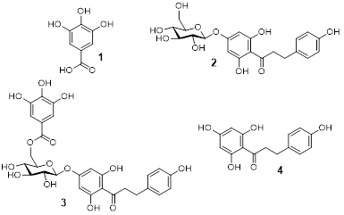
Figure 1 Chemical structures of gallic acid (1), trilobatin (2), yanangdaengin (3), and phloretin (4).
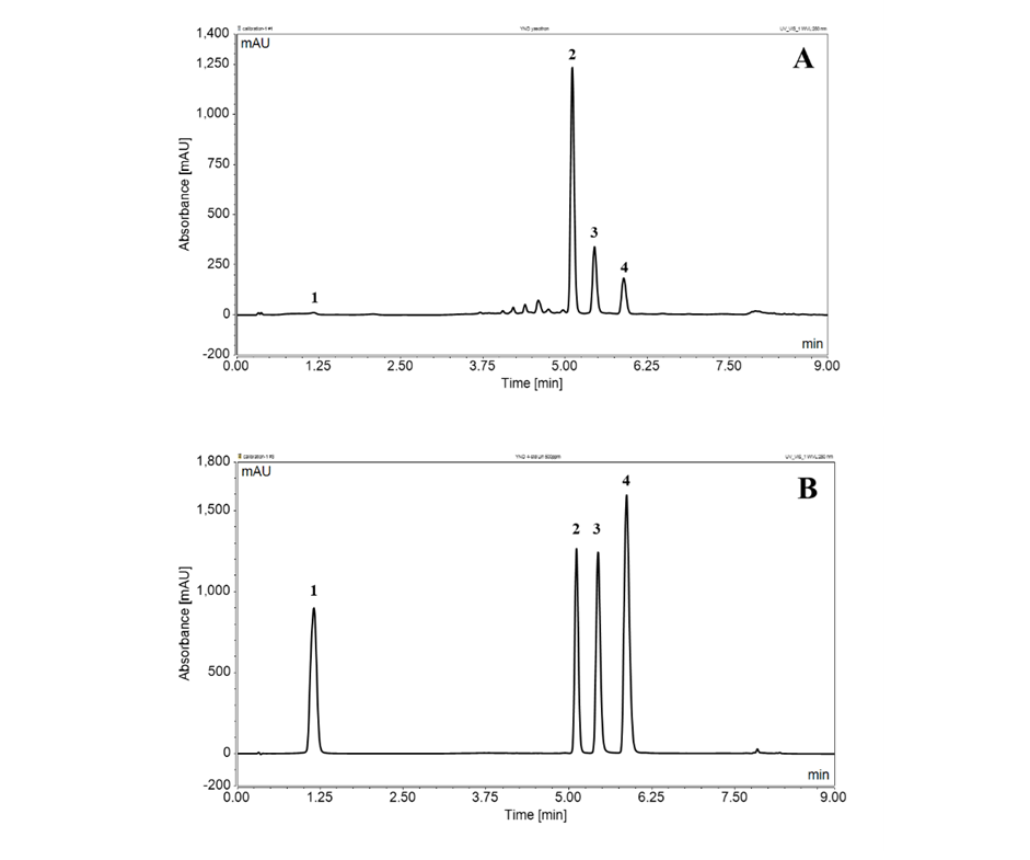 Figure
2
UHPLC chromatograms at 280 nm of (A) P.
strychnifolia leaf
extract and (B) authentic standards. Peak identification: 1=
gallic acid, 2=
trilobatin, 3=
yanangdaengin, and 4=
phloretin.
Figure
2
UHPLC chromatograms at 280 nm of (A) P.
strychnifolia leaf
extract and (B) authentic standards. Peak identification: 1=
gallic acid, 2=
trilobatin, 3=
yanangdaengin, and 4=
phloretin.
Regarding the α-glucosidase inhibitory activity, the extract obtained with 50% ethanol using sonication and heating reflux showed highest α-glucosidase inhibitory activity with IC50 of 3.70 and 3.76 µg/mL, respectively, followed by ethanol (heating reflux), ethanol (sonication), water (sonication), water (reflux), ethyl acetate, and n-hexane which exhibited IC50 values of 5.08, 5.37, 16.12, 22.62, 356.25, and >1,000 µg/mL, respectively. Yanangdaengin (3) showed potent α-glucosidase inhibitory activity with IC50 of 6.13 µg/mL (10.42 µM) followed by trilobatin (1) and phloretin (4) which showed IC50 values of 41.35 and 410.04 µg/mL (150.77 and 939.60 µM), respectively. Gallic acid (1) and also d-pinitol did not exhibit notable α-glucosidase inhibitory activity (IC50 >1,000 µg/mL). The standard drug acarbose exhibited an inhibitory activity of 212.46 µg/mL (329.09 µM).
The kinetic parameters of α-glucosidase inhibition by yanangdaegin (3) were determined using Lineweaver-Burk plots. In the absence of the inhibitor, the Vmaxwas 505.09 µmol/min and the Kmwas 2.77 mM. In the presence of the yanangdaengin (3) at a concentration range of 2–17 µM, the Vmax decreased while Km remained unchanged. The plots of 1/V versus 1/[pNPG] gave a series of straight lines with intersections close to the x-axis, as shown in Figure 3(A). This pattern suggests that yanangdaengin (3) inhibited α-glucosidase in a non-competetive mode. The inhibition constant (Ki) for yanangdaengin (3) was calculated from the Dixon plot, yielding a value of Ki= 18.38 µM (Figure 3(B)).
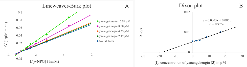
Figure 3 Kinetic study of the α-glucosidase inhibition. (A) Lineweaver-Burk plot analysis of kinetics of α-glucosidase inhibition by yanangdaengin (3) and (B) Dixon plot of slope and concentration of yanangdaengin (3).
Molecular docking was performed to predict the binding interactions of yeast α-glucosidase with gallic acid (1), trilobatin (2), yanangdaengin (3), phloretin (4) and acarbose. Active amino acid regions, including ASP69, HIS112, ARG213, ASP215, GLU277, HIS351, ASP352, and ARG442, were designated as active sites. The docking protocol was validated by redocking the co-crystallized ligand, α-d-glucopyranose, yielding an RMSD value of 0.5321 Å, which confirms the reliability of the method (RMSD < 2 Å). The calculated binding energies and interacting amino acid residues are presented in Table 6. Yanangdaengin (3) exhibited the most potent binding affinity with the lowest binding energy (−10.7 kcal/mol). This was followed by trilobatin (2) (−9.4 kcal/mol), phloretin (4) (−7.8 kcal/mol), and gallic acid (1) (−6.2 kcal/mol). The known inhibitor acarbose showed a binding energy of −8.2 kcal/mol. The 2D interaction diagrams for acarbose and yanangdaengin (3) are depicted in Figure 4.
Table 6 Docking studies of α-glucosidase inhibitors and the four major constituents of Phanera strychnifolia leaf extracts with the catalytic portion of yeast α-glucosidase.
Compound |
Chemical structure |
Binding energy (kcal/mol) |
No. of H-bond interaction |
Amino acid residues |
Yanangdaengin (3) |
|
−10.7 |
10 |
ASP69, ASP215, SER240, ASP242, GLU277, ARG315, GLN353, ARG442 |
Trilobatin (2) |
|
−9.4 |
4 |
LYS156, SER157, GLN279, LEU313 |
Acarbose |
|
−8.2 |
8 |
LYS156, SER157, TYR158, GLU277, HIS280, LEU313, GLU411 |
Phloretin (4) |
|
−7.8 |
4 |
ASP69, HIS112, GLN182, ASP307 |
Gallic acid (1) |
|
−6.2 |
3 |
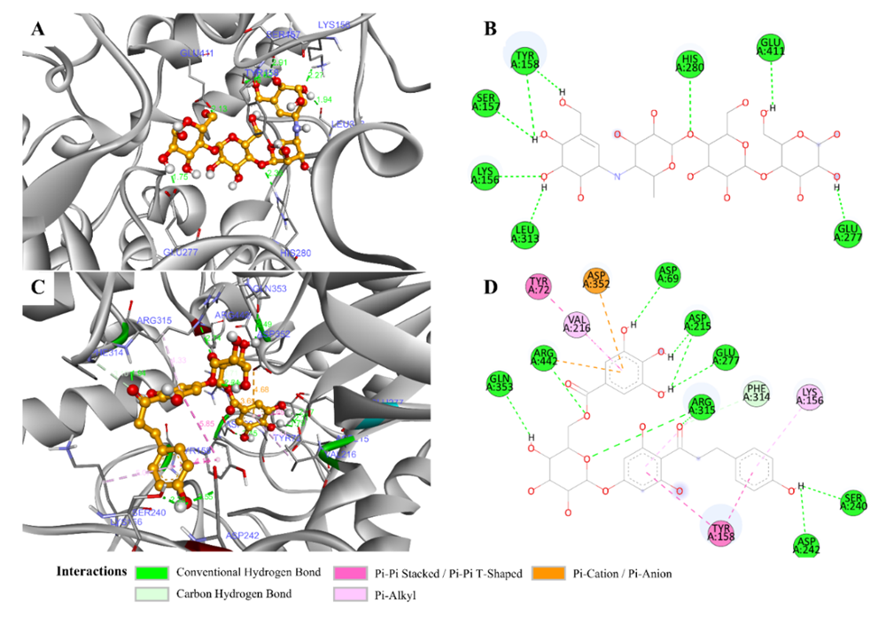
Figure 4 The binding modes of acarbose (A, B) and yanangdaengin (3) and (C, D) in the active site of α-glucosidase. Green and pink dashed lines represent hydrogen bonds and hydrophobic interactions, respectively. This visualization was created with Discovery Studio Visualizer v.21.1.0.20298.
Discussion
The control of hyperglycemia is one of the main strategies used in the treatment of diabetes, and this can be achieved using substances that can inhibit enzymes involved in digestive metabolism. Postprandial hyperglycemia results from the hydrolysis of starch by pancreatic -amylase and the uptake of glucose by intestinal through microvilli. The inhibition of these enzymes is essential to reduce the hydrolysis and absorption of carbohydrates and help the control of postprandial hyperglycemia [4]. Alpha-glucosidase is a significant enzyme that catalyzes the last step in carbohydrate digestion. The inhibition of this enzyme can reduce glucose absorption in the small intestine, leading to the control of blood glucose level [21]. The sodium-glucose co-transporters (SGLTs) are crucial for reabsorbing glucose from the filtered blood back into the circulation. The P. strychnifolia aqueous leaf extract showed inhibition of the intestinal glucose uptake and downregulation of SGLT1 and GLUT2 mRNA expressions [9]. Trilobatin (2) was identified as a non-selective inhibitor of SGLT1/2 in a previous study [22]. Phloretin (4) was known to inhibit the glucose transporter (GLUT-1) expressed in erythrocytes and blood-brain barrier [23]. Gallic acid (1) inhibits the intestinal glucose transporters SGLT1 and GLUT-2, which helps to reduce the blood glucose levels by slowing glucose absorption in the gut [24].
In this work we investigated leaves of P. strychnifolia, a rarely studied ethnomedicinally used plant [8]. To date, only a few phytochemical studies (including the plant synonyms Bauhinia strychnifolia Craib and Lysiphyllum strychnifolium (Craib) A.Schmitz) were published from the plant species investigated. d-pinitol, gallic acid (1), and the dihydrochalcones trilobatin (2), yanangdaengin (3), and phloretin (4) were identified as main and active constituents of [11]. Latter, d-pinitol has been shown as more or less inactive [12]. In this work we were able to show that extraction with 50% (v/v) aqueous ethanol led to a high yield of the above-mentioned compounds. The developed UHPLC method showed acceptable validation parameters. LOD and LOQ were improved from 0.03 and 0.1 to 0.02 and 0.06 µg/mL for all analytes, respectively [25]. The specificity of the method was also verified using stress testing method. This stress test exhibited enhanced degradation at pH values >8. Light and oxidative conditions might only slightly affect the stability of the tested compounds.
The extract obtained with 50% aqueous ethanol showed the highest α-glucosidase and α-amylase inhibitory effect. For the α-glucosidase inhibitory activity, yanangdaengin (3) exhibited the highest activity followed by phloretin (4) and trilobatin (2). Gallic acid (1) and d-pinitol were inactive in the conducted in this assay. Yanangdaengin (3) exhibited an about 30 times higher activity than the standard drug acarbose. These results are in line with the previous report from Kidruangphokin et al. [12]. The more potent α-glucosidase inhibitory activity of yanangdaengin (3) was also confirmed by computerized molecular docking. The low binding energy of yanangdaengin (3) was caused by 10 strong hydrogen bonds, 2 electrostatic interaction and 6 hydrophobic interactions, resulting in potent α-glucosidase inhibitory activity with IC50 of 6.13 µg/mL, while acarbose had binding energy of −8.2 kcal/mol. This was caused by 8 strong conventional hydrogen bonds, It resulted in a weaker α-glucosidase inhibitory activity with IC50 of 212.46 µg/mL. Based on the molecular docking analysis, yanangdaengin (3) exhibited the highest affinity for α-glucosidase.
Structurally, yanangdaengin (3) is identified as a hydroxylated dihydrochalcone glucoside with a galloyl ester moiety attached on C-6 of the glucose moiety (Figure 1). Consequently, strong hydrogen-bond interactions are established between these specific polar moieties. However, despite these polar characteristics, its orientation fits well into the hydrophobic pocket due to the flexibility of this chemical structure. Crucially, the interactions of the phenyl ring of the dihydrochalcone moiety with the amino acid residues LYS156 and TYR158 also lead to strong hydrophobic relations. A comparison of the amino acid residues involved in the binding of acarbose and yanangdaengin (3) revealed one identical residue, GLU277, which is an active-site amino acid residue. Significantly, yanangdaengin (3) interacts with the amino acid residues ASP69, ASP215, GLU277, ASP352, and ARG442. These specific residues are all located within the active binding site of α-glucosidase, which is considered a critical region for drug-enzyme interaction and binding affinity [17,18]. The other compounds identified in this study do not exhibit the above-mentioned structural features, or only exhibit them partially, so no strong interaction similar to that of yanangdaengin (3) is to be expected. Similar results were reported recently by Songoen et al. [26] where hydroxylated phenolic compounds also exhibited strong interactions with α-glucosidase.
Compounds 2–4 together with d-pinitol were inactive in the α-amylase assay, and gallic acid (1) caused false positive results by reacting with the DSNA reagent in this assay. This result also indicates that the strong α-amylase inhibitory effect of the crude extract is due to compounds or synergistic effects not identified in this work.
Conclusions
In this work, we investigated in detail the main constituents of Phanera strychnifolia. Our work revealed reveale ethanol/water (1:1, v/v) as most efficient extraction solvent and developed a simple, quick, and reliable UHPLC method for qualitative and quantitative analyses of the main constituents gallic acid (1), and the dihydrochalcones trilobatin (2), yanangdaengin (3), and phloretin (4) over a concentration range of 2–500 µg/mL. Yanangdaengin (3) exhibited potent α-glucosidase inhibitory activity with an IC50 of 6.13 µg/mL (10.42 µM), approximately 30 times more active than the commercial drug acarbose. The efficacy of 3 was confirmed by computerized molecular docking experiments. The results show that P. strychnifolia possess a potential as herbal medicine, but further research is required to assess the most effective and safe dosage and the optimal administration route for clinical use.
Acknowledgements
This study was supported by Thammasat University Research Fund, Thailand Contract No. TUFT 21/2567. We thank Drug Discovery and Development Center, Office of Advanced Science and Technology, Thammasat University, Thailand for the research facilities.
Declaration of Generative AI in Scientific Writing
The authors declare that generative AI and AI-assisted technologies were not used in the writing process.
CRediT Author Statement
Sumet Kongkiatpaiboon: Conceptualization, Funding acquisition, Investigation, Methodology, Project administration, Resources, Supervision, Validation, Writing-original draft, Writing-review and editing. Ngampuk Tayana: Conceptualization, Investigation, Methodology, Validation, Writing-review and editing. Boonwiset Seaho: Conceptualization, Investigation, Methodology, Validation, Writing-original draft. Johann Schinnerl: Conceptualization, Methodology, Formal analysis, Writing-review and editing. Nongnaphat Duangdee: Conceptualization, Formal analysis, Validation, Writing-review and editing. Wichayasith Inthakusol: Conceptualization, Investigation, Methodology. Chawannuch Mudjupa: Conceptualization, Data curation, Formal analysis, Validation, Writing-review and editing. Savita Chewchinda: Conceptualization, Data curation, Formal analysis, Validation, Writing-review and editing.
References
[1] SA Antar, NA Ashour, M Sharaky, M Khattab, NA Ashour, RT Zaid, EJ Roh, A Elkamhawy and AA Al-Karmalawy. Diabetes mellitus: Classification, mediators, and complications; A gate to identify potential targets for the development of new effective treatments. Biomedicine & Pharmacotherapy 2023; 168, 115734.
[2] U Galicai-Garcia, A Benito-Vicente, S Jebari, A Larrea-Sebal, H Siddiqi, KB Uribe, H Ostolaza and C Martin. Pathophysiology of Type 2 Diabetes Mellitus. International Journal of Molecular Sciences 2020; 21, 6275.
[3] GBD 2021 Diabetes Collaborators. Global, regional, and national burden of diabetes from 1990 to 2021, with projections of prevalence to 2050: A systematic analysis for the Global Burden of Disease Study 2021. Lancet 2023; 402, 203-234.
[4] DP Farias, FF de Araujo, IA Neri-Numa and GM Pastore. Antidiabetic potential of dietary polyphenols: A mechanistic review. Food Research International 2021; 145, 110383.
[5] K Shapiro and WC Gong. Natural Products Used for Diabetes. Journal of the American Pharmaceutical Association 2002; 42, 217-222.
[6] KW Jiang, SR Gu, SJ Li, TY Tu and DX Zhang. Three new combinations in the genus Phanera (Fabaceae: Cercidoideae) including the lectotypification of Bauhinia strychnifolia. Phytotaxa 2023; 584, 52-54.
[7] R Pooma and S Suddee. Thai plant names. Tem Smitinand revised edition 2014. Office of the Forest Herbarium, Department of National Park, Wildlife and Plant Conservation, Bangkok, Thailand, 2014.
[8] W Wuttidhamavej. Thai Traditional Medicine. Odean Store Press, Bangkok, Thailand, 1997.
[9] AS Goli, VH Sato, H Sato, S Chewchinda, J Leanpolchareanchai, J Nontakham, J Yahuafai, T Thilavech, P Meesawatsom and M Maitree. Antihyperglycemic effects of Lysiphyllum strychnifolium leaf extract in vitro and in vivo. Pharmaceutical Biology 2023; 61(1), 189-200.
[10] VH Sato, S Chewchinda, N Nuamnaichati, S Mangmool, B Sungthong, P Lertsatitthanakorn, S Ohta and H Sato. Pharmacological mechanisms of the water leaves extract of Lysiphyllum strychnifolium for its Anti-Inflammatory and Anti-Hyperuricemic actions for gout treatment. Pharmacognosy Magazine 2019; 14(60), 98.
[11] S Kongkiatpaiboon, N Duangdee, N Tayana, J Schinnerl, M Bacher and S Chewchinda. Yanangdaengin, a dihydrochalcone glucoside galloyl ester as active antioxidative agent from leaves of Lysiphyllum strychnifolium (syn. Bauhinia strychnifolia). Chinese Herbal Medicine 2020; 12(2), 452-455.
[12] M Kidruangphokin, N Suphrom and S Boonphong. -Glucosidase Inhibitory and Antioxidant Activities of Ethanolic Extracts of Different Parts of Lysiphyllum strychnifolium and Their Constituents. Journal of Herbs, Spices & Medicinal Plants 2023; 29(3), 274-287.
[13] Validation of Analytical Procedures Q2(R2). Complete Revision of Guideline. 38. Available at: https://database.ich.org/sites/default/files/ICH_Q2-R2_Document_Step2_Guideline_2022_0324.pdf, accessed January 2026.
[14] S Kongkiatpaiboon, N Duangdee, S Chewchinda, O Poachanukoon and K Amnuaypattanapon. Development and validation of stability indicating HPLC method for determination of adrenaline tartrate. Journal of King Saud University-Science 2019; 31(1), 48-51.
[15] S Chewchinda, S Kongkiatpaiboon and P Sithisarn. Evaluation of antioxidant activities, total phenolics and total flavonoid contents of aqueous extracts of leaf, stem, and root of Aerva lanata. Chiang Mai University Journal of Natural Sciences 2019; 18(3), 345-357.
[16] S Chewchinda, N Leakaya, H Sato and VH Sato. Antidiabetic effects of Maclura cochinchinensis (Lour.) Corner heartwood extract. Journal of Traditional and Complementary Medicine 2021; 11(1), 68-74.
[17] T Limcharoen, P Chaniad, P Chonsut, C Punsawad, T Juckmeta, A Konyanee, IR Rais and S Sangkaew. Alpha-Glucosidase inhibition, antioxidant activities, and molecular docking study of krom luang chumphon khet udomsak, a Thai Traditional Remedy. Advances in Pharmacological and Pharmaceutical Science 2024; 2024, 1322310.
[18] K Yamamoto, H Miyake, M Kusunoki and S Osaki. Crystal structures of isomaltases from Saccharomyces cerevisiae and in complex with its competitive inhibitor maltose. The FEBS Journal 2010; 277(20), 4205-4214.
[19] J Eberhardt, D Santos-Martins, AF Tillack and S Forli. AutoDock Vina 1.2.0: New docking methods, expanded force field, and python bindings. Journal of Chemical Information and Modeling 2021; 61(8), 3891-3898.
[20] O Trott and AJ Olson. AutoDock Vina: Improving the speed and accuracy of docking with a new scoring function, efficient optimization, and multithreading. Journal of Computational Chemistry 2010; 31(2), 455-461.
[21] K Thongra-Ar, P Rojsanga, S Chewchinda, S Mangmool and P Sithisarn. Antioxidant, α-glucosidase and α-amylase inhibitory activities of Persicaria odorata. Chiang Mai University Journal of Natural Sciences 2021; 20(3), e2021051.
[22] L Wang, M Liu, F Yin, Y Wang, X Li, Y Wu, C Ye and J Liu. Trilobatin, a novel SGLT1/2 inhibitor, selectively induces the proliferation of human hepatoblastoma cells. Molecules 2019; 24(18), 3390.
[23] M Lunder, M Janic, M Sabovic and A Janez. SGLT-2 inhibitors: A novelty in the treatment of type 2 diabetes. Zdravniski Vestnik 2018; 87, 493-505.
[24] H Wang, MI Fowler, DJ Messenger, JJ Ordaz-Ortiz, X Gu, S Shi, LA Terry, MJ Berry, G Lian and S Wang. Inhibition of the intestinal postprandial glucose transport by gallic acid and gallic acid derivatives. Food & Function 2021; 12(12), 5399-5406.
[25] S Kongkiatpaiboon, N Tayana, W Inthakusol, N Duangdee, S Chewchinda and V Keeratinijakal. HPLC quantification of chemical markers from Lysiphyllum strychnifolium. Revista Brasileira de Farmacognosia 2022; 32, 266-272.
[26] W Songoen, W Phanchai, J Schinnerl, L Brecker, M Thabpho, S Tharamak, W Pluempanupat, S Sukkhaeng and S Chansuthep. Identification of specialized metabolites from Artocarpus lacucha as potent alpha-glucosidase and acetylcholinesterase inhibitors: Enzyme kinetic, in vitro and in silico study. Journal of Natural Medicines 2025; 79(4), 896-912.