
Trends
Sci.
2026; 23(9):
12797
Antihypertensive Effects of Compound A-42 via Regulation of Calcium-Dependent Ion Transport Systems
Shodiyakhon Sodiqova2,*, Anvar Zaynabiddinov2, Izzatullo Abdullaev1,
Ulugbek Gayibov1 and Ziyodullokh Ziyoyiddinov3
1A.S. Sadykov Institute of Bioorganic Chemistry of the Science Academy of Uzbekistan “Laboratory of Plant Cytoprotectors, Tashkent, Uzbekistan
2Andijan State University, Andijan Region, Uzbekistan
3National University of Uzbekistan, Toshkent, Uzbekistan
(*Corresponding author’s e-mail: [email protected])
Received: 25 November 2025, Revised: 21 December 2025, Accepted: 28 December 2025, Published: 20 March 2026
Abstract
Hypertension is a complex cardiovascular disorder associated with impaired calcium (Ca²⁺) regulation in vascular smooth muscle and cardiac tissues. The present study investigated the antihypertensive potential of a new bioactive compound, A-42, through a combined in silico and in vivo approach. Molecular docking analysis demonstrated that A-42 interacts with several calcium-regulating proteins, including L-type and R-type Ca²⁺ channels, SERCA, RyR2, Ca²⁺-ATPase, Na⁺/Ca²⁺ exchanger (NCX), and renin. The compound exhibited notable binding affinities, with binding energies ranging from –5.3 to –6.2 kcal/mol. The strongest affinities were observed for the L-type Ca²⁺ channel (–6.2 kcal/mol), SERCA (–6.0 kcal/mol), and NCX (–6.0 kcal/mol). Key amino acid interactions included hydrogen bonds and π–alkyl or π–anion interactions with residues such as ARG A:593, PHE A:587, LEU F:269, LYS A:158, THR A:230, and ASP A:829, indicating a stable ligand–protein complex formation and potential calcium-channel-modulating activity. The in vivo studies, performed using the tail-cuff method, confirmed the hypotensive effects of A-42 in rats. Intravenous administration at doses of 10, 20, and 30 mg/kg led to a dose-dependent reduction in systolic and diastolic blood pressure. The 20 mg/kg dose produced the most pronounced and stable antihypertensive effect, significantly lowering blood pressure (p-value < 0.05) and preventing the sharp rise in pressure induced by adrenaline in the hypertensive model. In the adrenaline-induced hypertension model, the systolic and diastolic pressures in A-42–treated rats decreased from 138.3 ± 13.6 / 102.8 ± 10.1 mmHg to 103.8 ± 11.2 / 73.5 ± 8.7 mmHg, respectively, within the first hour of administration. The combined in silico and in vivo results indicate that compound A-42 acts as a multi-target modulator of calcium homeostasis, affecting both membrane and intracellular Ca²⁺ transport systems. These interactions likely contribute to its antihypertensive mechanism by reducing intracellular Ca²⁺ influx, enhancing Ca²⁺ sequestration, and restoring vascular tone.
Keywords: A-42 compound, L-type Ca²⁺ channel, Molecular docking, Adrenaline-induced hypertension, Tail-cuff method, Antihypertensive activity
Introduction
Hypertension remains one of the leading causes of cardiovascular morbidity and mortality worldwide, affecting over one billion individuals and accounting for nearly half of all deaths due to heart disease and stroke. Despite the availability of numerous antihypertensive
agents—including calcium channel blockers, angiotensin-converting enzyme inhibitors, β-adrenergic antagonists, and diuretics—many patients continue to experience suboptimal blood pressure control, adverse side effects, or therapeutic resistance. These limitations underscore the urgent need for the development of novel agents that act through multi-target mechanisms to restore vascular and cardiac homeostasis with improved efficacy and safety [1,2].
Among the physiological regulators of vascular tone and cardiac contractility, calcium (Ca²⁺) signaling plays a central role. The contraction of vascular smooth muscle and the excitation–contraction coupling of cardiomyocytes are tightly controlled by intracellular Ca²⁺ dynamics. Several membrane and sarcoplasmic proteins—including L-type (Caᵥ1.2) and R-type (Caᵥ2.3) Ca²⁺ channels, the sarcoplasmic/endoplasmic reticulum Ca²⁺-ATPase (SERCA), ryanodine
receptors (RyR2), and the Na⁺/Ca²⁺ exchanger (NCX)—coordinate Ca²⁺ influx, sequestration, and efflux to maintain homeostasis. Dysregulation of these systems contributes to elevated intracellular Ca²⁺ concentrations, sustained vascular constriction, and progressive hypertensive pathology. Consequently, selective modulation of these transport mechanisms represents a promising therapeutic strategy for blood pressure control [3,4].
Recent advances in computational pharmacology have facilitated the identification of bioactive molecules capable of interacting with multiple ion-transport targets. Molecular docking and in silico modeling approaches enable the prediction of binding affinities and conformational interactions between small molecules and target proteins, thus guiding experimental design prior to in vivo evaluation. Complementary in vivo methods—such as the tail-cuff technique—allow the assessment of real-time hemodynamic effects, providing a translational link between molecular interaction and systemic response [5,6].
In this context, the present study investigates the antihypertensive potential of compound A-42, a newly synthesized bioactive molecule, using an integrated in silico and in vivo framework. Computational docking analyses were performed to evaluate its interaction with key calcium-handling proteins, including L-type and R-type Ca²⁺ channels, SERCA, RyR2, Ca²⁺-ATPase, NCX, and renin. Furthermore, in vivo experiments were conducted in adrenaline-induced hypertensive rats to assess the compound’s effects on systolic and diastolic blood pressure across multiple doses [7,8].
By correlating molecular docking results with physiological outcomes, this study aims to elucidate the mechanistic basis of A-42’s antihypertensive action and to determine its potential as a multi-target modulator of Ca²⁺ homeostasis in cardiovascular regulation. The findings contribute to a broader understanding of how calcium-related signaling pathways can be pharmacologically targeted for the prevention and management of hypertension [9,10].
Materials and methods
Animal ethics
All preoperative and experimental procedures were reviewed and approved by the Institutional Committee for Animal Use and Care. Animals were housed in a controlled vivarium under standardized conditions, including a relative humidity of 55% - 65%, an ambient temperature of 22 ± 2 °C, and free access to standard laboratory chow and water. All animal-handling procedures were conducted in full compliance with the European Directive 2010/63/EU on the protection of animals used for scientific purposes. Ethical approval for the study was granted by the Animal Ethics Committee of the Institute of Bioorganic Chemistry, Academy of Sciences of the Republic of Uzbekistan (Protocol No. 133/1a/h, dated 4 August 2016).
Chemical and pharmacological characterization of compound A-41
Compound A-42 is chemically identified as 3,4,5-trihydroxybenzoic acid methyl ester (methyl gallate), with a molecular formula of C₈H₈O₅ and a molecular weight of 184.15 g/mol. Its structure comprises three phenolic hydroxyl (–OH) groups and one methyl ester (–COOCH₃) group attached to a benzene ring (Figure 1). These functional groups provide strong hydrogen-donating capacity and electron delocalization, which underlie the compound’s pronounced antioxidant and potential cytoprotective activities.
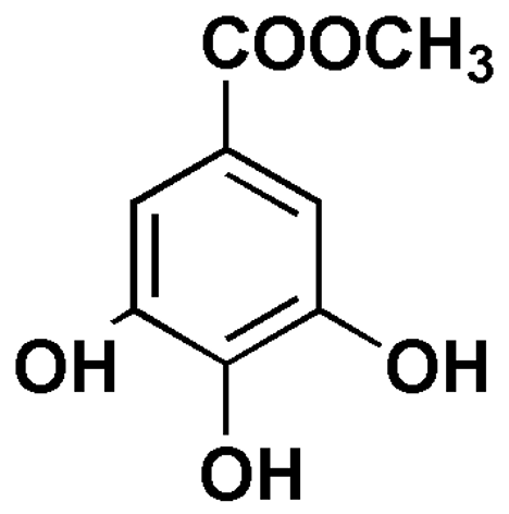
Figure 1 Chemical structure of compound A-42 (methyl gallate).
Compound A-42 belongs to the class of phenolic esters. Its moderate lipophilicity facilitates penetration through biological membranes and enables interactions with protein active sites, including calcium-dependent ion transport systems. The phenolic hydroxyl groups contribute to reactive oxygen species (ROS) scavenging and membrane stabilization, whereas the ester moiety improves solubility and metabolic stability, potentially enhancing bioavailability and pharmacodynamic efficacy [11].
Data collection and software
Publicly available structural databases and open-source computational tools were employed in this study. Three-dimensional crystal structures of calcium-regulating and ion-transport proteins were obtained from the Protein Data Bank (PDB), including Ca²⁺-ATPase (PDB IDs: 6JJU, 1HNY), Ca²⁺ R-type channel (PDB ID: To be specified), Ca²⁺ L-type channel (PDB ID: 6JP5), Na⁺/Ca²⁺ exchanger (NCX) (PDB ID: 8SGI), ryanodine receptor (RyR2) (PDB ID: 5C33), angiotensin–renin complex (PDB ID: 2REN), and sarcoplasmic/endoplasmic reticulum Ca²⁺-ATPase (SERCA) (PDB ID: 2ZOX).
Pharmacological and physicochemical properties of compound A-42 were retrieved from the PubChem database. Protein visualization and structural preparation were performed using PyMOL v1.2. Molecular docking simulations were carried out using AutoDock 4.2 and AutoDock Tools (ADT) [12,13], while ligand–protein interaction analysis and visualization were conducted with Discovery Studio Visualizer v4.5.
Measurement of blood pressure in rats using the tail-cuff method
Systolic blood pressure was measured using a non-invasive tail-cuff plethysmography system (Model Sistola AcqKnowledge, Neurobotics, Russia). Experiments were performed under controlled environmental conditions (22 - 24 °C) to minimize stress-induced variability. Prior to measurements, animals were acclimatized to the restraining device for three consecutive days to reduce handling-related stress and ensure reproducibility of the results [14].
Experimental animals and group allocation
Adult male Wistar rats weighing 180 - 220 g were used in the study. Animals were randomly divided into experimental groups, with equal numbers of rats in each group. The experimental design included the following groups: Intact control group – rats receiving saline only and not subjected to hypertension induction. Hypertensive control group – rats with adrenaline-induced hypertension receiving saline treatment. A-42 (10 mg/kg) group – hypertensive rats treated with compound A-42 at a dose of 10 mg/kg. A-42 (20 mg/kg) group – hypertensive rats treated with compound A-42 at a dose of 20 mg/kg. A-42 (30 mg/kg) group – hypertensive rats treated with compound A-42 at a dose of 30 mg/kg. Compound A-42 was administered intravenously. Blood pressure was measured before and after treatment using the tail-cuff method at predetermined time points. Animals were randomly assigned to groups to minimize selection bias.
Results and discussion
Binding characteristics of compound A-42 with Ca²⁺ L-Type Ion channels
Prior to performing in vitro and in vivo experiments, the potential biological activity of the tested compounds was initially assessed through in silico molecular docking. These analyses focused on evaluating the binding affinity of the compounds toward Ca²⁺ L-type (Caᵥ) ion channels, which are known to play a central role in hypertension development [15,16].
In this work, the interaction between compound A-42 and Ca²⁺ L-type channels was examined using computational docking methods. The coordinates of the active binding pocket were identified with Discovery Studio, while the three-dimensional (3D) ligand structures were built in Avogadro. Docking simulations were carried out using AutoDock Vina, allowing the calculation of binding energies for each ligand [17,18].
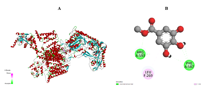
Figure 2 Interaction of compound A-42 with Ca²⁺ L-type ion channels. (A) Overall view of the protein structure. (B) Detailed visualization of ligand–amino acid interactions.
Compound A-42 demonstrated a binding energy of –6.2 kcal/mol and formed several key interactions, including conventional hydrogen bonds with ARG A:593 and PHE A:587, as well as π–alkyl interactions with LEU F:269. These findings helped pinpoint the critical amino acid residues involved in Ca²⁺ L-type channel blockade. Effective inhibition of the channel is largely associated with interactions involving LEU F:269, SER F:265, ASP:598, ARG A:593, PHE A:587, and TYR A:585. The interplay between these residues and the ligands offers valuable insight into the molecular mechanisms that govern Ca²⁺ channel modulation and inhibition (Figure 2).
Interaction of compound A-42 with Ca²⁺ R-Type Ion channels
Voltage-gated Ca²⁺ R-type (Caᵥ) channels can be influenced by a range of bioactive molecules, including several therapeutic agents used to manage hypertension and other cardiovascular disorders. Additionally, structural analyses using cryo-electron microscopy (cryo-EM) have revealed detailed conformations of Caᵥ1.1 channels bound to both antagonists and agonists, offering important insights into their ligand-binding behavior [21,22].
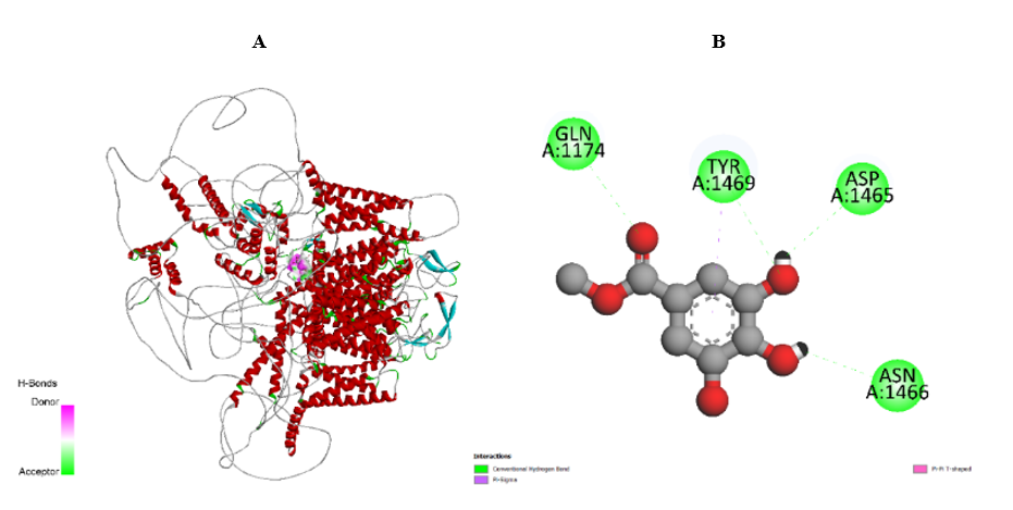
Figure 3 Interaction of compound A-42 with Ca²⁺ R-type ion channels. (A) Overall structure of the protein. (B) Ligand–amino acid interactions within the binding site.
In this work, the interaction between compound A-42 and Ca²⁺ R-type channels was examined through in silico molecular docking. The coordinates of the protein’s active site were defined using Discovery Studio, and the three-dimensional (3D) ligand structures were prepared in Avogadro. Docking simulations were conducted with AutoDock Vina, and the binding affinities of each ligand were obtained (Figure 3).
In the subsequent stage of analysis, compound A-42 demonstrated a binding energy of –5.5 kcal/mol, confirming a stable interaction with the Ca²⁺ R-type channel. The residues contributing to this binding included GLN A:1174, TYR A:1469, ASP A:1465, and ASN A:1466, all forming conventional hydrogen bonds. These results enabled the identification of critical amino acids involved in A-42 recognition by R-type channels.
Additionally, the observed differences in amino acid interaction profiles among ligands provide valuable insight into their potential modulatory effects on ion channel function. Building on these findings, future studies will aim to investigate the pharmacological significance and therapeutic prospects of these ligands in cardiovascular regulation [19,20].
Binding energetics of compounds with intracellular Ca²⁺ channels
Following the evaluation of compound interactions with Ca²⁺ L-type and R-type channels, it became important to assess their affinity toward a key intracellular Ca²⁺ transport system—the sarcoplasmic/endoplasmic reticulum Ca²⁺-ATPase (SERCA). SERCA is essential for preserving intracellular Ca²⁺ balance by actively pumping Ca²⁺ from the cytosol into the sarcoplasmic reticulum. Its proper activity is vital for maintaining Ca²⁺ homeostasis and supporting normal contractile function in cardiac and smooth muscle cells. For this reason, the binding energies and amino acid interactions between compound A-42 and the SERCA protein were examined in detail [21,22].
Binding of compound A-42 with SERCA
Docking simulations demonstrated that compound A-42 binds to SERCA with a calculated binding energy of –6.0 kcal/mol, indicating the formation of a stable protein–ligand complex. The compound established several key interactions within the active site: LYS A:158, ASN A:39, and GLY A:227 formed conventional hydrogen bonds that strongly anchored the ligand; LEU A:41 engaged in π-alkyl interactions, supporting hydrophobic stabilization within the protein’s lipophilic region; and THR A:230 contributed donor–donor hydrogen bonding, further reinforcing the ligand’s orientation inside the binding pocket.
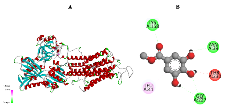
Figure 4 Molecular docking visualization of compound A-42 with SERCA. (A) Overall structure of the SERCA protein. (B) Interaction of the ligand with amino acid residues at the active site.
Although the binding pattern of A-42 was similar to that of compound A-41, the specific residues involved suggest potential differences in their pharmacological behavior and functional modulation of SERCA activity (Figure 4). Future investigations will focus on exploring the cellular and physiological effects of these ligands in greater detail, particularly their role in modulating Ca²⁺ homeostasis and their therapeutic potential under cardiovascular and cellular stress conditions [23,24].
Binding energetics of compounds with the RyR2 receptor
Alongside SERCA, ryanodine receptors (RyR2) are essential regulators of intracellular Ca²⁺ balance. Abnormal RyR2 function—especially under hypertensive or other cardiovascular pathological states—can disrupt Ca²⁺ signaling and contribute to disease development. For this reason, the interaction of compound A-42 with the RyR2 receptor was examined, and its binding energetics were assessed through in silico molecular docking.
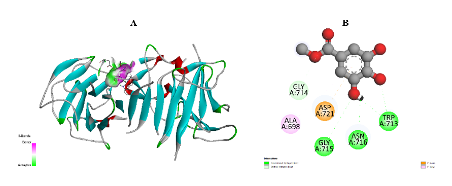
Figure 5 Interaction of compound A-42 with the RyR2 receptor. (A) Overall structure of the RyR2 protein. (B) Detailed view of ligand–residue interactions within the active site.
Binding of compound A-42 with the RyR2 receptor
Compound A-42 showed a binding energy of –5.8 kcal/mol with the RyR2 receptor, indicating moderate binding stability. The ligand formed conventional hydrogen bonds with GLY A:715, ASN A:716, and TRP A:713, effectively anchoring it within the receptor’s active pocket. ALA A:698 contributed π-alkyl interactions, supporting hydrophobic stabilization, while GLY A:714 formed carbon–hydrogen bonds aiding ligand positioning. Additionally, ASP A:721 engaged in π-anion interactions, providing electrostatic stabilization. Although the binding affinity for RyR2 was slightly lower compared with its interaction with SERCA and L-type channels, the presence of hydrogen-bonding, hydrophobic, and electrostatic contacts suggests that A-42 may exert modulatory effects on RyR2 activity (Figure 5).
Interaction of ligands with Ca²⁺-ATPase protein channels
The Ca²⁺-ATPase (SERCA) and ryanodine receptor (RyR2) protein channels play essential roles in maintaining intracellular calcium homeostasis. In particular, Ca²⁺-ATPase is critically involved in cardiac muscle contraction and blood pressure regulation. Dysfunction of these protein channels may lead to impaired Ca²⁺ signaling, contributing to the development of hypertension and other cardiovascular diseases. Therefore, in the next phase of the study, the binding energetics of ligands with Ca²⁺-ATPase were analyzed using in silico molecular docking [25].
Binding of compound A-42 with Ca²⁺-ATPase
Compound A-42 exhibited a binding energy of –5.9 kcal/mol with Ca²⁺-ATPase, indicating the formation of a stable and energetically favorable complex. The ligand interacted with the following amino acid residues at the enzyme’s active site: ASP A:351, THR A:353, and ARG A:489 – conventional hydrogen bonds contributing to strong stabilization of the ligand within the active pocket of SERCA; ARG A:559 – π-cation interactions that reinforced ligand anchoring within the binding region (Figure 6).
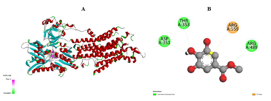
Figure 6 Molecular docking visualization of compound A-42 with Ca²⁺-ATPase. (A) Overall 3D structure of the Ca²⁺-ATPase protein. (B) Ligand–residue interactions at the enzyme’s active site.
These results confirm that compound A-42 displays a relatively strong affinity toward Ca²⁺-ATPase and may modulate its activity. The interaction pattern supports the hypothesis that A-42 could influence intracellular Ca²⁺ transport and thus exert potential cardioprotective and antihypertensive effects through regulation of Ca²⁺ homeostasis.
Interaction of ligands with the Na⁺/Ca²⁺ exchanger
In the development of hypertension, calcium homeostasis and ion transport mechanisms play a crucial role. In particular, ion transport proteins such as SERCA, RyR2, and the Na⁺/Ca²⁺ exchanger (NCX) are key regulators of cardiac and vascular functions. In this study, the binding energetics of ligands with NCX were evaluated to assess their potential modulatory activity.
Binding of compound A-42 with NCX
Molecular docking analysis revealed that compound A-42 exhibited a binding energy of –6.0 kcal/mol with the Na⁺/Ca²⁺ exchanger, suggesting a stable interaction with the protein (Figure 7). The ligand formed the following key interactions: ASP A:829 and THR A:172 – conventional hydrogen bonds that stabilized the ligand within the NCX binding site; VAL A:99 – π-alkyl interactions contributing to hydrophobic stabilization within the lipophilic environment of the protein.
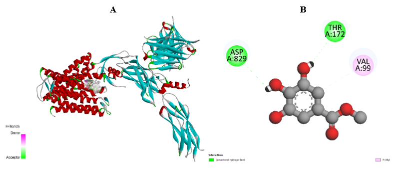
Figure 7 Interaction of compound A-42 with the Na⁺/Ca²⁺ exchanger (NCX). (A) Overall structure of the NCX protein. (B) Visualization of ligand–amino acid interactions within the active site.
Interaction analysis of ligands with the Renin–protein complex
During this study, special attention was given to the roles of angiotensin and renin, as these signaling pathways are critically involved in the regulation and elevation of blood pressure. Accordingly, the interaction of compound A-42 with the renin protein was analyzed through in silico molecular docking.
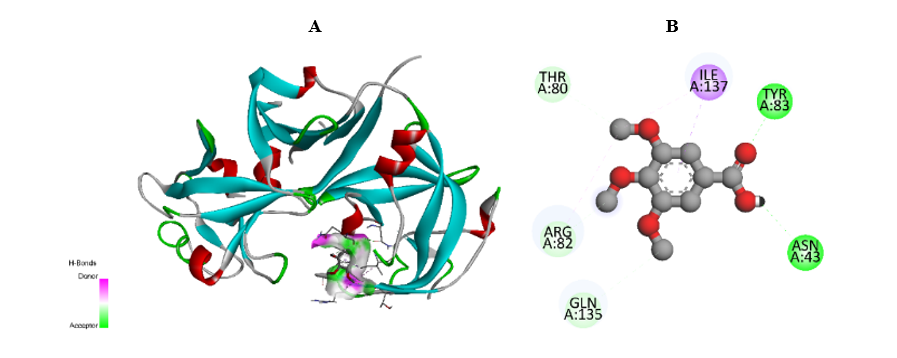
Figure 8 Molecular docking interaction of compound A-42 with the renin protein complex. (A) Overall structure of the renin protein. (B) Visualization of ligand–amino acid interactions within the binding site.
The docking results revealed that compound A-42 exhibited a binding energy of –5.3 kcal/mol with the renin protein, indicating a moderately stable interaction. The ligand formed the following key contacts: THR A:85 and SER A:84 — conventional hydrogen bonds stabilizing the ligand within the active site; TYR A:83 — π-sigma interaction, contributing to additional hydrophobic and aromatic stabilization (Figure 8).
In Vivo effects of compound A-42 on blood pressure
The dose-dependent effects of compound A-42 on arterial blood pressure were evaluated in vivo using the tail-cuff method. According to the literature, several in vivo techniques can be applied to assess the physiological activity of bioactive compounds; among them, intravenous administration is one of the most effective for investigating direct cardiovascular responses, as it allows rapid delivery of the compound into the circulatory system [26].
Table 1 Dose-dependent in vivo antihypertensive activity of compound A-42 (M ± m).
|
mg/kg |
Baseline |
1 hour |
2 hours |
3 hours |
||||
SBP |
DBP |
SBP |
DBP |
SBP |
DBP |
SBP |
DBP |
||
A42 |
10 |
130.5 ± 13.1 |
98.0 ± 9.6 |
112.3 ± 11.1 |
91.5 ± 8.9 |
84.3 ± 8.2 |
62.0 ± 6.1 |
106.3 ± 10 |
86.0 ± 8.4 |
20 |
131.0 ± 13.0 |
92.3 ± 9.1 |
105.0 ± 10.0 |
73.5 ± 7.2 |
108.8 ± 10.7 |
85.0 ± 8.3 |
86.0 ± 8.5 |
53.3 ± 5.2 |
|
30 |
118.3 ± 11.7 |
81.5 ± 7.9 |
94.8 ± 9.2 |
56.8 ± 5.4 |
126.0 ± 12.3 |
92.0 ± 9.0 |
126.8 ±12 |
91.8 ± 9.1 |
|
Before experimentation, rats were randomly divided into control and experimental groups. For each compound, animals were allocated into four groups (n = 3 per group): Control rats received physiological saline (0.9% NaCl), while experimental rats received A-42 at doses of 10, 20, or 30 mg/kg. Baseline blood pressure values (0 h) were recorded prior to administration. The compounds were then administered intravenously, and systolic and diastolic blood pressures were monitored every hour for 3 h to assess cardiovascular responses31. The findings demonstrated that A-42 exhibited no strictly linear dose-dependent trend. At lower doses (10 mg/kg), a marked hypotensive effect was observed, while some responses were more pronounced at 20 mg/kg. However, higher doses (30 mg/kg) did not produce significant changes. These observations indicate that A-42 exerts complex, dose-specific cardiovascular effects, likely associated with its individual pharmacodynamic profile. Further experimental evaluation is required to elucidate the underlying mechanisms of action.
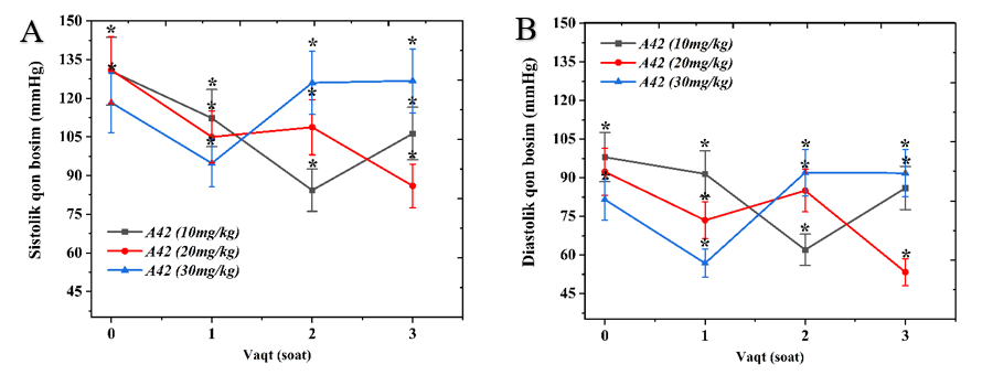
Figure 9 Administration of compound A-42 at doses of 10, 20, and 30 mg/kg led to a dose-dependent decrease in systolic and diastolic blood pressure. The observed results indicate that the antihypertensive effect of A-42 increases proportionally with the administered dose (n = 4, p-value < 0.05).
In the control group, the systolic pressure was 130.5 ± 13.1 mmHg, and the diastolic pressure was 98.0 ± 9.6 mmHg (Figure 9). In the 10 mg/kg group, after 1 h systolic pressure decreased to 112.3 ± 11.1 mmHg and diastolic to 91.5 ± 8.9 mmHg; after 2 h it dropped significantly to 84.3 ± 8.2 and 62.0 ± 6.1 mmHg, respectively; after 3 h partial recovery occurred, with systolic pressure 106.3 ± 10.2 mmHg and diastolic 86.0 ± 8.4 mmHg (Table 1). In the 20 mg/kg group, the control pressure values were 131.0 ± 13.0 and 92.3 ± 9.1 mmHg. After one hour, systolic and diastolic pressures fell to 105.0 ± 10.0 and 73.5 ± 7.2 mmHg; after 2 h they reached 108.8 ± 10.7 and 85.0 ± 8.3 mmHg; and after 3 h they further declined to 86.0 ± 8.5 and 53.3 ± 5.2 mmHg. In the 30 mg/kg group, baseline pressures were 118.3 ± 11.7 and 81.5 ± 7.9 mmHg. After administration, systolic and diastolic pressures were 94.8 ± 9.2 and 56.8 ± 7.7 mmHg at one hour; 126.0 ± 12.3 and 92.0 ± 9.0 mmHg at 2 h; and 126.8 ± 12.4 and 91.8 ± 9.1 mmHg at 3 h, respectively. The results indicate that A-42 produced variable blood pressure responses depending on the dose. Significant reductions were observed at 10 and 20 mg/kg, while the 30 mg/kg dose exhibited a mild hypertensive tendency. The most effective and stable dose was 20 mg/kg, showing marked hypotension followed by stabilization, and was thus considered the optimal therapeutic dose for subsequent experiments33. After determining the most effective dose, the antihypertensive activity of A-42 was examined in a rat model of adrenaline-induced hypertension using the tail-cuff method. Rats were divided into healthy control, hypertensive control (adrenaline-treated), and experimental (adrenaline + A-42, 20 mg/kg) groups. All animals were adult male Wistar rats weighing 300 - 350 g.
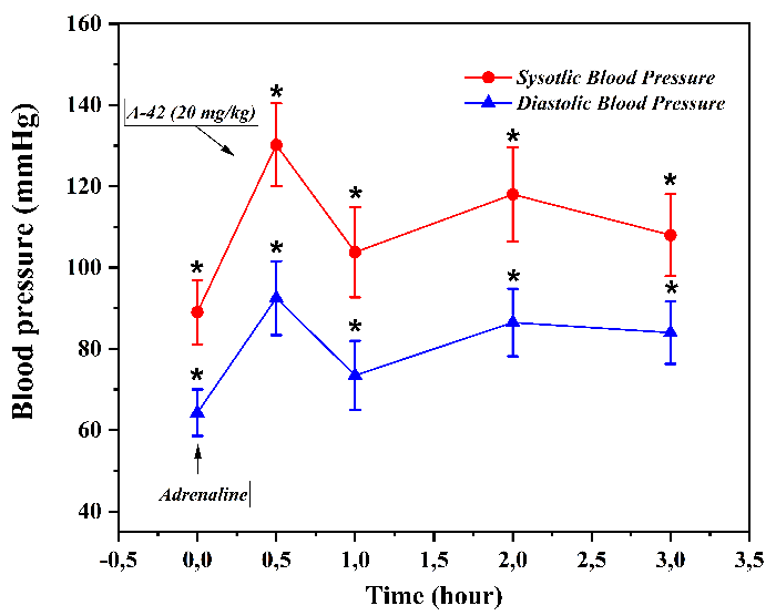
Figure 10 (A) Stepwise changes in systolic and diastolic blood pressure observed after intravenous administration of compound A-42 at a dose of 10 mg/kg following adrenaline injection. The antihypertensive effect of A-42 was statistically significant, with p-value < 0.05 (n = 4).
Baseline systolic (SBP) and diastolic (DBP) pressures were measured before treatment: In the control group SBP was 93.3 ± 8.3 mmHg and DBP 68.3 ± 5.7 mmHg, while in the A-42 group SBP was 89.0 ± 7.9 mmHg and DBP 64.3 ± 5.7 mmHg. After intravenous administration of adrenaline hydrochloride (except for the healthy controls), blood pressure increased sharply in the hypertensive model to SBP 138.3 ± 13.6 mmHg and DBP 102.8 ± 10.1 mmHg. However, in the A-42 pretreated group, the rise was attenuated, reaching SBP 130.3 ± 10.2 mmHg and DBP 92.5 ± 9.1 mmHg. Subsequent hourly measurements following A-42 administration showed that after one hour SBP and DBP were 103.8 ± 11.2 and 73.5 ± 8.7 mmHg; after 2 h 118.0 ± 11.5 and 86.5 ± 8.3 mmHg; and after 3 h 108.0 ± 10.1 and 84.0 ± 7.7 mmHg. In the adrenaline-only control group, blood pressure remained elevated: After one hour SBP was 133.3 ± 14.9 and DBP 95.5 ± 9.3 mmHg; after 2 h 118.8 ± 10.0 and 90.8 ± 9.1 mmHg; and after 3 h 114.8 ± 6.6 and 83.8 ± 6.4 mmHg (Figure 10). These findings demonstrate that A-42 at a dose of 20 mg/kg effectively attenuates adrenaline-induced hypertension, producing a significant and sustained hypotensive effect compared with untreated hypertensive controls.
Discussion
The present study evaluated the antihypertensive potential of compound A-42 using a combined in silico and in vivo approach, with particular emphasis on its interaction with Ca²⁺ transport systems and its hemodynamic effects in an adrenaline-induced hypertension model. Dysregulation of intracellular Ca²⁺ homeostasis is a well-established contributor to increased vascular smooth muscle tone and elevated blood pressure, making calcium-handling proteins key pharmacological targets in hypertension therapy [1-3]. In this context, the obtained results indicate that A-42 acts as a multi-target modulator of Ca²⁺ homeostasis, which may underlie its antihypertensive efficacy.
Molecular docking and mechanistic insights
Molecular docking analyses demonstrated that A-42 forms stable complexes with both L-type and R-type Ca²⁺ channels, with binding energies comparable to those reported for several calcium channel–blocking agents [4,5]. L-type Ca²⁺ channels play a dominant role in mediating Ca²⁺ influx in vascular smooth muscle cells and are the primary targets of clinically used antihypertensive drugs such as dihydropyridines and benzothiazepines [6,7]. The interaction of A-42 with key residues involved in channel gating and ion permeation suggests that it may partially inhibit Ca²⁺ entry, thereby reducing vascular contractility.
R-type Ca²⁺ channels, although less studied, have been implicated in fine-tuning vascular tone and sympathetic neurotransmission [8]. The observed binding of A-42 to functionally relevant residues of the R-type channel suggests an additional mechanism through which the compound may attenuate Ca²⁺ influx, complementing L-type channel modulation and contributing to overall antihypertensive activity.
Beyond membrane channels, A-42 showed notable affinity for intracellular Ca²⁺-handling proteins, including SERCA, Ca²⁺-ATPase, and RyR2. SERCA-mediated Ca²⁺ reuptake into the sarcoplasmic reticulum is essential for lowering cytosolic Ca²⁺ levels and promoting vascular relaxation [9]. The stabilization of SERCA through hydrogen bonding and polar interactions observed in this study suggests that A-42 may enhance Ca²⁺ sequestration, a mechanism reported for several cardioprotective and vasorelaxant agents [10].
RyR2 is responsible for Ca²⁺ release from the sarcoplasmic reticulum and plays a critical role in excitation–contraction coupling [11]. Moderate binding of A-42 to RyR2 residues may indicate a modulatory rather than inhibitory effect, potentially preventing excessive Ca²⁺ release without disrupting physiological signaling. Such balanced modulation of Ca²⁺ cycling has been proposed as a favorable strategy to reduce vascular tone while maintaining cardiac function [12].
In addition, A-42 exhibited strong binding affinity toward the Na⁺/Ca²⁺ exchanger (NCX), a key determinant of Ca²⁺ extrusion in vascular and cardiac cells [13]. Facilitation of NCX-mediated Ca²⁺ efflux could further contribute to the reduction of intracellular Ca²⁺ concentration, reinforcing the antihypertensive effect. The combined targeting of Ca²⁺ influx, sequestration, release, and extrusion pathways highlights the multi-level regulation of Ca²⁺ homeostasis by A-42, which may distinguish it from single-target calcium channel blockers.
The moderate interaction observed between A-42 and the renin protein suggests a potential auxiliary influence on the renin–angiotensin–aldosterone system (RAAS). Although weaker than its interactions with Ca²⁺-handling proteins, partial modulation of renin activity could complement calcium-dependent mechanisms, as combined Ca²⁺ channel and RAAS modulation has been shown to provide enhanced antihypertensive efficacy in clinical settings [14,15].
In vivo hemodynamic effects
The in vivo findings are consistent with the docking results and support the proposed mechanism of action. A-42 produced significant, dose-dependent reductions in systolic and diastolic blood pressure, confirming its antihypertensive activity in experimental animals. The observation that the 20 mg/kg dose elicited the most pronounced and stable effect suggests the existence of an optimal therapeutic window, a phenomenon also reported for other vasorelaxant and calcium-modulating compounds [16].
In the adrenaline-induced hypertension model, pretreatment with A-42 effectively attenuated catecholamine-driven increases in blood pressure. Adrenaline-induced hypertension is known to involve enhanced Ca²⁺ influx through voltage-dependent Ca²⁺ channels and increased intracellular Ca²⁺ availability [17]. The ability of A-42 to blunt this pressor response supports the hypothesis that its antihypertensive action is mediated primarily through modulation of Ca²⁺ transport systems.
Pharmacological implications
Taken together, these findings suggest that A-42 exerts its antihypertensive effects through integrated, multi-target regulation of Ca²⁺ homeostasis, affecting both membrane and intracellular Ca²⁺ transport mechanisms. Such a multi-mechanistic profile may offer advantages over conventional antihypertensive agents that act on a single target, potentially resulting in improved efficacy and reduced compensatory responses. Furthermore, the possible secondary influence on the RAAS pathway suggests that A-42 may combine vascular and hormonal mechanisms of blood pressure control, a strategy that aligns with modern approaches to antihypertensive drug development [18,19].
Conclusions
Compound A-42 exhibits consistent antihypertensive activity in an adrenaline-induced hypertension model and demonstrates favorable interactions with key Ca²⁺-handling proteins in in silico analyses. While the observed concordance between computational binding profiles and in vivo blood pressure reduction supports the involvement of calcium-dependent mechanisms, the present findings should be interpreted as indicative rather than definitive evidence of multi-target calcium modulation. The computational results primarily serve as a hypothesis-generating framework, suggesting potential interactions of A-42 with L-type and R-type Ca²⁺ channels, SERCA, RyR2, and the Na⁺/Ca²⁺ exchanger. Experimental confirmation of these interactions will require direct functional validation, including electrophysiological recordings of Ca²⁺ currents, isolated vascular smooth muscle contractility assays under Ca²⁺-controlled conditions, and targeted biochemical analyses of intracellular Ca²⁺ handling. Future studies should therefore focus on mechanism-oriented experiments, such as patch-clamp analyses to assess channel subtype selectivity, calcium imaging to quantify intracellular Ca²⁺ dynamics, and extended safety evaluations to determine long-term cardiovascular effects. These investigations will be essential to substantiate the proposed mechanisms and to define the translational potential of A-42 as an antihypertensive agent.
Declaration of Generative AI in Scientific Writing
Only minimal assistance was used from QuillBot for paraphrasing selected sentences. All scientific content, interpretation, and conclusions were developed independently by the authors.
CRediT Author Statement
Rohatoy Sayidaliyeva: Conceptualization, Methodology, Data curation, Writing – Original Draft. Anvar Zaynabiddinov: Formal analysis, Validation, Visualization. Izzatullo Abdullaev and Ziyoyiddinov Ziyodullokh: Investigation, Laboratory experiments, Data collection. Ulugbek Gayibov: Supervision.
References
S Sodiqova, S Kadirova, A Zaynabiddinov, I Abdullaev, L Makhmudov, U Gayibov, M Yuldasheva, M Xolmirzayeva, R Rakhimov, A Mutalibov and H Karimjonov. Channelopathy activity of A-41(Propyl Ester of Gallic Acid): Experimental and computational study of antihypertensive activity. Trends in Sciences 2025; 22(9), 10496.
TF Aripov and UG Gayibov. Antiradical and antioxidant activity of the preparation “Rutan” from Rhus coriaria L. Journal of Theoretical Medicine 2023; 4, 164-170.
R Sayidaliyeva, S Kadirova, A Zaynabiddinov, I Abdullaev, L Makhmudov, U Gayibov, M Yuldasheva, M Kholmirzayeva, R Rakhimov, A Mutalibov and H Karimjonov. A-51 as a natural calcium channel blocker: An integrative study targeting hypertension. Trends in Sciences 2025; 22(11), 10760.
M Zaripova, I Abdullaev, A Bogbekov, U Gayibov, S Omonturdiev, R Makhmudov, N Ergashev, G Jabbarova, S Gayibova and T Aripov. In Vitro and in Silico studies of Gnaphalium U. Extract: Inhibition of α-amylase and α-glucosidase as a potential strategy for metabolic syndrome regulation. Trends in Sciences 2025; 22(8), 10098.
Y Mirzayeva, I Abdullaev, U Gayibov, S Omonturdiev, Z Allaniyazova, S Umrkulova and P Usmanov. In vitro and computational evaluation of 1-O-benzoylkarakoline on vascular calcium transport. Trends in Sciences 2026; 23(3), 11850.
AV Mahmudov, OS Abduraimov, SB Erdonov, UG Gayibov and LY Izotova. Bioecological features of nigella sativa L. in different conditions of Uzbekistan. Plant Science Today 2022; 9(2), 421-426.
U Gayibov, I Abdullaev, F Sobirova, S Omonturdiev, A Abdullaev, S Gayibova and T Aripov. Plant-derived and synthetic antihypoxic agents in cardiovascular diseases: Mechanisms, key pathways and therapeutic potential. Plant Science Today 2025. https://doi.org/10.14719/pst.7810
D Inomjonov, I Abdullaev, S Omonturdiev, A Abdullaev, L Maxmudov, M Zaripova, M Abdullayeva, D Abduazimova, S Menglieva, S Gayibova, Ma Sadbarxon, U Gayibov and T Aripov. In vitro and in vivo studies of crategus and inula helenium extracts: Their effects on rat blood pressure. Trends in Sciences 2025, 22(3), 9158.
I Abdullaev, U Gayibov, S Omonturdiev, S Fotima, S Gayibova and T Aripov. Molecular pathways in cardiovascular disease under hypoxia: Mechanisms, biomarkers, and therapeutic targets [J]. The Journal of Biomedical Research 2025; 39(3), 254-269.
A Abdullaev, I Abdullaev, A Bogbekov, U Gayibov, S Omonturdiev, S Gayibova, M Turahodjayev, K Ruziboev and T Aripov. Antioxidant potential of rhodiola heterodonta extract: Activation of Nrf2 pathway via integrative in vivo and in silico studies. Trends in Sciences 2025; 22(5), 9521.
MR Zaripova, SN Gayibova, RR Makhmudov, AA Mamadrahimov, Nl Vypova, UG Gayibov, SM Miralimova and TF Aripov. Characterization of Rhodiola heterodonta (Crassulaceae): Phytocomposition, antioxidant and antihyperglycemic activities. Preventive Nutrition and Food Science 2024; 29(2), 135-145.
AV Mahmudov, OS Abduraimov, SB Erdonov, AL Allamurotov, OT Mamatqosimov, UG Gayibov and L Izotova. Seed productivity of Linum usitatissimum L. in different ecological conditions of Uzbekistan. Plant Science Today 2022; 9(4), 1090-1101.
U Gayibov, SN Gayibova, MK Pozilov, FS Tuxtaeva, UR Yusupova, GMK DJabbarova, ZA Mamatova, NA Ergashev and TF Aripov. Influence of quercetin and dihydroquercetin on some functional parameters of rat liver mitochondria. Journal of Microbiology, Biotechnology and Food Sciences 2021; 11(1), e2924.
S Omonturdiev, I Abdullaev, A Khasanov, A Abdullaev, D Inomjanov, U Gayibov, S Gayibova, K Baratov, Y Mirzaeva, M Allamuratov, P Usmanov and T Aripov. Molecular mechanisms and experimental protocols in the study of vasorelaxant activity in aortic smooth muscle cells. Trends in Sciences 2026; 23(2), 11549.
MK Pozilov, UG Gayibov, MI Asrarov, MI Asrarov, NG Abdulladjanova, HS Ruziboev and TF Aripov. Physiological alterations of mitochondria under diabetes condition and its correction by polyphenol gossitan. Journal of Microbiology, Biotechnology and Food Sciences 2022; 12(2), e2224.
AG Vakhobjonovna, KE Jurayevich, AIZ Ogli, EN Azamovich, MR Rasuljonovich and AM Islomovich. Tannins as modulators in the prevention of mitochondrial dysfunction. Trends in Sciences 2025; 22(8),10436.
I Abdurazakova, A Zaynabiddinov, I Abdullaev, L Makhmudov, U Gayibov, S Omonturdiev, G Abdullayev, M Xolmirzayeva and S Zhurakulov. Pharmacological evaluation of F-45 on the cardiovascular system using in vitro, in vivo models and molecular dockings. Trends in Sciences 2025; 22(12), 10924.
M Mamajanov, I Abdullaev, G Sotimov, S Mavlanova, Q Niyozov, M Mirzaolimov, A Najimov, E Mirzaolimov, M Raximberganov and U Abdullayev. Mitochondrial and pharmacokinetic insights into 3,5,7,2′,6′-pentahydroxyflavanone: Respiratory modulation, calcium handling, and membrane stability. Trends in Sciences 2025; 22(12), 10984.
Z Shakiryanova, R Khegay, U Gayibov, A Saparbekova, Z Konarbayeva, A Latif and O Smirnova. Isolation and study of a bioactive extract enriched with anthocyanin from red grape pomace (Cabernet sauvignon). Agronomy Research 2023; 21(3), 1293-1303.
UG Gayibov, SN Gayibova, HM Karimjonov, AA Abdullaev, et al. Antioxidant and cardioprotective properties of polyphenolic plant extract of Rhus glabra L. Plant Science Today 2024. https://doi.org/10.14719/pst.3442
O Gaibullayeva, A Islomov, D Abdugafurova, B Elmurodov, B Mirsalixov, L Mahmudov, I Adullaev, K Baratov, S Omonturdiev and S Sa’Dullayeva. Inula helenium L. root extract in sunflower oil: Determination of its content of water-soluble vitamins and immunity-promoting effect. Biomedical Pharmacology Journal 2024; 17(4), 2729-2737.
Y Umidakhon, B Erkin, G Ulugbek, N Bahadir and A Karim. Correction of the mitochondrial NADH oxidase activity, peroxidation and phospholipid metabolism by haplogenin-7-glucoside in hypoxia and ischemia. Trends in Sciences 2022; 19(21), 6260.
O Sirojiddin, A Izzatullo, U G lugbek, I Dolimjon, G Sabina, M Rustamjon, A Takhir, Eİ Torunoğlu and EC Aytar. Cardiovascular effects of Matricaria chamomilla extract: Calcium channel modulation and vasorelaxant activity. Naturwissenschaften 2026; 113(1), 14.
M Li, M Zhang, AT Fatixovich, GU Gapparjanovich and H Du. Green-synthesized Zn²⁺-polyphenol networks (CGA/RA) for enhanced multifunctional food preservation. Food Measure 2025. https://doi.org/10.1007/s11694-025-03764-y
UG Gayibov, SN Gayibova, HM Karimjonov, AA Abdullaev, DS Abduazimova, RN Rakhimov, HS Ruziboev, MA Xolmirzayeva, AE Zaynabiddinov and TF Aripov. Antiradical and antioxidant activity of the preparation “Rutan” from Rhus coriaria L. Journal of Theoretical and Clinical Medicine 2023; 4, 164-170.
AQQ Azimova, AX Islomov, SA Maulyanov, D Gulyamovna Abdugafurova, LU Mahmudov, IZiyoyiddin Abdullaev, AS Ishmuratova, SQ Siddikova and IR Askarov. Determination of vitamins and pharmacological properties of Vitis vinifera L. plant fruit part (mixed varieties) syrup-honey. Biomedical and Pharmacology Journal 2024; 17(4), 2779-2786.