
Trends
Sci.
2026;
23(9):
13044
Genotype-dependent In Vitro Floral Induction and Reversion in Moroccan Cannabis sativa L. Varieties under Zeatin, Gibberellic Acid, and Thidiazuron Treatments
Meryem Tahtah1,2,*, Toufik Ibrahim2, Fathellah Hachimi2,3,
Karima Samir1 and Aicha Nordine4
1Department of biology, Laboratory of Ecology and Environment, Faculty of Sciences Ben M'sik, University Hassan II, Casablanca, Morocco
2Department of Fundamental Sciences, Laboratory of In Vitro Culture, National School of Agriculture of Meknes, Meknes, Morocco
3Department of Biology, Laboratory of Biotechnology and Bioresource Valorization, Faculty of Sciences,
Moulay Ismail University, Meknes, Morocco
4Department of Biology, Laboratory of Biology and Health, Faculty of Sciences Ben M'sik, University Hassan II, Casablanca, Morocco
(*Corresponding author’s e-mail: [email protected])
Received: 15 December 2025, Revised: 12 January 2026, Accepted: 20 January 2026, Published: 30 March 2026
Abstract
Micropropagation of Cannabis sativa L. offers an efficient way to produce uniform plants while enabling precise control of vegetative growth and flowering in vitro, where induction and reversion are shaped by genotype and plant growth regulators. Two Moroccan Cannabis sativa varieties Beldia and Khardala were cultured in vitro on MS/2 medium supplemented with macronutrients to assess the effects of ZEN, GA₃, and TDZ (0, 0.25, 0.5, 0.75 mg L⁻¹) on vegetative growth, floral induction, and floral reversion. Significant genotype-dependent responses were observed. Vegetative growth was optimized in Beldia under ZEN and GA₃, reaching 9.17 nodes, whereas Khardala responded best to low TDZ concentrations with 7.21 nodes. Floral induction under GA₃ reached 50% in Beldia and 42% in Khardala, indicating a genotype-specific sensitivity to growth regulators. In contrast, floral reversion was most pronounced in Khardala, with 75% reversion associated. Rooting was maximized with 1 mg L⁻¹ IBA, achieving 95.8% rooting in Beldia and 87.5% in Khardala, while survival rate after acclimatization exceeded 94%. Phytohormone type and concentration were shown to strongly influence genotype-specific growth and flowering in Cannabis sativa, providing a practical basis for developing in vitro propagation and floral induction protocols for Moroccan varieties.
Keywords: Micropropagation, In vitro flowering, Floral reversion, Zeatin, Gibberellic acid, Thidiazuron, Cannabis sativa, Genotype response, Phytohormone signaling
Introduction
Cannabis sativa L. is an annual herbaceous plant widely cultivated for industrial, pharmaceutical, and recreational purposes due to its ability to accumulate a wide range of secondary metabolites, notably cannabinoids, terpenes, and flavonoids, which are recognized for their biological and medicinal properties [1-3]. Among these compounds, Δ9-tetrahydrocannabinol (THC) and cannabidiol (CBD) have attracted increasing pharmacological interest because of their analgesic, anti-inflammatory, and neuroprotective effects [4,5]. These metabolites are primarily biosynthesized in the glandular trichomes of inflorescences, a process tightly regulated by genetic and environmental factors [6]. However, conventional production of C. sativa remains constrained by genetic heterogeneity, agro-climatic variability, and susceptibility to diseases, which has driven the development of biotechnological approaches, particularly in vitro culture, to obtain healthy and genetically uniform plants [7,8]. Micropropagation thus represents an efficient strategy for rapid clonal multiplication, while allowing precise control of growth conditions and facilitating the investigation of developmental and metabolic processes [9,10]. Although several advances have been reported in the optimization of in vitro cannabis propagation, including the use of temporary immersion systems [11], alternative cytokinins such as meta-topolin [12], and ethylene-related regulators to improve regeneration [13,14], most protocols still rely on Murashige and Skoog (MS) medium, whose efficiency may decline during prolonged culture periods [15].
In vitro flowering of C. sativa remains a relatively underexplored phenomenon, despite its relevance as a model for investigating floral transition and floral reversion under hormonally controlled conditions [16,17]. Previous studies have shown that certain growth substances, including gibberellins, zeatin, and thidiazuron, can induce floral transition in vitro [18,19]; however, the mechanisms governing floral reversion and their dependence on genotype remain largely unresolved. This knowledge gap limits a comprehensive understanding of the developmental plasticity of C. sativa under in vitro conditions. This issue is particularly relevant in the Moroccan context, where two major C. sativa types are cultivated. The local landrace Beldia, long adapted to Moroccan agro-climatic conditions, exhibits a vegetative architecture and secondary metabolism distinct from those of Khardala, an improved variety selected for enhanced vigor and yield potential. These morpho-physiological and biochemical differences suggest differential sensitivity to growth regulators, making these two genotypes a suitable biological model for analyzing genotype-dependent phytohormonal responses, particularly with respect to flowering and floral reversion [20-22].
The present study introduces a novel aspect by highlighting the potential specific role of zeatin in the regulation of floral reversion, a process that has been rarely addressed in Cannabis sativa. This study hypothesizes that zeatin interacts with endogenous growth regulators to modulate floral induction and reversion, and aims to compare vegetative growth and floral responses in two Moroccan Cannabis sativa varieties to support the development of genotype-adapted in vitro protocols.
Materials and methods
Plant material and culture conditions
In the summer of 2023, we collected seeds from two types of Cannabis sativa in the Larache region of northern Morocco: Beldia and Khardala. We kept them in a refrigerator at 4 °C until it was time to use them. To disinfect the seeds, we immersed them in 70% (v/v) ethanol for 1 min and then treated them with 1% hydrogen peroxide H₂O₂ for 2 min. Finally, we soaked them in 4.6% sodium hypochlorite for 15 min [23]. Then, we washed the seeds three times in sterile distilled water and placed them on sterile filter paper to air dry.
Sterilized seeds were germinated in glass jars with 25 mL of half-strength MS medium (MS/2) [24], added to 15 g L⁻¹ sucrose, 0.5% MS vitamins and solidified with 8 g L⁻¹ agar, without plant growth regulators. Per jar three seeds were sown. The pH of the medium was adjusted to 5.7 before autoclaving (121 °C, 100 kPa, for 20 min). Cultures were grown in a growth at 25 ± 2 °C with a photoperiod of 16 h (16/8 h light/dark) 60 µmol m⁻² s⁻¹ and relative humidity of 64%.
Culture medium, and experimental design
One month old Cannabis sativa plantlets from seed germination were used to excise nodal segments (≤ 2 cm, leafless). Each nodal segment was individually cultured in test tubes with 10 mL half-strength (MS/2) [24] supplemented with macronutrients [25]: 830.5 mg L⁻¹ anhydrous calcium chloride, 451.8 mg L⁻¹ anhydrous magnesium sulfate, and 425 mg L⁻¹ monobasic potassium phosphate. The media was further supplemented with 30 g L⁻¹ sucrose, 0.5% MS vitamins and 8 g L⁻¹ agar (Difco). Afterward, the medium pH was set to 5.7 before autoclaving (121 °C for 20 min at 100 kPa).
To test their influence on the induction of adventitious buds and subsequent shoot growth, cytokinins (thidiazuron or gibberellic acid or zeatin) were each tested at 0, 0.25, 0.5 and 0.75 mg L⁻¹ concentrations individually [26,27]. For each variety and each treatment, 48 explants were used as biological replicates. Each individually cultured explant represented an experimental unit. Cultures were randomly arranged in the growth chamber to minimize positional effects. The experiment was independently repeated, yielding comparable results.
Floral induction and reversion
Young Axillary buds derived from young in vitro plantlets, as well as floral buds formed on in vitro-developed shoots, were excised and cultured in sterile 90-mm Petri dishes containing 25 mL of modified half-strength Murashige and Skoog basal medium enriched with adjusted macronutrients, namely 830.5 mg L⁻¹ anhydrous calcium chloride (CaCl₂), 451.8 mg L⁻¹ anhydrous magnesium sulfate (MgSO₄), and 425 mg L⁻¹ monobasic potassium phosphate (KH₂PO₄) [25]. The medium was supplemented with 30 g L⁻¹ sucrose and 0.5% MS vitamins, solidified with 8 g L⁻¹ agar, and the pH was adjusted to 5.7 prior to sterilization. Gibberellic acid (GA₃), zeatin (ZEN), or thidiazuron (TDZ) was added separately to the culture medium at concentrations of 0, 0.25, 0.5, and 0.75 mg L⁻¹) [27,28]. However, TDZ was not used in floral reversion experiments.
Floral induction was operationally defined as the visible development and maintenance of floral organs, particularly calyces and pistillate structures, on cultured explants during the culture period. Floral reversion was defined as the loss of floral identity accompanied by the emergence of vegetative structures, such as leaf primordia or the direct appearance of leaves, originating from previously floral tissues. Upon the appearance of flowers, explants were transferred to sterile test tubes containing 15 mL of the same culture medium to allow completion of development and to ensure adequate elongation of floral structures.
Evaluations were performed after 30 days of culture. Floral induction and reversion responses were recorded in a binary manner (presence or absence) and expressed as percentages. To minimize potential Petri dish effects and reduce pseudo-replication, explants were randomly distributed among culture dishes and spatially separated during the experiment. Observations were based on direct morphological examination under sterile conditions and were documented by photographic records. Each treatment consisted of 48 biological replicates, with four explants per Petri dish per variety, each explant being considered an independent experimental unit.
Rooting and acclimatization of plantlets
Rooting was initiated on in vitro derived shoots (~3 cm in height) obtained from the elongation of axillary buds. These shoots were transferred to basal MS/2 medium (previously described, supplemented with 30 g L⁻¹ sucrose and adjusted to pH 5.7). To induce rooting, different concentrations of indole-3-butyric acid (IBA; 0.5, 1.0, and 2.0 mg L⁻¹) were tested, along with a hormone-free control. After 4 weeks of culture, rooting efficiency was evaluated based on the percentage of rooted shoots and the mean root length.
In vitro–rooted plantlets (3 - 5 cm) were carefully removed from culture vessels, and residual agar was gently rinsed off with sterile distilled water to reduce the risk of microbial contamination. Plantlets were then transplanted into transparent plastic cups containing a sterilized substrate mixture of peat moss, perlite, and vermiculite (1:1:1, v/v/v). Cups were at first sealed to ensure high relative humidity (~90%) to help the plant acclimate from in vitro to ex vitro conditions. Irrigation was carried out regularly to ensure adequate substrate moisture without waterlogging. The survival rate (%) was recorded as the primary parameter to assess acclimatization success.
Culture conditions
All cultures were maintained at 25 ± 1 °C under a 16 h photoperiod with a light intensity of 2,500 lux. These standard conditions also provided consistent growth and the opportunity of evaluating at the same time vegetative multiplication, floral induction or reversion responses.
Data analysis
All experimental data were analyzed using OriginPro 2025b. Data normality and homogeneity of variances were verified prior to analysis to meet the assumptions of parametric tests. A two-way ANOVA was performed to assess the effects of genotype, treatment, and their interaction, considering each explant as an independent experimental unit with 48 biological replicates per treatment and variety. When significant differences were observed, mean separation was carried out using Tukey’s HSD test at p ≤ 0.05. Pearson’s correlation coefficients were calculated to descriptively examine relationships between plant growth regulators and growth parameters within each variety and were interpreted cautiously in light of known physiological responses. Pairwise associations were visualized using a lower-triangular scatter matrix, and all graphs and statistical illustrations were generated using OriginPro 2025b.
Results and discussion
Micropropagation
The evaluation of different concentrations of ZEN, GA₃, TDZ in the Cannabis sativa L. varieties Beldia and Khardala revealed contrasting morphological and physiological responses, reflecting a strong dependence on the interaction between genotype and phytohormone type. Statistical analysis showed that all observed differences were highly significant (p-values < 0.001), supporting the robustness and reliability of the variations recorded among treatments and varieties.
The Beldia variety exhibited a pronounced association between ZEN- and GA₃-based treatments, particularly at 0.5 mg L⁻¹, and enhanced vegetative growth levels. Under these conditions, node number reached 9.17, leaf number reached 21.13 with GA₃, branch number reached 3.04, and stem length reached 9.01 cm with ZEN and 9.48 cm with GA₃ (Figure 1). In contrast, Khardala displayed more moderate responses to ZEN and GA₃, but showed a stronger association with TDZ treatments. At 0.25 mg L⁻¹ TDZ, leaf number reached 6.85, regeneration rate reached 100%, and branch number peaked at 1.50. Node production was highest under ZEN at 0.5 (7.21), while stem length was optimized under ZEN with a value of 6.78 cm. In Beldia, TDZ at 0.5 mg L⁻¹ was statistically associated with higher values of node number (8.96), leaf number (12.61), and stem length (8.47 cm), suggesting a combined response related to shoot multiplication and elongation. In Khardala, the same TDZ concentration was associated with a more moderate growth response (3.71 nodes, 6.18 leaves, 4.27 cm stem length), while maintaining a high regeneration rate (83%) (Figures 1 - 5).
Pearson correlation analysis revealed significant positive associations between ZEN- and GA₃-based treatments and several vegetative growth parameters in both cultivars, including node number (r-values = 0.95), leaf number (r-values = 0.97), branch number (r-values = 0.96), and stem length (r-values = 0.95) (Figures 6 and 7). These correlations represent statistically coherent relationships.
The present results confirm that Cannabis sativa micropropagation is strongly dependent on the interaction between genotype and the applied plant growth regulator. The use of zeatin in cannabis micropropagation remains relatively underexplored compared with other commonly used cytokinins. Zeatin is a naturally occurring isoprenoid cytokinin which, although more expensive than BAP, kinetin, or TDZ, has frequently been associated with high shoot multiplication while maintaining continuous and vigorous growth in certain plant species [29], making it a regulator of interest for the optimization of micropropagation protocols. Moreover, the combination of GA₃ and TDZ has been reported in the literature to be associated with improved shoot growth and plantlet formation [30,31], including in Humulus lupulus, a species closely related to Cannabis sativa. GA₃ is also commonly associated with in vitro regeneration and elongation of conductive tissues, while the cytokinin-GA₃ balance is recognized as a key factor in tissue differentiation processes [32-34]. Regarding TDZ, pioneering studies by Wang et al. [34] and Lata et al. [30] demonstrated its effectiveness in promoting shoot induction in Cannabis sativa, observations that have been confirmed by subsequent investigations [35]. Wang et al. [34] notably reported high regeneration rates (3.22 axillary shoots per explant) using 0.2 mg L⁻¹ TDZ. In the present study, even higher regeneration rates were observed under certain conditions, which may be related to varietal-specific sensitivity and fine adjustments of culture conditions. Nevertheless, the use of TDZ is not without limitations, as hyperhydricity remains a frequent issue in in vitro culture, often leading to morphological abnormalities, developmental disorders, and acclimatization difficulties. These contrasting responses highlight the major influence of genetic diversity and the complexity of interactions between endogenous and exogenous phytohormones [36,37].
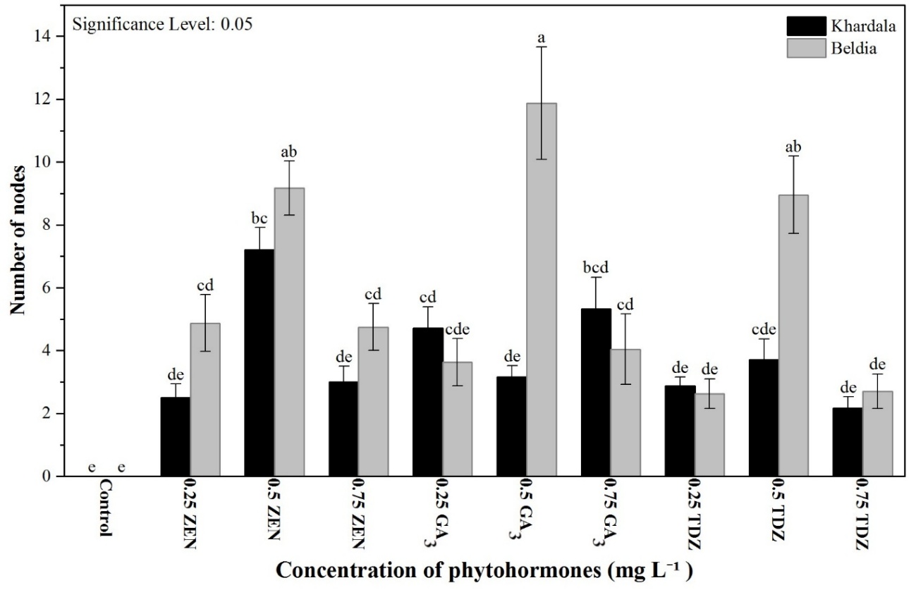
Figure 1 Number of nodes formed per explant as a function of different phytohormones in two Cannabis sativa varieties.
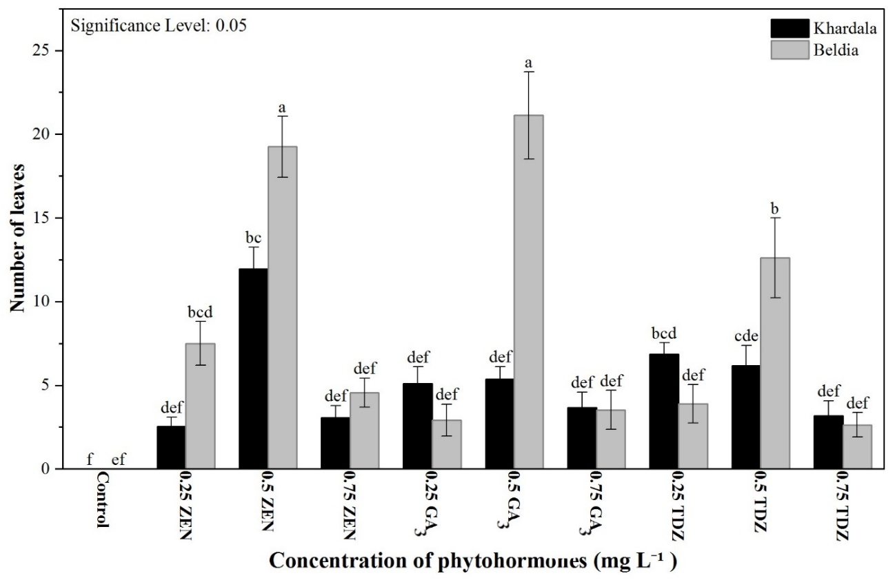
Figure 2 Number of leaves formed per explant as a function of different phytohormones in two Cannabis sativa varieties.
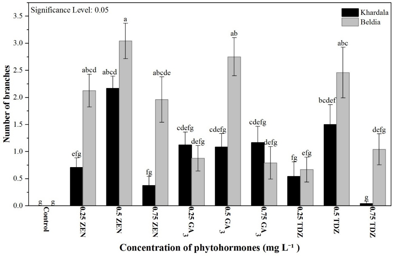
Figure 3 Number of branches formed per explant as a function of different phytohormones in two Cannabis sativa varieties.
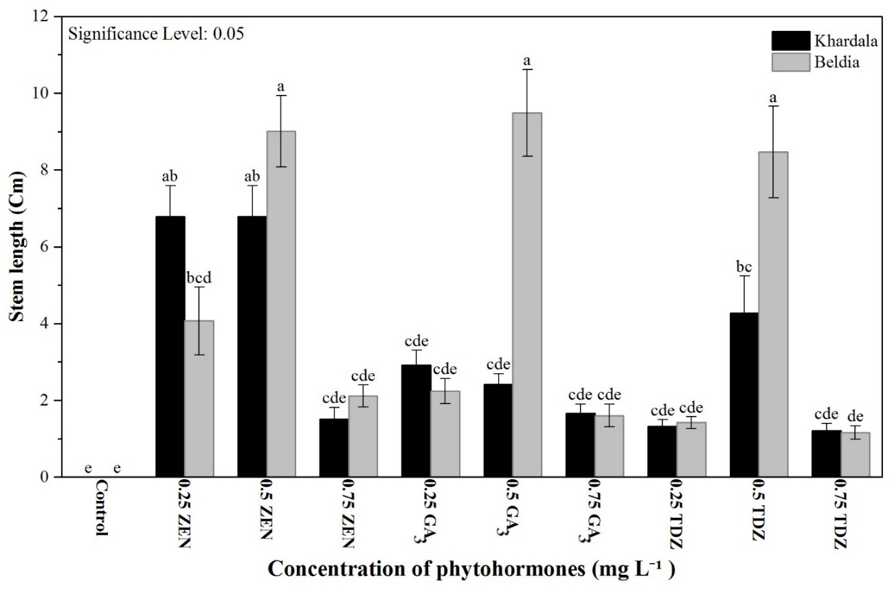
Figure 4 Stem length (cm) as a function of different phytohormones in two Cannabis sativa varieties.
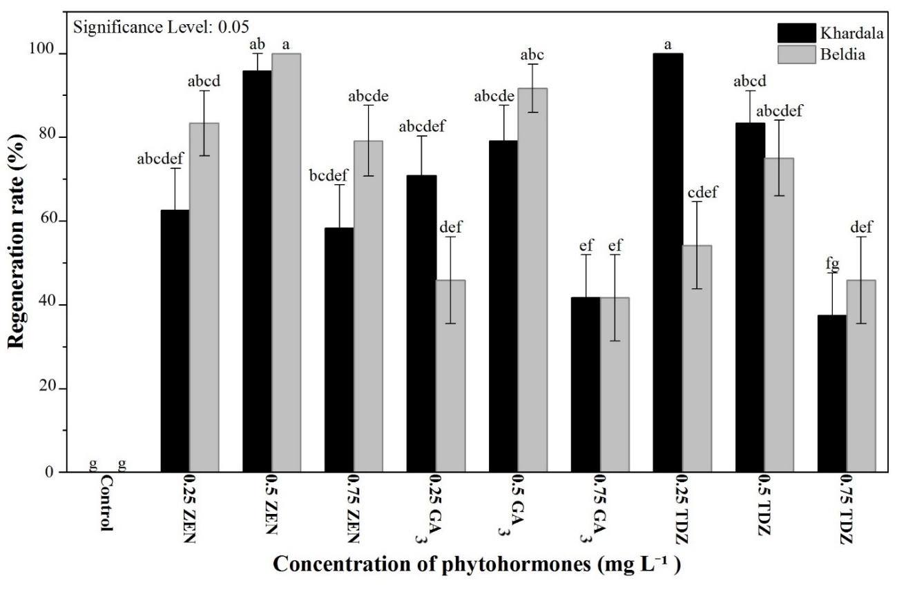
Figure 5 Regeneration rate (%) as a function of different phytohormones in two Cannabis sativa varieties.
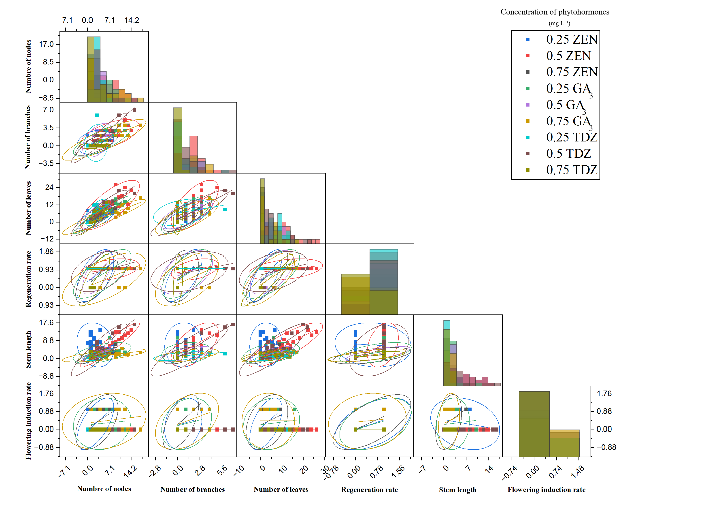
Figure 6 Correlations between growth parameters of the Khardala variety and the applied phytohormones (ZEN, TDZ, GA3).
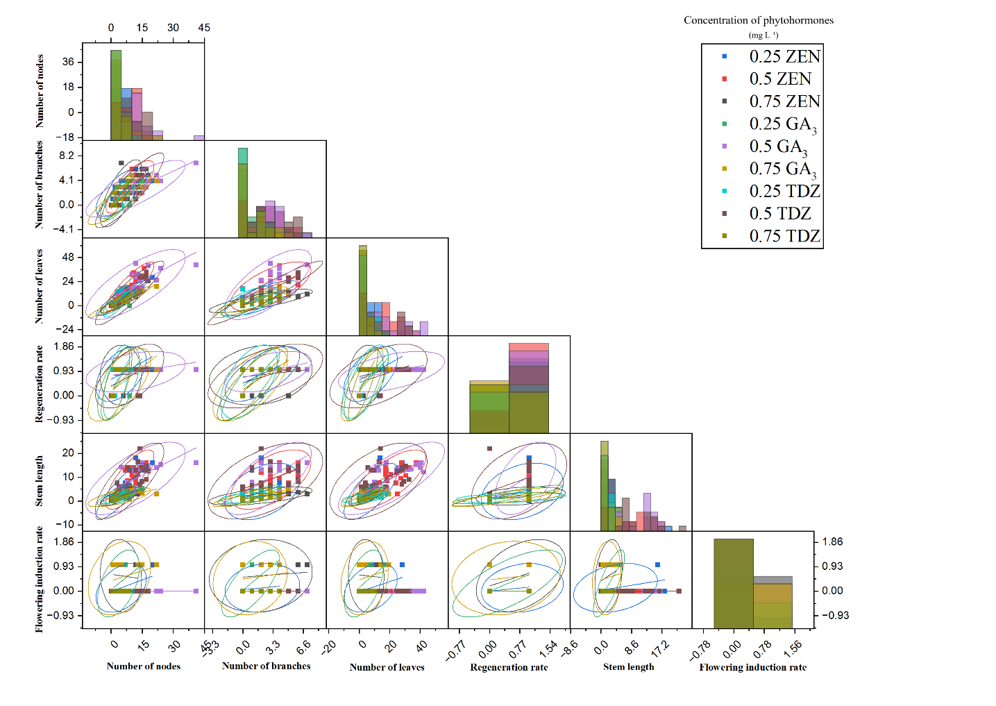
Figure 7 Correlations between growth parameters of the Beldia variety and the applied phytohormones (ZEN, TDZ, GA3).
Floral induction and reversion
In vitro flowering induction was highly significant (p-values < 0.001) and strongly dependent on both type and concentration of phytohormones. No flowering occurred in the controls, confirming the necessity of exogenous growth regulators. At 0.25 mg L⁻¹, ZEN induced moderate flowering, with a stronger response in Khardala (29%) than in Beldia (13%). In contrast, no flowering was observed at 0.5 mg L⁻¹ in either variety, indicating an inhibitory effect at this intermediate concentration (Figure 8). At 0.75 mg L⁻¹, flowering increased sharply, reaching 38% in Khardala and 58% in Beldia, indicating a biphasic response. GA₃ showed a similar trend: 0.25 mg L⁻¹ induced comparable flowering in both varieties (29%), while 0.5 mg L⁻¹ resulted in no induction, and 0.75 mg L⁻¹ stimulated flowering to 42% in Khardala and 50% in Beldia. TDZ did not trigger flowering at any concentration (Figures 9(A) and 9(B)). The analysis of in vitro flowering highlights a highly significant effect (p-values < 0.001) of both the type and concentration of phytohormones on floral reversion and proliferation in the two Cannabis sativa varieties (Table 1). Regarding zeatin effect (ZEN), a clear dose-response relationship was observed: Increasing concentrations enhanced both floral reversion and proliferation. At 0.75 mg L⁻¹, reversion rates reached 75% in Khardala and 71% in Beldia, while proliferation rates were 88% and 79%, respectively. These results indicate that higher zeatin concentrations promote floral transition and floral structure multiplication, with stronger reversion in Khardala and greater proliferation in Beldia. (Figures 9(E) and 9(D)). In contrast, responses to GA₃ were more variable and less linear. At a low concentration (0.25 mgL⁻¹), both varieties exhibited moderate but significant levels of reversion and proliferation (38% and 46% in Khardala; 50% and 63% in Beldia). However, at 0.5 mg L⁻¹, a marked decline was recorded, especially in floral proliferation in Beldia (17%), suggesting a potential inhibitory effect at intermediate doses. At 0.75 mg L⁻¹, the responses increased again, reaching 42% and 54% in Khardala, and 79% and 83% in Beldia, indicating a bell-shaped dose-response pattern where only higher concentrations provide significant stimulation, particularly in Beldia. Overall, comparison of the two regulators indicates that zeatin produced more consistent responses in floral reversion and shoot proliferation, while genotype-dependent differences were observed, with Beldia responding more strongly to GA₃ and Khardala to zeatin. Our findings appear promising compared with previous studies aimed at optimizing floral induction protocols. For instance, the use of DKW basal medium supplemented with 2 mg L⁻¹6-BA, applied exclusively during the vegetative phase, has been reported to promote meristem enlargement. Moreover, supplementation with 1 - 5 mg L⁻¹ 6-BA during the floral phase enabled the evaluation of its effect on floral induction [19]. Cytokinins play a pivotal role in this process: they directly regulate meristem size, thereby enhancing floral production [38], and promote the transition from vegetative to floral meristems in vitro [39]. Other studies have reported that treatment with PBZ at a concentration of 2.94 mg L⁻¹ resulted in the highest number of flowers (7.95) compared to other treatments [17]. This strategy offers multiple advantages, including a reduced life cycle and an increase in cannabinoid content. However, its efficiency remains genotype-dependent, requiring optimization of culture media for each cultivar [8]. In this context, our results highlight the relevance this approach for addressing challenges related to cannabis breeding and secondary metabolite production. Piunno et al. [40] reported that floral reversion occurred inconsistently and was limited to a subset of the cultivars tested, suggesting variability among genotypes. In their study, reversion was mainly observed in association with pre-existing meristematic tissues rather than new organogenesis. Comparable results were obtained with the application of 0.225 mg L⁻¹ BAP to flower pairs, which resulted in an average reversion rate of 69%. Similarly, treatments with meta-topolin (0.241 and 2) induced analogous responses, with flower pairs exhibiting approximately 2.5-fold higher reversion rates (70% - 81%) [16].
Floral reversion is well documented as an efficient strategy for the micropropagation of species recalcitrant to in vitro culture and represents a valuable tool in conservation programs for threatened species [41-43]. Histological observations revealed the presence of meristematic structures at the base of freshly dissected flowers. A vegetative meristem associated with adjacent floral meristems located near the ovary base and bracts was consistently observed in reverting explants. Floral reversion was accompanied by a progressive transition from reproductive to vegetative development over an approximately two-week period, after which normal vegetative growth was established. During this transitional phase, the emergence of additional floral structures along the elongating axis was occasionally observed before the stabilization of exclusively vegetative development [16]. At the molecular and physiological levels, the vegetative-floral transition in Cannabis sativa is regulated by genetic networks associated with shoot apical meristem activity. Exogenous sugars, particularly sucrose supplied in MS medium, function both as an energy source and as signaling compounds. Through the trehalose-6-phosphate pathway, sucrose has been reported to influence the regulation of flowering-related genes, including FLOWERING LOCUS T (FT), as demonstrated in other plant species [44,45]. The FT protein functions as a mobile florigen, translocating to the meristem where it promotes the initiation and differentiation of floral primordia [18]. Phytohormones represent another central regulatory layer of floral induction. Gibberellins (e.g., GA₃) promote the degradation of DELLA repressors, thereby enabling the activation of floral integrator genes such as SOC1 and LFY, both of which play pivotal roles in floral morphogenesis [46,47]. Cytokinins regulate the auxin/cytokinin balance within the meristem, and depending on concentration, can reprogram meristematic cells toward a floral fate [48,49]. A biphasic cytokinin response was observed, with low and high concentrations promoting floral induction, while intermediate levels (e.g., 0.5 mg L⁻¹) favored vegetative development. This pattern reflects dose-dependent regulation of meristem activity and has been reported in other species, where cytokinin signaling does not follow a linear response [16]. This dynamic reflects tightly regulated hormonal control of the vegetative-to-reproductive transition. Mineral nutrients contribute indirectly: Ca²⁺ acts as a signaling messenger, K⁺ supports sucrose transport to the meristem, and Mg²⁺ sustains photosynthesis and carbohydrate supply required for meristem reprogramming [50-53]. In vitro conditions influence developmental responses. Artificial photoperiods, controlled light intensity, limited gas exchange, and moderate oxidative stress define the culture microenvironment [54,55]. These factors are perceived by the meristem as changes in developmental context. In C. sativa, in vitro flowering reflects the interaction of hormonal, metabolic, mineral, and environmental signals. This response highlights the plasticity of the shoot apical meristem. Cannabis therefore represents a useful model for studying flowering regulation in agriculturally and pharmaceutically relevant species.
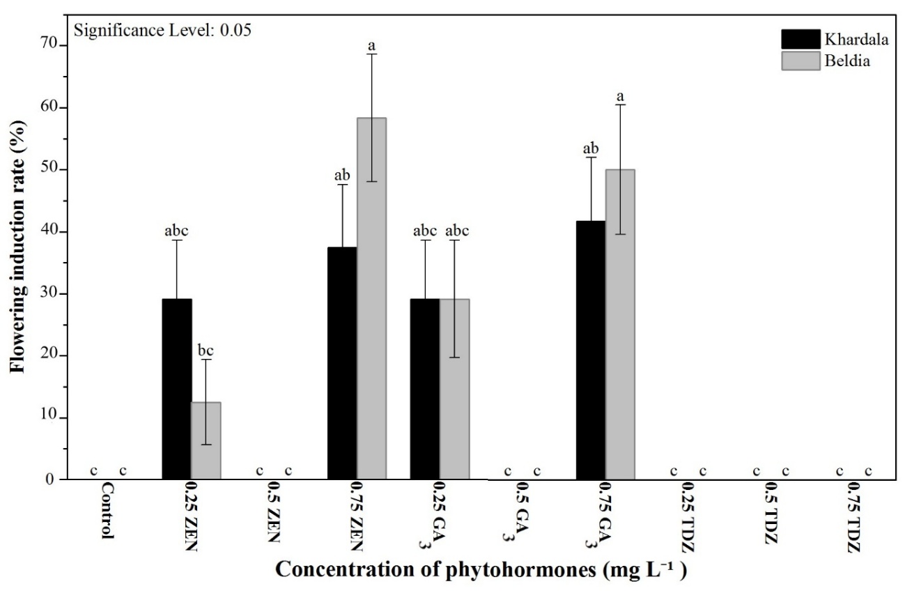
Figure 8 Flowering induction rate (%) from axillary buds as a function of different phytohormones in two Cannabis sativa varieties.
Table 1 Floral reversion and proliferation rates in Cannabis sativa varieties under different in vitro treatments.
Varieties |
Treatment |
Floral reversion rate (%) ± S.E. |
Floral proliferation rate (%) ± S.E. |
Khardala |
Control |
0.00 ± 0.00e |
20.83 ± 0.08bc |
0.25 mg L⁻¹ ZEN |
16.66 ± 0.08de |
25.00 ± 0.09bc |
|
0.5 mg L⁻¹ ZEN |
16.66 ± 0.08de |
33.33 ± 0.10bc |
|
0.75 mg L⁻¹ ZEN |
75 ± 0.09ab |
87.5 ± 0.07a |
|
0.25 mg L⁻¹ GA3 |
37.5 ± 0.10bcde |
45.83 ± 0.10abc |
|
0.5 mg L⁻¹ GA3 |
20.83 ± 0.08de |
29.16 ± 0.09bc |
|
0.75 mg L⁻¹ GA3 |
41.66 ± 0.10abcd |
54.16 ± 0.10abc |
|
Beldia |
Control |
0.00 ± 0.00e |
25 ± 0.09bc |
0.25 mg L⁻¹ ZEN |
20.88 ± 0.08de |
33.33 ± 0.10bc |
|
0.5 mg L⁻¹ ZEN |
33.33 ± 0.10cde |
45.83 ± 0.10abc |
|
0.75 mg L⁻¹ ZEN |
70.83 ± 0.09abc |
79.16 ± 0.08a |
|
0.25 mg L⁻¹ GA3 |
50 ± 0.10abcd |
62.50 ± 0.10ab |
|
0.5 mg L⁻¹ GA3 |
29.16 ± 0.09de |
16.66 ± 0.08c |
|
0.75 mg L⁻¹ GA3 |
79.16 ± 0.08a |
83.33 ± 0.08a |
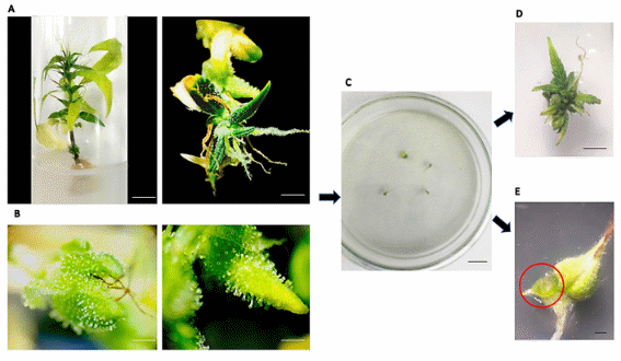
Figure 9 Floral induction and reversion from young flowers: (A) In vitro shoot proliferation from floral explants; (B) Flowers and leaves rich in glandular trichomes observed under a stereomicroscope (objective×1); (C) Culture medium containing sterile young flowers; (D) Floral reversion with emergence of leafy shoots; (E) Proliferation of newly formed flowers observed under a stereomicroscope (objective×1).
Rooting and acclimatization of plantlets
The assessment of in vitro rooting in Cannabis sativa plantlets indicated that responses differed based on both variety and IBA concentration. For Khardala variety, the highest rooting rate (87.5%) and longest root length (4.25 cm) were observed at 1 mg L⁻¹IBA, whereas the lowest concentration (0.5 mg L⁻¹) resulted in very poor rooting (12.5%) and extremely short roots (0.09 cm). Increasing the concentration to 2 mg L⁻¹reduced rooting efficiency (70.83%) and produced intermediate root lengths (1.57 cm). Survival after acclimatization remained high, reaching 94%. For Beldia variety, a similar pattern was observed, with slightly higher values: The optimal rooting rate (95.83%) and maximum root length (6.55 cm) were also obtained at 1 mg L⁻¹IBA. The 0.5 mg L⁻¹concentration resulted in low rooting (16.66%) and short roots (0.13 cm), while 2 mg L⁻¹significantly reduced rooting (50%) and produced relatively short roots (0.95 cm). Plantlet survival after acclimatization was similarly high at 95% (Table 2).
Our results suggest that, for both varieties, an intermediate IBA concentration (1 mg L⁻¹) is ideal for enhancing both rooting efficiency and root development. The high survival rates after acclimatization demonstrate that the roots formed in vitro were functional and well-adapted for transfer to ex vitro conditions (Figure 10). Currently, indole-3-butyric acid (IBA) and indole-3-acetic acid (IAA) are among the most commonly used plant growth regulators for root induction. Similar results were reported by Wróbel et al. [35], who tested different concentrations of IBA and IAA (0.25 - 0.75 mg L⁻¹) and observed effective rooting responses. Likewise, Stephen et al. [56], using the same MS-based medium as Lata et al. [57] supplemented with IBA, recorded a high rooting response in medium containing 1 mg L⁻¹IBA.The success of micropropagation therefore largely depends on the efficient transition of in vitro-grown plantlets to ex vitro conditions, which is critical for their survival and subsequent growth [58]. Excess IBA can inhibit root initiation by disrupting the optimal auxin gradient required for root primordia formation. It may also promote callus formation at the expense of organized root development.
Table 2 Effect of variety and phytohormone concentration on rooting rate, root length, and survival plantlets rate of Cannabis sativa.
Varieties |
Survival rate (%) ± S.E. |
Treatment |
Rooting rate (%) ± S.E. |
Root length (cm) ± S.E. |
Khardala |
94 ± 0.02a |
Control 0.5 mg L⁻¹IBA |
0.00 ± 0.00c 12.5 ± 0.07c |
0.00 ± 0.00c 0.09 ± 0.05c |
1 mg L⁻¹ IBA |
87.5 ± 0.07a |
4.25 ± 0.79b |
||
2 mg L⁻¹IBA |
70.83 ± 0.09ab |
1.57 ± 0.36c |
||
Beldia |
95 ± 0.03b |
Control 0.5 mg L⁻¹ IBA |
0.00 ± 0.00c 16.66 ± 0.08c |
0.00 ± 0.00c 0.13 ± 0.06c |
1 mg L⁻¹IBA |
95.83 ± 0.04a |
6.55 ± 0.90a |
||
2 mg L⁻¹IBA |
50 ± 0.10b |
0.95 ± 0.27c |
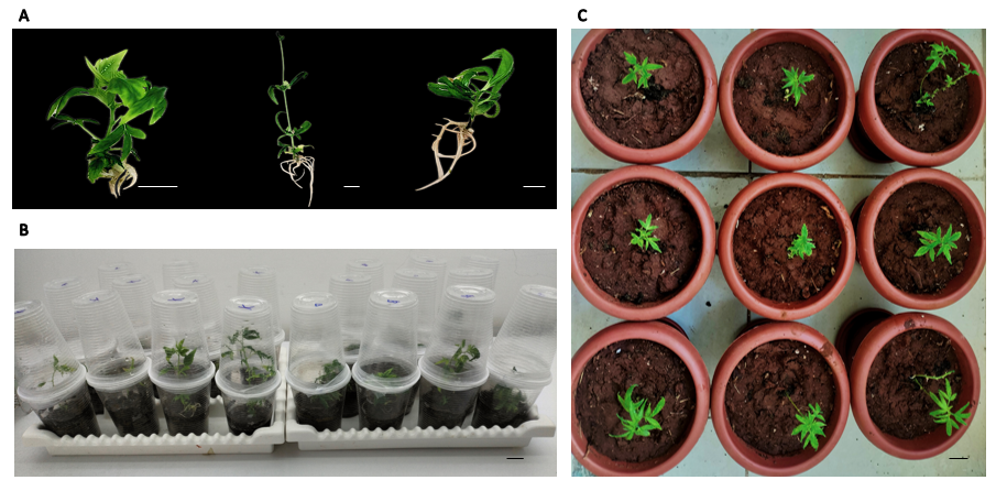
Figure 10 Rooting and acclimatization stages: (A) Rooting of different plants; (B) Acclimatization of C. sativa in vitro plantlets; (C) Transfer of Cannabis sativa plants to ex vitro conditions.
Conclusions
This study demonstrates that in vitro vegetative growth, floral induction, reversion, and rooting in Cannabis sativa are strongly genotype- and phytohormone-dependent. The distinct responses of the Moroccan varieties Beldia and Khardala to zeatin and GA₃ refine existing micropropagation protocols by enabling stage-specific and genotype-adapted regulation rather than generalized approaches. The identification of biphasic hormonal responses provides practical guidance for maintaining vegetative growth or inducing reproductive transitions under controlled conditions. These findings have direct implications for accelerating breeding cycles and supporting standardized plant material production, with potential applications in research and pharmaceutical contexts.
Acknowledgements
This work was carried out in collaboration between the Faculty of Sciences Ben M’Sik and the National School of Agriculture of Meknes. The authors would like to express their sincere gratitude to Mr. Amine Samih for his valuable assistance with the statistical analysis, and to Ms. Ilham Belkoura for her careful correction and revision of the manuscript.
CRediT author statement
Meryem Tahtah: Conceptualization; Methodology; Software; Writing - Original Draft. Toufik Ibrahim: Methodology; Data curation; Validation; Supervision. Fathellah Hachimi: Visualization; Writing - Review & Editing. Karima Samir: Supervision; Writing - Review & Editing. Aicha Nordine: Supervision; Writing - Reviewing and Editing.
Declaration of generative AI in scientific writing
The authors acknowledge the use of generative AI tools (e.g., ChatGPT by OpenAI) in the preparation of this manuscript, specifically for language editing and grammar correction. No content generation or data interpretation was performed by AI. The authors take full responsibility for the content and conclusions of this work.
References
[1] CM Andre, JF Hausman and G Guerriero. Cannabis sativa: The plant of the thousand and one molecules. Frontiers in Plant Science 2016; 7, 19.
[2] EB Russo. The case for the entourage effect and conventional breeding of clinical cannabis: No “strain,” no gain. Frontiers in Plant Science 2018; 9, 01969.
[3] JJ Zager, I Lange, N Srividya, A Smith and BM Lange. Gene networks underlying cannabinoid and terpenoid accumulation in cannabis. Plant Physiology 2019; 180(4), 1877-1897.
[4] H Lowe, N Toyang, B Steele, J Bryant and W Ngwa. The endocannabinoid system: A potential target for the treatment of various diseases. International Journal of Molecular Sciences 2021; 22(17), 9472.
[5] SE Turner, CM Williams, L Iversen and BJ Whalley. Molecular Pharmacology of Phytocannabinoids. 1st ed. Vol 103. Pogrchem, Germany, 2017, p. 61-101.
[6] SJ Livingston, TD Quilichini, JK Booth, DC Wong, KH Rensing, J Laflamme‐Yonkman, SD Castellarin, J Bohlmann, JE Page and AL Samuels. Cannabis glandular trichomes alter morphology and metabolite content during flower maturation. The Plant Journal 2020; 101(1), 37-56.
[7] H Lata, S Chandra, IA Khan and MA ElSohly. High frequency plant regeneration from leaf derived callus of high Δ9-tetrahydrocannabinol yielding Cannabis sativa L. Planta Medica 2010; 76(14), 1629-1633.
[8] AS Monthony, SR Page, M Hesami and AMP Jones. The past, present and future of Cannabis sativa tissue culture. Plants 2021; 10(1), 185.
[9] S Chandra, H Lata and MA ElSohly. Propagation of cannabis for clinical research: An approach towards a modern herbal medicinal products development. Frontiers in Plant Science 2020; 11, 00958.
[10] X Zhang, G Xu, C Cheng, L Lei, J Sun, Y Xu, C Deng, Z Dai, Z Yang, X Chen, C Liu, Q Tang and J Su. Establishment of an Agrobacterium-mediated genetic transformation and CRISPR/Cas9-mediated targeted mutagenesis in Hemp (Cannabis sativa L.). Plant Biotechnology Journal 2021; 19(10), 1979-1987.
[11] B Wróbel, M Hryniewicz, I Kulkova, K Mazur, Z Jakubowska, K Borek, J Dobrzyński, A Konieczna, A Miecznikowski, K Piasecka-Jóźwiak and A Fabiszewska. Fermentation quality and chemical composition of industrial hemp (Cannabis sativa L.) silage inoculated with bacterial starter cultures: A pilot study. Agronomy 2023; 13(5), 1371.
[12] SC Baek, SY Jeon, YJ Choi, BH Byun, DH Kim, GR Yu, H Kim and Lim DW. Establishment of an in vitro micropropagation system for Cannabis sativa ‘Cheungsam’. Horticulturae 2024; 10(10), 1060.
[13] SM Ahsan, M Injamum-Ul-Hoque, AK Das, S Shaffique, M Hasan, SM Kang, IJ Lee and HW Choi. Tuning up in vitro growth and development of Cannabis sativa: Recent advances in micropropagational approach. Applied Biosciences 2025; 4(1),12.
[14] M Deguchi, S Kane, S Potlakayala, H George, R Proano, V Sheri, WR Curtis and S Rudrabhatla. Metabolic engineering strategies of industrial hemp (Cannabis sativa L.): A brief review of the advances and challenges. Frontiers in Plant Science 2020; 11, 580621.
[15] M Tahtah, T Ibrahim, F Hachimi, K Samir and A Nordine. Enhancement of Cannabis sativa micropropagation for two Moroccan cultivated varieties using a multifactorial experimental approach. International Journal of Agriculture and Biology 2026; 35, 350107.
[16] AS Monthony, S Bagheri, Y Zheng and AMP Jones. Flower power: Floral reversion as a viable alternative to nodal micropropagation in Cannabis sativa. In Vitro Cellular & Developmental Biology-Plant 2021; 57, 1018-1030.
[17] L Ajdanian, H Arouiee, AMP Jones, M Hesami, H Nemati and M Pepe. Investigating the impact of paclobutrazol and tannic acid on floral development of in vitro-grown cannabis plantlets. Heliyon 2024; 10(17), 36768.
[18] CA Dowling, J Shi, JA Toth, MA Quade, LB Smart, PF McCabe, S Schilling and R Melzer. A Flowering Locus T ortholog is associated with photoperiod-insensitive flowering in hemp (Cannabis sativa L.). The Plant Journal 2024; 119(1), 383-403.
[19] O Lavie, K Buxdorf and LE Williams. Optimizing cannabis cultivation: An efficient in vitro system for flowering induction. Plant Methods 2024; 20, 141.
[20] F Bachir, M Eddouks, M Arahou and M Fekhaoui. Origin, early history, cultivation, and characteristics of the traditional varieties of Moroccan Cannabis sativa L. Cannabis and Cannabinoid Research 2022; 7(5), 603-615.
[21] C Benkirane, M Charif, CM Müller, Y Taaifi, F Mansouri, M Addi, M Bellaoui, H Serghini-Caid, A Elamrani and M Abid. Population structure and genetic diversity of Moroccan cannabis (Cannabis sativa L.) germplasm through simple sequence repeat (SSR) analysis. Genetic Resources and Crop Evolution 2024; 71, 2037-2051.
[22] PA Chouvy and K Afsahi. Hashish revival in Morocco. International Journal of Drug Policy 2014; 25(3), 416-423.
[23] M Pepe, M Hesami and AMP Jones. Machine learning-mediated development and optimization of disinfection protocol and scarification method for improved in vitro germination of cannabis seeds. Plants 2021; 10(11), 2397.
[24] T Murashige and F Skoog. A revised medium for rapid growth and bio assays with tobacco tissue cultures. Physiologia plantarum 1962; 15(3). 473-497.
[25] JD Lubell-Brand, LE Kurtz and MH Brand. An in vitro–ex vitro micropropagation system for hemp. HortTechnology 2021; 31(2), 199-207.
[26] A Trajkovska, O Tusevski, VS Gjorgievska, IC Karanfilova, I Davkova, M Karapandzova, G Stefkov, S Kulevanova and SG Simic. Cannabinoid production in various Cannabis sativa L. in vitro cultures. Plant Cell, Tissue and Organ Culture (PCTOC) 2025; 160, 10.
[27] T Aftab. Auxins, Cytokinins and Gibberellins Signaling in Plants. Springer Cham, Switzerland, 2022, p. 47-123.
[28] S Sudhakaran, JA Teixeira da Silva and S Sreeramanan. Test Tube Bouquets: In Vitro Flowering. Vol II. Global Science Books, London, 2006, p. 336-346.
[29] D Sun, M Wang, P Zhao, Y Ding, B Huang and J Tang. Evaluation of different types of cytokinins as zeatin replacement on in vitro proliferation of Vaccinium dunalianum. Pakistan Journal of Botany 2023; 55(1), 135-140.
[30] H Lata, S Chandra, I Khan and MA ElSohly. Thidiazuron-induced high-frequency direct shoot organogenesis of Cannabis sativa L. In Vitro Cellular & Developmental Biology – Plant 2009; 45, 12-19.
[31] T Hirakawa and S Tanno. In vitro propagation of Humulus lupulus through the induction of axillary bud development. Plants 2022; 11(8), 1066.
[32] ME Eriksson, M Israelsson, O Olsson and T Moritz. Increased gibberellin biosynthesis in transgenic trees promotes growth, biomass production and xylem fiber length. Nature Biotechnology 2000; 18, 784-788.
[33] TT Sekioka and JS Tanaka. Differentiation in callus-cultures of cucumber (Cucumis sativa L.). HortScience 1981; 16, 451-451.
[34] R Wang, LS He, B Xia, JF Tong, N Li and F Peng. A micropropagation system for cloning of hemp (Cannabis sativa L.) by shoot tip culture. Pakistan Journal of Botany 2009; 41(2), 603-608.
[35] T Wróbel, M Dreger, K Wielgus and R Słomski. Modified nodal cuttings and shoot tips protocol for rapid regeneration of Cannabis sativa L. Journal of Natural Fibers 2022; 19(2), 536-545.
[36] RC Clarke and MD Merlin. Cannabis domestication, breeding history, present-day genetic diversity, and future prospects. Critical Reviews in Plant Sciences 2016; 35(5-6), 293-327.
[37] K Ioannidis, I Tomprou, V Mitsis and P Koropouli. Genetic evaluation of in vitro micropropagated and regenerated plants of Cannabis sativa L. using SSR molecular markers. Plants 2022; 11(19), 2569.
[38] Y Han, H Yang and Y Jiao. Regulation of inflorescence architecture by cytokinins. Frontiers in Plant Science 2014; 5, 00669.
[39] NJ Taylor, ME Light and JV Staden. In vitro flowering of Kniphofia leucocephala: Influence of cytokinins. Plant Cell, Tissue and Organ Culture 2005; 83, 327-333.
[40] KF Piunno, G Golenia, EA Boudko, C Downey and AMP Jones. Regeneration of shoots from immature and mature inflorescences of Cannabis sativa. Canadian Journal of Plant Science 2019; 99, 556-559.
[41] MR Appleton, GD Ascough and J Van Staden. In vitro regeneration of Hypoxis colchicifolia plantlets. South African Journal of Botany 2012; 80, 25-35.
[42] NA Reshi, MS Sudarshana and BP Nandini. In vitro propagation from inflorescence explantsof Anisochilus carnosus- an ethno-medicinal herb. International Journal of Pharmaceutical Sciences and Research 2014; 5(6), 2423-2427.
[43] S Kumar, N Sahu and A Singh. High-frequency in vitro plant regeneration via callus induction in a rare sexual plant of Cenchrus ciliaris L. In Vitro Cellular & Developmental Biology – Plant 2015; 51, 28-34.
[44] CM Figueroa and JE Lunn. A tale of two sugars: Trehalose 6-phosphate and sucrose. Plant Physiology 2016; 172(1), 7-27.
[45] LH Cho, R Pasriga, J Yoon, JS Jeon and G An. Roles of sugars in controlling flowering time. Journal of Plant Biology 2018; 61, 121-130.
[46] C Schwechheimer. Gibberellin signaling in plants-the extended version. Frontiers in Plant Science 2012; 2, 00107.
[47] J Lee and I Lee. Regulation and function of SOC1, a flowering pathway integrator. Journal of Experimental Botany 2010; 61(9), 2247-2254.
[48] GE Schaller, A Bishopp and JJ Kieber. The yin-yang of hormones: Cytokinin and auxin interactions in plant development. The Plant Cell 2015; 27(1), 44-63.
[49] YH Su, YB Liu and XS Zhang. Auxin-cytokinin interaction regulates meristem development. Molecular Plant 2011; 4(4), 616-625.
[50] N Tuteja and S Mahajan. Calcium signaling network in plants: An overview. Plant Signaling & Behavior 2007; 2(2), 79-85.
[51] X Li, L Chen, L Yao, J Zou, J Hao and W Wu. Calcium-dependent protein kinase CPK32 mediates calcium signaling in regulating Arabidopsis flowering time, National Science Review 2022; 9(1), 180.
[52] BG Ayre. Membrane-transport systems for sucrose in relation to whole-plant carbon partitioning. Molecular Plant 2011; 4(3), 377-394.
[53] M Ishfaq, Y Wang, M Yan, Z Wang, L Wu, C Li and X Li. Physiological essence of magnesium in plants and its widespread deficiency in the farming system of China. Frontiers in Plant Science 2022; 13, 802274.
[54] K Takeno. Stress-induced flowering: The third category of flowering response. Journal of Experimental Botany 2016; 67(17), 4925-4934.
[55] TP Pasternak and D Steinmacher. Plant growth regulation in cell and tissue culture in vitro. Plants 2024; 13(2), 327.
[56] C Stephen, VA Zayas, A Galic and MP Bridgen. Micropropagation of Hemp (Cannabis sativa L.). HortScience 2023; 58(3), 307-316.
[57] H Lata, S Chandra, IA Khan and MA ElSohly. Propagation through alginate encapsulation of axillary buds of Cannabis sativa L.an important medicinal plant. Physiology and Molecular Biology of Plants 2009; 15(1), 79-86.
[58] Š Mestinšek-Mubi, S Svetik, M Flajšman and J Murovec. In vitro tissue culture and genetic analysis of two high-CBD medical cannabis (Cannabis sativa L.) breeding lines. Genetika 2020; 52(3), 925-941.