
Trends
Sci.
2026;
23(9):
12912
Circadian Rhythm Disruption Modulates Plasma Melatonin, Aortic Senescence, and Blood Pressure Homeostasis in Rats
Andi Noor Kholidha Syarifin¹,², Ani Retno Prijanti³,⁴,
Wawaimuli Arozal⁵ and Sri Widia Azraki Jusman³,⁴,*
1Doctoral Program in Biomedical Science, Faculty of Medicine, Universitas Indonesia, Jakarta 10430, Indonesia
2Department of Biochemistry, Faculty of Medicine, Universitas Halu Oleo, Kendari 93231, Indonesia
3Department of Biochemistry and Molecular Biology, Faculty of Medicine, Universitas Indonesia,
Jakarta 10430, Indonesia
4Center of Hypoxia and Oxidative Stress Studies, Faculty of Medicine, Universitas Indonesia, Jakarta 10430, Indonesia
5Department of Pharmacology and Therapeutic, Faculty of Medicine, Universitas Indonesia, Jakarta 10430, Indonesia
(*Corresponding author’s email: [email protected])
Received: 7 December 2025, Revised: 21 January 2026, Accepted: 5 February 2026, Published: 30 March 2026
Abstract
Exposure to light at night disrupts circadian homeostasis and suppresses endogenous melatonin production. Melatonin is crucial for blood pressure regulation and exhibits potent antioxidant properties. Sustained oxidative stress in the aorta contributes to endothelial dysfunction and premature aging. This study investigated the effect of nighttime light exposure on plasma melatonin levels, blood pressure, and molecular markers of vascular stress and senescence in rats. Sprague–Dawley rats were assigned to five groups: Control, Dim light (DL), DL with melatonin (DL + Mel), continuous light exposure (CLE), and CLE plus Melatonin (CLE + Mel). Melatonin (10 mg/kg) was administered orally. ELISA quantified plasma melatonin concentrations, and aortic tissue was analyzed to measure glutathione (GSH) content, intercellular adhesion molecule-1 (ICAM-1), interleukin-6 (IL-6), senescence-associated β-galactosidase (SA-β-Gal) activity, and melatonin receptor type 1 (MT1) gene expression. Nighttime light exposure was associated with a marked reduction in circulating melatonin and an elevation in blood pressure. Melatonin supplementation partially restored melatonin levels and attenuated both systolic and diastolic hypertension. In the aorta, DL and CLE exhibited increased ICAM-1 and IL-6 expression, enhanced SA-β-Gal activity, upregulated MT1 expression, and depleted GSH levels. These molecular alterations were mitigated by melatonin treatment. Collectively, these findings demonstrate that nighttime light exposure induces circadian disruption that drives hypertension, endothelial dysfunction, and premature vascular senescence, whereas melatonin supplementation confers significant protective effects.
Keywords: Circadian rhythm disruption, Melatonin, blood pressure, Oxidative stress, Vascular senescence
Introduction
Circadian rhythms regulate a wide range of physiological processes [1], and their disruption by light at night (LAN) has significant implications for endocrine function, metabolic regulation, and cardiovascular health [2,3]. LAN suppresses melatonin production and alters circadian gene expression in both central and peripheral tissues, contributing to hormonal imbalance and oxidative stress. Although the effects of
LAN on melatonin regulation are well established, its impact on vascular aging and blood pressure regulation remains insufficiently defined [4,5].
Reduced melatonin increases reactive oxygen species (ROS), promoting chronic vascular oxidative stress, endothelial dysfunction, and premature arterial aging—key contributors to hypertension [6,7] Epidemiological studies further show that night-shift workers experience substantially higher rates of hypertension and cardiometabolic disease, underscoring the clinical importance of circadian misalignment [8-12].
Melatonin plays a multifaceted role in cardiovascular homeostasis, acting through receptor-mediated pathways and potent antioxidant effects [6,13,14]. Disrupted melatonin secretion can disturb the normal circadian pattern of blood pressure, while elevated ROS induces ICAM-1 expression, inflammation, and senescence-associated phenotypes in vascular cells [15-17].
Despite growing evidence, the mechanistic links between circadian disruption, oxidative injury, and vascular aging remain incompletely understood. This study, therefore, evaluated blood pressure, plasma melatonin, IL-6, and ICAM-1 expression, senescence-associated β-galactosidase (SA-β-Gal) activity, and the GSH/GSSG ratio in rats exposed to nighttime light to elucidate early molecular markers of vascular dysfunction.
Material and methods
Experimental design
This is an in vivo experimental study that employed a post-test–only group design. Adult male Sprague–Dawley rats (8 - 10 weeks old, 200 - 250 g) were obtained from the Indonesian National Agency of Drug and Food Control of Indonesia (BPOM Indonesia). The animals were randomly assigned to 5 groups: The control group (C); Dim light–at–night group (DL); Dim light with melatonin group (DL + Mel); Continuous light exposure group (CLE); and the CLE with melatonin group (CLE + Mel). Each group consists of five rats, based on Federer’s formula [18].
The study was conducted at the Animal Research Facility of the Indonesian Medical Education and Research Institute (ARF-IMERI) and the Department of Biochemistry and Molecular Biology, Faculty of Medicine, Universitas Indonesia. All procedures were approved by the Health Research Ethics Committee, Faculty of Medicine, Universitas Indonesia (Approval No. KET-278/UN2.F1/ETIK/PPM.00.02/2024).
Chemicals and reagents
Melatonin powder (SIGMA Aldrich M5250), Ethanol 96 %, NaCl 0.9 %, Ketamin-Xylazine (Ket-A-Xyl®), Phospat Buffer Saline (PBS) 0.01 M pH = 7.4 (SolarBio P1020), T-GSH/Oxidized Glutathione (GSSG) Colorimetric Assay Kit (Elabscience E-BC-K097-M), Rat Melatonin ELISA Kit (Elabscience E-EL-R0031), Rat IL-6 ELISA Kit (Finetest ER0042), Rat ICAM-1 ELISA Kit (Finetest ER0028), β-Galactosidase Activity Assay Kit (Elabscience E-BC-K631-M), Tripure Reagent (AMT Bio ATB2700), DEPC Treated Water (Himedia ML024), SensiFAST SYBR No.-ROX One-Step Kit (Bioline BIO-72005).
Melatonin preparation
Melatonin solution was prepared by weighing 36.4 mg of melatonin powder and dissolving it in a total volume of 35 mL of Aquadest, after first dissolving it in 365 μL of 96% ethanol. Oral melatonin treatment was performed at 10 mg/kg of body weight, delivered in an average administration volume of 2.5 mL. The procedure of melatonin preparation was carried out in dim conditions to avoid exposure to light. The solution was stored in a dark bottle. Rats in DLAN + Mel and CLE + Mel groups were given oral treatment of melatonin at a dose of 10 mg/kg/BW every day at 5:30 p.m. using an oral gavage [19].
Experimental protocols
Light exposure at night was provided for 5 weeks to the treatment group. The control group received normal light, which was 12 h of light (200 lx) and 12 h of darkness (0 lx). Meanwhile, the treatment group was exposed to light at night: Dim light (5 lx) and bright light (200 lx). The other two treatment groups were dim- and bright-light exposure, each accompanied by daily oral administration of melatonin. This procedure was based on the method proposed by Verma et al. [20] with modifications.
Each cage, measuring 54×41×33 cm3, contains three rats. It was placed in a temperature-controlled room with free access to food and water (ad libitum). The lights were switched off at 6:00 a.m and back on at 6.00 p.m. and turned on at 6:00 p.m. All rats were placed in an acclimatization cage for a week before the experiment started. Light was emitted by dimmable LED strip/downlight lamps placed on the upper wall on a shelf facing the cages. The dimmer switch is used to control the intensity of light provided inside the cage. Light intensity was measured with a luxmeter.
Blood pressure measurement
Blood pressure was measured at the end of the experimental period using a non-invasive tail-cuff system (CODA®, Kent Scientific, USA). This method is widely used in rodents and is appropriate for detecting relative differences between experimental groups. Even though it remains susceptible to restraint-induced disruption compared with the telemetry-based method. Rats were gently restrained in acrylic holders and acclimatized for 10 - 15 min before measurement to minimize stress-related variability [21,22]. The tail was positioned through an occlusion cuff equipped with a volume-pressure recording sensor. For each animal, 3 - 4 consecutive measurements were obtained, and the average value was used to determine systolic and diastolic blood pressure.
Blood sampling and organ collection
Blood sampling was obtained from the retroorbital plexus, 2 - 3 mL, in the morning, and stored in an Ethylenediaminetetraacetic acid (EDTA) tube. Plasma separation was performed after 30 min of standing in EDTA tubes. The samples were then centrifuged (4 °C, 15 min, 1,000 g). The plasma fraction was collected and tested immediately or aliquoted and stored at –80 °C. Blood plasma samples were used for melatonin hormone measurement using the Enzyme-Linked Immunosorbent Assay (ELISA) method.
After blood sampling, the abdominal aorta was isolated and weighed. The tissue was used for several biochemical analyses. Tissue to be used for biochemical analysis was stored at –80 °C until needed for subsequent procedures.
Measurement of plasma melatonin
Melatonin hormone levels were measured using a Rat Melatonin ELISA testing kit (Elabscience E-EL-R003), according to the manufacturer’s instructions. This ELISA kit uses the competitive ELISA principle. The micro ELISA plate provided in this kit has been pre-coated with Rat MT. During the reaction, Rat MT in samples or Standard competes with a fixed amount of Rat MT on the solid phase supporter for sites on the Biotinylated Detection Ab specific to Rat MT [23]. Sample absorbance was read using Thermo Scientific Varioskan LUX Multimode Microplate Reader at a wavelength of 450 nm. Hormone levels were determined by interpolating the sample absorbance values against a melatonin standard curve [24].
Homogenate preparation
Tissue homogenate was prepared separately for each biochemical parameter test of the tissue. The tissue was homogenized using different solvents based on the manufacturer’s instructions. As much as 60 mg of aortic tissue was homogenized with 540 μL solvent, which is recommended in the kit.
Homogenates for ICAM-1 and IL-6 protein assay were prepared with PBS. It was centrifuged at a speed of 5,000 g at a temperature of 4 °C for 5 min. The homogenate for the SA-β-Gal enzyme activity assay was prepared with the buffer solution from the kit. Meanwhile, the homogenate for GSH/GSSG ratio measurement was prepared with protein precipitator solution. The homogenate was then centrifuged at 10,000 g for 10 min at 4 °C. The supernatant was separated, aliquoted, and kept at –80 °C until being used for further testing.
Measurement of ICAM-1 and IL-6
The ICAM-1 and IL-6 levels of the aortic tissue samples were then measured using ELISA methods, which were a double-antibody sandwich ELISA detection kit and had a 4-hour assay duration. The microplate included with this kit has been pre-coated with anti-ICAM-1 (Finetest ER0028) and anti IL-6 antibodies (Finetest ER0042) [25]. The protein content of the supernatant was determined using the Bradford Method [27].
Measurement of SA β-galactosidase enzyme activity
β-Galactosidase enzyme activity as a marker of cellular senescence was measured using the β-Galactosidase Activity Assay kit (Elabscience E-BC-K631-M). In the enzyme activity test, 20 µL of standard or sample was placed in a well. 30 µL of substrate working solution was added to the sample well, while in the standard and control wells, 30 µL of buffer solution was added. 10 µL of activator agent was then added to each well and homogenized using a shaker incubator at 37 °C for 10 min, avoiding light exposure. After incubation, the optical density (OD) of the sample was measured using a Thermo Scientific Varioskan LUX Multimode Microplate Reader at a wavelength of 400 nm. The absorbance of the sample would be interpolated with the standard curve, and with calculations using the formula from the procedure kit, the β-Galactosidase activity contained in 1 g of protein per hour in hydrolyzing the substrate into 1 µmol p-nitrophenol at a temperature of 37 °C is defined as 1 unit of enzyme activity [28]. Protein levels in the samples were determined using the Bradford Method [27].
T-GSH and GSH/GSSG ratio assay
T-GSH content and GSH/GSSG ratio assay using the Total Glutathione (T-GSH/Oxidized Glutathione (GSSG) Colorimetric Assay Kit, 96T) (Elabscience E-BC-K097-M). The supernatant was neutralized and divided for measurement of total GSH and free GSH. For total GSH, samples were reacted with DTNB in the presence of glutathione reductase and NADPH, and absorbance at 412 nm was recorded. Free GSH was measured directly under the same conditions, whereas GSSG was determined by first derivatizing GSH with 2-vinylpyridine, followed by enzymatic reduction and colorimetric detection. The concentrations were derived from standard curves, and the GSH/GSSG ratio was established as an indication of cellular redox homeostasis [29].
Quantitative real-time polymerase chain reaction
Total RNA was extracted from frozen aorta tissue samples using TriPure reagent. The relative gene expression level of aorta melatonin receptor type 1, MT1 (Accession number NM 053676.1), was determined using SensiFAST TM SYBR® No-ROX One-Step Kit on a Thermo Cycler (Bio-Gener, Gene Amp PCR System, Q3202) according to the manufacturer’s instructions. GAPDH (Accession number NM_017008.4) was used as a housekeeping gene for normalization. The relative gene quantification was performed using Livak’s method [30]. Table 1 lists the primers used for RT-qPCR.
Table 1 List of primers [31].
Gene of interest |
Primer sequences |
GAPDH |
F: 5’- TGCTGAGTAYGTCGTGGAGTC -3’ |
|
R: 5’- TTGGTGGTGCAGGAKGCATTGC-3’ |
MT1 |
F: 5’- CTACATTTGCCACAGTCTC -3’ |
|
R: 5’- CATATCCTTAAGTAGCAGAAAG-3’ |
Statistical analysis
All parameters were analyzed and displayed in tables, graphs, and figures using GraphPad Prism 10 (GraphPad Software Inc.). Descriptive statistics were shown as mean ± SEM. Bivariate analysis was used to evaluate hypotheses, with a one-way ANOVA test followed by Tukey’s HSD post hoc test if the data were normally distributed. The data normality is tested using Saphiro-wilk test. Kruskal-Wallis test followed by Dunn’s test if the data were not. p < 0,05 indicated statistical significance.
Results and discussion
Our current study demonstrated that exposure to light at night, either DL or bright continuous light, induces an elevation in blood pressure in rats. The animals exhibited circadian rhythm disruption, characterized by increased body weight and reduced circulating melatonin. The upregulation of MT1 gene expression under circadian disruption was associated with a vasoconstrictive response. Since the role of melatonin is as an endogenous antioxidant, the decline in plasma melatonin induced oxidative stress, evidenced by reduced T-GSH levels and a lower GSH/GSSG ratio. Notably, these alterations were ameliorated in rats receiving oral melatonin supplementation. The oxidative stress of aortic tissue due to reduced melatonin levels appeared to correlate with increased ICAM-1 expression, indicative of endothelial dysfunction. Furthermore, elevated IL-6 expression of protein and relative mRNA levels, accompanied by enhanced SA-β-galactosidase activity in rats exposed to light at night, support the occurrence of cellular senescence associated with circadian rhythm disruption.
Rat body weight profiles
During the experiment, the rat’s weight was measured weekly. The data showed an increase in weight except for the 2 groups, the DL+ Mel and CLE + Mel groups. These two groups that received melatonin treatment showed a stable body weight profile by the third week of the experiment. The changes in rat weight are illustrated in Figure 1. Our findings showed that disruption of the circadian rhythm is associated with a significant increase in body weight of the rats. Furthermore, melatonin administration inhibits weight gain in rats. While body weight increased in the control and light at night groups, melatonin administration prevented body weight gain from week 2 to week 5 in the group with melatonin supplementation. This finding was consistent with a study by Meng et al., who discovered that circadian rhythm disruption caused decreased muscle mass, protein quality, and melatonin production but increased visceral fat levels and insulin resistance in women aged 31 - 40 years, implying that disruption to the circadian rhythm may be a factor for increasing the risk of cardiovascular disease and diabetes in the age group [8]. Chronic circadian rhythm disruption for 6 months in rats causes adipocyte hypertrophy followed by inflammation and fibrosis in adipose tissue [32]. In another study, it was found that deletion of the Bmal1 gene, a gene that regulates circadian rhythms and plays a role in adipocyte fat metabolism, disrupts fat metabolism regulation and leads to obesity. This is similar to the condition of circadian rhythm disorders, where the condition can affect the expression pattern of the Bmal1 gene, thereby affecting target genes related to fat metabolism [33].
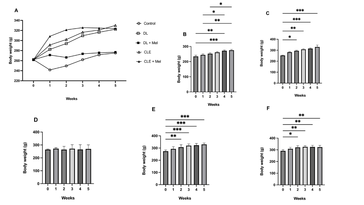
Figure 1 Body weight profile and mean body weight comparison in each treatment group per week. (A): Body weight profile, (B): Body weight of control group, (C): Body weight of DL Group, (D): Body weight of DL + Mel, (E): Body weight of CLE group, (F): Body weight of CLE + Mel. Data are shown as mean ± SEM and tested according to one-way ANOVA followed by Fisher’s LSD Test between groups. Significant difference * (p 0.033), ** (p 0.002), *** (p 0.001). Control: Group that was not exposed to light at night; DL: Group that was exposed to dim light (5 lx) at night; CLE: Continuous Light Exposure, Group that was exposed to bright light (200 lx) at night.
Plasma melatonin level and the expression of melatonin receptor type 1 (MT1) in the rat aortic tissue after circadian rhythm disruption
Low-intensity (5 lx) and high-intensity (200 lx) light exposure at night altered plasma melatonin profiles. Light exposure at night decreased melatonin production in the treatment groups relative to the control group. Figure 2(A) showed that plasma melatonin levels increased in the DL group given melatonin (DL + Mel) orally compared to the DL group only. However, this was not observed in the CLE group, where melatonin administration did not show a significant difference between the CLE and CLE + Mel groups.
Figure 2(B) shows a comparison of MT1 gene expression between the control group and the treatment group. The results showed that there is upregulated MT 1 expression in rat aortic tissue of the DL and CLE groups, with no significant difference compared to controls. On the other hand, the groups with melatonin administration showed a decreased MT1 gene expression, even though they did not elicit significant changes compared with the non-melatonin-treated DL and CLE groups. Remarkably, MT1 expression was lower in rats exposed to dim light with melatonin supplementation compared with those subjects under continuous light, regardless of melatonin administration status (Dunn’s test p < 0.05).
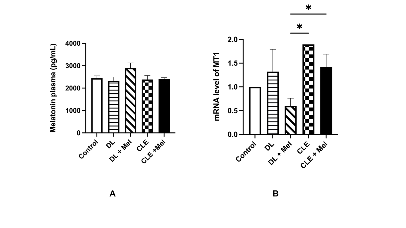
Figure 2 Melatonin concentration and MT1 gene expression. (A) Comparison of the melatonin plasma level between groups. (B) Comparison of melatonin receptor type 1 (MT1) expression in the aorta samples between groups. Significant difference * (p 0.033).
Our analysis of plasma melatonin revealed that circadian disruption alters plasma melatonin profiles. Exposure to light at night resulted in a lower plasma melatonin concentration than that observed in the control group. Melatonin production at night is regulated by the circadian rhythm controlled by the suprachiasmatic nucleus (SCN) in the hypothalamus [34]. Exposure to light at night disrupts the circadian rhythm and affects changes in the pattern of melatonin hormone production [35]. Insufficient melatonin production due to exposure to light at night can disrupt the circadian rhythm and affect the body’s metabolism.
Oral melatonin supplementation significantly elevated plasma melatonin levels in the DL + Mel group compared to the DL group. A comparable trend was observed in the CLE + Mel group, which demonstrated a significant increase relative to the DL group. Still, the difference remained non-significant when compared with both the control and CLE groups. Our findings showed that oral administration of exogenous melatonin (10 mg/kgBW/day) increased plasma melatonin levels in rats of the DL group compared with those that did not receive melatonin. This was in line with previous studies, which found that exogenous melatonin administration exerts a protective antioxidant effect in several animal models induced by oxidative stress [36-39]. However, while melatonin administration can increase plasma melatonin in rats with circadian rhythm disruption due to dim light exposure, it did not increase plasma melatonin in rats exposed to continuous light in the CLE group. Previous experimental studies support these mechanisms: Under continuous light conditions, even when melatonin was administered, plasma melatonin might not rise significantly due to complete suppression of endogenous pineal gland synthesis [40].
This study also found that there was an effect of nighttime light exposure on changes in gene expression of melatonin receptor type 1 (MT1) in aortic tissue. In this study, MT1 gene expression in aortic tissue showed an increase in the group exposed to light at night, where the relative MT1 expression in the CLE group was significantly higher than in the control group, although the DL group did not show a significant difference. The biological clock and melatonin levels influence the expression of MT1 by modulating circadian genes such as the bmal1 gene [41].
MT1 upregulation may represent a compensatory or adaptive response to reduced circulating melatonin under conditions of circadian disruption, aimed at preserving melatonin signaling sensitivity rather than initiating vascular dysfunction [42,43]. Meanwhile, in other hand, MT1 receptor activation has also been linked to vasoconstrictive responses, contributing to elevations in blood pressure [44]. Recent comprehensive reviews indicate that MT1 and MT2 melatonin receptors are heterogeneously expressed throughout the vascular system, with MT1 often linked with vasoconstrictive responses and MT2 with vasorelaxant effects [44]. Meanwhile, the functional outcomes of the receptors depend on vascular bed, receptor density, and context rather than the simple causality of expression alone [45]. These divergent vascular actions are attributed to the differential cellular distribution of melatonin receptors within the vasculature. Specifically, MT1-mediated modulation of vascular tone has been observed in multiple vessels, whereas MT2 activation is generally associated with endothelial nitric oxide-dependent vasodilation, suggesting diverse receptor roles related to changes in receptor expression at different tissue sites, with MT1 receptors primarily expressed in vascular smooth muscle cells and MT2 receptors predominantly localized to endothelial cells, particularly in the aorta [45,6].
Effects of circadian rhythm disruption on blood pressure
In systolic and diastolic data, there were significant differences between the control group, the DL group, and the CLE group, as well as between the CLE group and the group receiving oral melatonin treatment (Dunn’s test, p < 0.05). This indicates that exposure to light at night affects blood pressure profile in the treatment group compared to the control group, and with the administration of melatonin, it reduces the impact of light exposure on blood pressure.
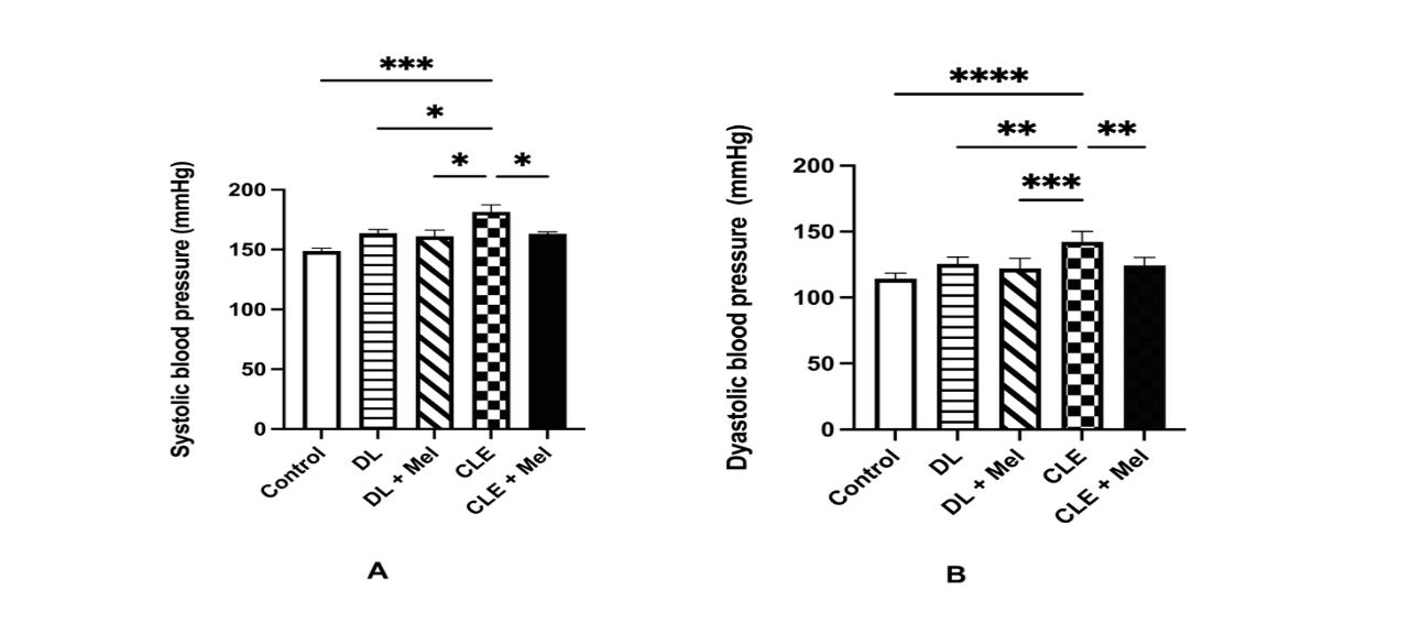
Figure 3 Comparison of systolic (A) and diastolic and (B) blood pressure between groups. Significant difference * (p 0.033), ** (p 0.002), *** (p 0.001).
There was a significant increase in blood pressure profiles, both in systolic and diastolic, in the groups
exposed to dim light (5 lx) and bright light (200 lx) compared to the control group (Figure 3). In this study, the result showed an increase in blood pressure in the group exposed to both DL (5 lx) and bright light (200 lx) compared to the control group. Meanwhile, the group exposed to light at night had decreased melatonin levels compared to the control group.
The antihypertensive effect of melatonin is significant, as evidenced by lower blood pressure in the melatonin-treated group (CLE + Mel) compared to the untreated group (CLE). In this study, the group that received oral melatonin treatment had lower blood pressure (both systolic and diastolic) than the group that did not. One of the physiological processes regulated by the circadian rhythm in the body is blood pressure in the cardiovascular system. This indicates indirect functional evidence, where blood pressure is regulated by melatonin based on the interaction of this hormone with its receptors in the blood vessel tissue [46,47]. Furthermore, with melatonin’s role as an antioxidant through its interaction with one of the melatonin receptors in the cytoplasm, the effect of increased blood pressure due to oxidative stress can be suppressed [46,47]. Additionally, MT1 provides a significant role in vasoconstriction. Higher MT1 gene expression increases vasoconstriction associated with increased blood pressure [48,49].
Antioxidant capacity profile in the aortic rat tissue after circadian rhythm disruption
Exposure to the DL and CLE at night showed to decrease the antioxidant capacity (T-GSH and GSH/GSSG ratio) in the aortic rat tissue compared to the control group (Dunn’s test, p < 0.05). Furthermore, in the groups given melatonin treatment (DL + Mel and CLE + Mel), both T-GSH content and GSH/GSSG ratio were higher than in the groups that did not receive melatonin treatment (DL and CLE). However, this was not found to be statistically significant (Dunn’s test, p > 0.05) (Figure 4).
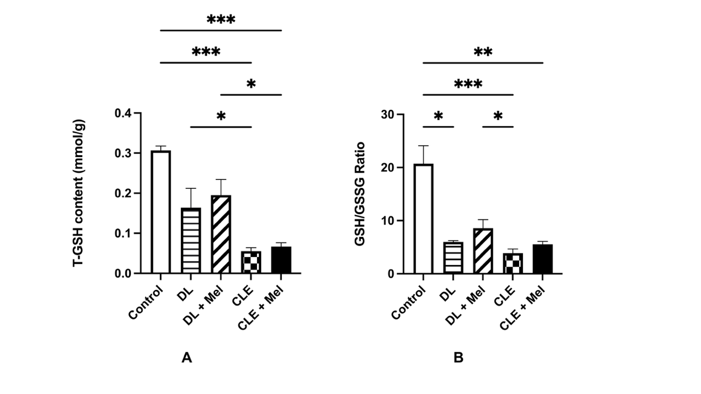
Figure 4 Antioxidant capacity profile of the rat aorta. (A) Comparison of T-GSH Level between groups. (B) Comparison GSH/GSSG ratio between groups. Significant difference * (p 0.033), ** (p 0.01) *** (p 0.001).
Our study provides evidence that disruption of circadian rhythm can modulate antioxidant capacity, reflected in both T-GSH levels and the GSH/GSSG ratio. These 2 variables showed identical trends, which had decreased after exposure to light at night and were lower than in the control group. Some previous studies indicate that circadian disruption, as in the fibrosis model rat, can affect oxidative stress, and that the time-restricted feeding and exercise models can increase proinflammatory markers [50-52]. In contrast, after administration of melatonin, T-GSH and GSH/GSSG ratio were increased compared to the group that did not receive the treatment. This indicated that melatonin’s antioxidant activity was working, although it could not yet match the control group’s antioxidant profile [6,37,53]. T-GSH represents the total glutathion pool available to combat oxidative stress, whereas the GSH/GSSG ratio shows the redox balance inside cells or tissues [54,55]. Oxidative stress and inflammation act primarily as upstream drivers of cellular senescence, triggering DNA damage, mitochondrial dysfunction, and other hallmarks [56]. Reactive oxygen species (ROS) generated during oxidative stress cause macromolecular damage and activate senescence pathways, often synergizing with pro-inflammatory cytokines like IL-6, IL-8, TNF-α, and IL-1β to accelerate this process [57,58].
Intercellular adhesion molecule-1 (ICAM-1) protein level in the aortic rat tissue after circadian rhythm disruption
Exposure to both DL and CLE at night induced an increase in ICAM-1 levels in aortic rat tissue. ICAM-1 levels in the CLE group were significantly higher than in the control group. Furthermore, very high levels of ICAM-1 were found in the CLE group, which was significantly different from the CLE group that received melatonin treatment (CLE + Mel) (Dunn’s test, p < 0.05) (Figure 5).
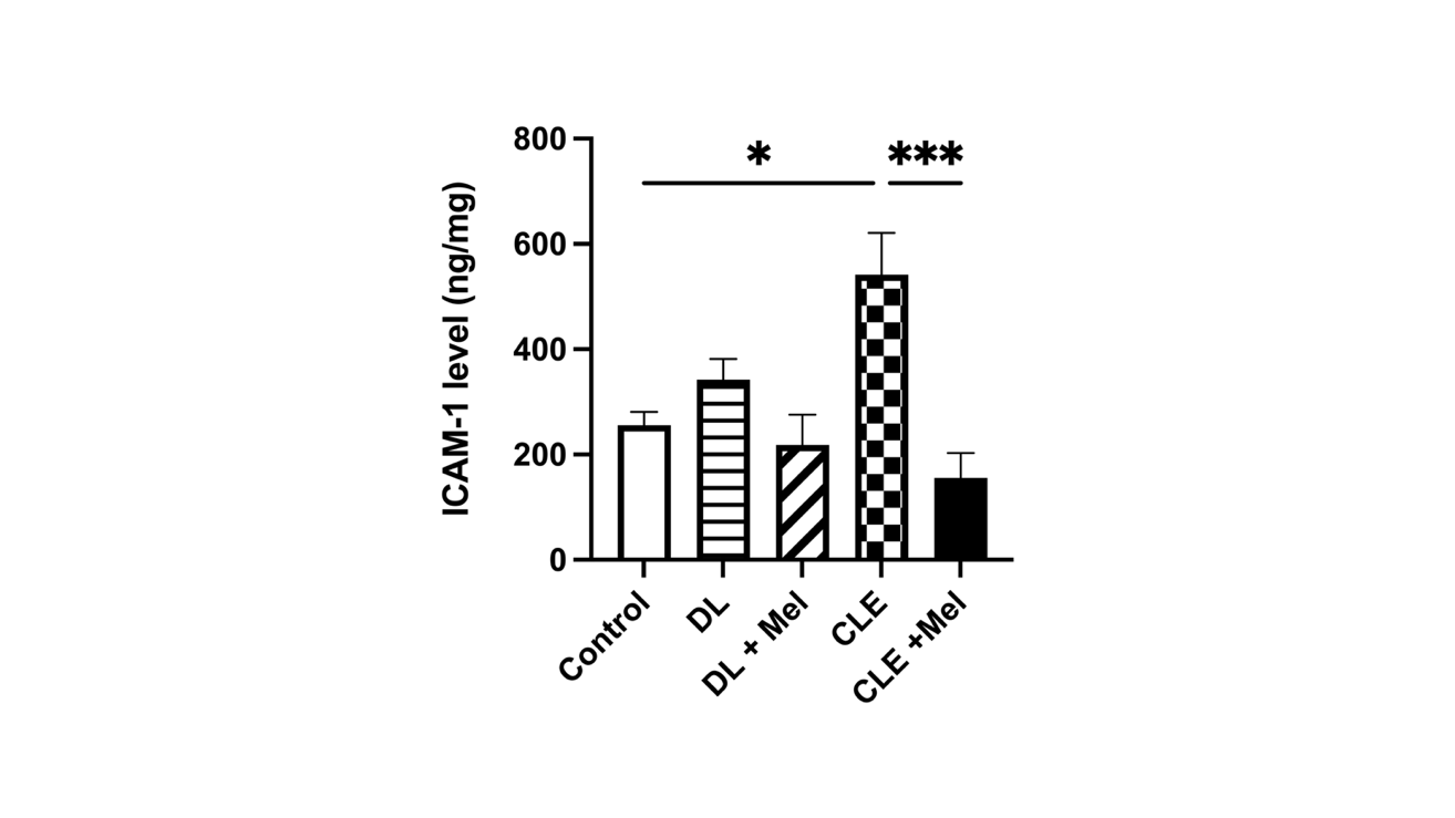
Figure 5 Comparison of Intercellular Cell Adhesion Molecule-1 (ICAM-1) between groups. Significant difference *(p 0.033), *** (p 0.001).
This finding provides indirect but biologically relevant functional evidence of vascular alterations associated with the change of circadian rhythm. Therefore, specific examinations of melatonin receptor function, such as vascular reactivity studies or pharmacological inhibition, will be important to conduct in the future. This clarification confirms that there is no overinterpretation of the research data on melatonin receptor expression, which is also a limitation of this study.
The recent study showed that the elevation in blood pressure was likely associated with vascular structural alterations, as indicated by increased ICAM-1 levels in both the DL and CLE group. Furthermore, melatonin administration improved blood vessel repair, as evidenced by decreased ICAM-1 protein levels in the melatonin-treated group. These findings were relevant to the role of melatonin as an antioxidant, whose decreased levels can lead to increased oxidative stress in cells and tissues [59-61]. A shortage of the melatonin hormone can cause increased inflammation, in which vascular adhesion molecules play an important role. Intercellular adhesion molecule-1 (ICAM-1) is an adhesion molecule found on the endothelial cell membrane that regulates leukocyte adhesion and migration to the vascular endothelium [62].
Senescence biomarker
SA β -Gal activity of the aortic rat tissue after circadian rhythm disruption
The DL group exhibited higher SA β-Gal activity than the control group. Activity of the enzyme decreased in the DL group with melatonin, compared to the DL group, although no significant difference. The current study found that the intensity of light exposure (5 and 200 lx) at night did not show different effects in the SA β-Gal activity. The DL group showed higher SA β-Gal activity than the CLE group; however, this difference was not statistically significant (Figure 6).
To assess the potential for aortic senescence, we measured SA β-Gal activity and IL-6 expression at protein and mRNA levels. The activity of the SA β-gal enzyme and the IL-6 profile are widely used as biomarkers of senescence in tissues and cell cultures. In our study, we found that SA β-Gal activity increased in the DL-exposed group compared to the control group, although the difference was not significant. Cells that age due to cellular aging accumulate in the arteries with aging, regardless of whether a person has age-related vascular disorders or not [63]. Stress during normal aging can induce cellular aging in cells associated with atherosclerosis [64]. Furthermore, the DL group that received melatonin treatment showed a decrease in SA β-Gal enzyme activity compared to those who did not receive melatonin. However, the CLE group did not show any significant difference in enzyme activity after receiving melatonin. Allegedly, the inability of melatonin treatment under continuous light exposure in rats to decrease the enzyme activity in the CLE group was primarily due to extreme suppression of endogenous melatonin and loss of its antioxidant role [40].
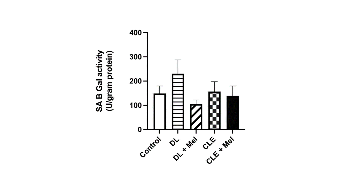
Figure 6 Comparison of senescence-associated β-galactosidase (SA β-Gal) enzyme activity between groups.
IL-6 expression in the aorta tissue after circadian rhythm disruption
The current study showed a non-significant reduction in IL-6 protein levels in rats with circadian rhythm disruption treated with melatonin, with ELISA results displaying a similar downward trend to that observed in relative IL-6 expression measured by real-time PCR. However, unlike the protein data, IL-6 mRNA levels were significantly reduced in the DL and CLE groups receiving melatonin compared to their untreated counterparts, indicating a more substantial melatonin effect at the transcriptional level (Dunn’s test, p < 0.05) (Figure 7).
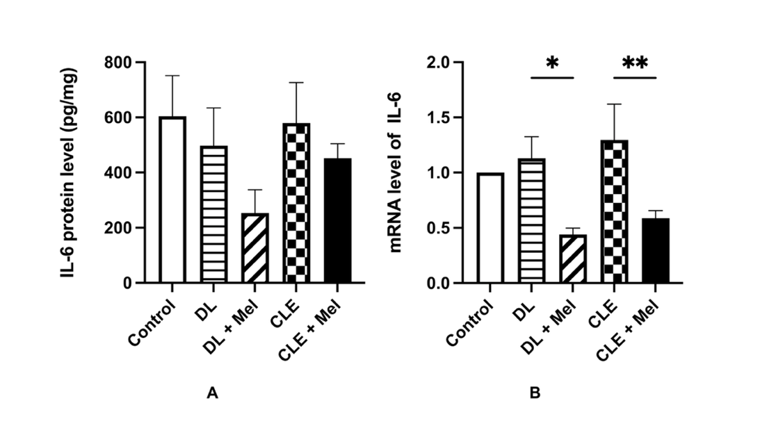
Figure 7 IL-6 Expression of the aortic rat tissue. (A) Comparison of IL-6 protein level and (B) Comparison of IL-6 relative mRNA level. Significant difference * (p 0.033), ** (p 0.01).
mRNA and protein levels of IL-6 increase in the group exposed to light, although only genetic expression differs significantly between the control and treatment groups. IL-6 is a well-established and robust biomarker of the senescence-associated secretory phenotype (SASP) [65,66]. However, in this study, we found that SA β-gal enzyme and the IL-6 protein levels are indirect or supportive indicators in senescence rather than definitive markers. Although increased IL-6 expression in aortic tissue in the current study represents an indirect indicator of cellular senescence rather than a primary functional determinant, melatonin administration markedly suppressed IL-6 mRNA expression, supporting a transcriptional anti-inflammatory effect of melatonin that may contribute to its senescence-modulating properties.
Melatonin exhibits well-documented anti-inflammatory effects, particularly through transcriptional suppression of pro-inflammatory cytokines like IL-6 [67]. Melatonin suppression of IL-6 occurs during chronic inflammation phases, without broadly inhibiting acute healing responses [68]. In a vascular context, this ties to reduced oxidative stress and senescence markers via pathways like SIRT/Nrf2, which could explain its protective role against circadian disruption-induced endothelial dysfunction [69]. Inhibition of IL-6 transcription by melatonin supports broader aging-modulating effects, including mitochondrial preservation and anti-aging in vascular smooth muscle cells. It places melatonin as a promising therapy for hypertension models involving circadian rhythm imbalance [68,70].
In this study, we used animal models to evaluate the effects of circadian rhythm disruption on phenotypic alterations, specifically the elevation of blood pressure, which is hypothesized to occur via oxidative stress and inflammatory pathways. Exposure to light at night induced increases in blood pressure, oxidative stress, endothelial dysfunction biomarkers, and senescence supportive indicators. We examined the effects of nighttime light exposure at two intensities—DL and CLE—and evaluated whether exogenous melatonin administration could mitigate the pathological outcomes associated with circadian disruption. The direct assessment of aortic tissue further supports the relevance of this study to blood pressure–related phenotypes. The absence of histological and complete senescence marker analyses was the limitation of this study. Therefore, definitive confirmation of vascular senescence will be important for future studies incorporating established senescence markers and histopathological assessments.
Conclusions
In conclusion, this study demonstrated that nighttime light exposure, whether dim light or continuous light exposure, elevated blood pressure in rats due to circadian disruption, increased body weight, and reduced circulating melatonin. Upregulation of MT1 expression is a compensatory response to reduced melatonin availability rather than a direct contributor to vasoconstriction, while melatonin decline induced oxidative stress, evidenced by decreased T-GSH levels and a lower GSH/GSSG ratio. Oral melatonin supplementation mitigated these effects. Oxidative stress in aortic tissue correlated with increased ICAM-1, indicating endothelial dysfunction. Elevated IL-6 expression and enhanced SA-β-galactosidase activity were indirect evidence and supported the induction of cellular senescence associated with circadian disruption. Melatonin treatment was effective in the DL group but showed limited efficacy in the bright light (CLE) group, due to suppression of endogenous melatonin synthesis by high-intensity light.
Acknowledgments
We would like to sincerely thank the technicians at ARF-IMERI, Pharmacokinetics, Medical Chemistry, and Biochemistry Laboratories, Faculty of Medicine, Universitas Indonesia, Indonesia for their assistance in animal care and biomedical measurements. The great appreciation also goes to The Indonesian Education Scholarship, The Center for Higher Education Funding and Assessment, and The Indonesian Endowment Fund for Education for providing support in funding, and the Doctoral Program in Biomedical Science, Faculty of Medicine, Universitas Indonesia, Indonesia for providing facilities that support the research in completing the necessary data.
Declaration of Generative AI in Scientific Writing
The authors acknowledge the use of generative AI tools (e.g., Grammarly) in the preparation of this manuscript, specifically for language editing and grammar correction. AI performed no content generation or data interpretation. The authors take full responsibility for the content and conclusions of this work.
CRediT Author Statement
Andi Noor Kholidha Syarifin: Conceptualization, Methodology, Investigation (in-vivo experiments), Data Curation, Formal Analysis, Writing – Original Draft, Writing – Review & Editing, Visualization. Sri Widia Azraki Jusman: Resources, Supervision, Conceptualization, Methodology, Data Curation, Writing – Review & Editing. Visualization. Ani Retno Prijanti: Supervision, Conceptualization, Methodology, Formal Analysis, Validation, Writing – Review & Editing. Wawaimuli Arozal: Resources, Supervision, Conceptualization, Methodology, Software, Data Curation, Writing – Review & Editing.
References
[1] MZ Danish and M Saleem. A literature review on ― biological. World Journal of Pharmaceutical Research 2020; 9(5), 2135-2148.
[2] D Stewart and U Albrecht. Beyond vision: Effects of light on the circadian clock and mood-related behaviours. npj Biological Timing and Sleep 2025; 2(1), 12.
[3] A Andreadi, S Andreadi, F Todaro, L Ippoliti, A Bellia, A Magrini, GP Chrousos and D Lauro. Modified cortisol circadian rhythm: The hidden toll of night-shift work. International Journal of Molecular Sciences 2025; 26(5), 2090.
[4] J Qi, F Pu, J Wang, Q Xu, Q Tang, J Li, B Wei, Q Yang, C Che, C Han, J Wang, L Li and H Liu. Effects of different light intensities on the transcriptome changes of duck retina and pineal gland. Poultry Science 2022; 101(6), 101819.
[5] VS Rumanova, M Okuliarova and M Zeman. Differential effects of constant light and dim light at night on the circadian control of metabolism and behavior. International Journal of Molecular Sciences 2020; 21(15), 5478.
[6] L Mendes, M Queiroz and CM Sena. Melatonin and vascular function. Antioxidants 2024; 13(6), 747.
[7] Y Xie, D Lou and D Zhang. Melatonin alleviates Age-associated endothelial injury of atherosclerosis via regulating telomere function. Journal of Inflammation Research 2021; 14, 6799-6812.
[8] R Meng, Y Cao, Y Kong, K Wang, Z Yang, Y Jia, C Dong, H Duan and M Han. Effects of circadian rhythm disorder on body composition in women aged 31 - 40 years. Annals of Palliative Medicine 2021; 10(1), 340-349.
[9] B Toffoli, F Tonon, F Giudici, T Ferretti, E Ghirigato, M Contessa, M Francica, R Candido, M Puato, A Grillo, B Fabris and S Bernardi. Preliminary study on the effect of a night shift on blood pressure and clock gene Expression. International Journal of Molecular Sciences 2023; 24(11), 9309.
[10] HS Debora and B Widanarko. The association between night shift work and hypertension among workers at a construction company in Jakarta. Malaysian Journal of Medicine and Health Sciences 2020; 16(3), 100-105.
[11] Z Xiao, C Xu, Q Liu, Q Yan, J Liang, Z Weng, X Zhang, J Xu, D Hang and A Gu. Night shift work, genetic risk, and hypertension. Mayo Clinic Proceedings 2022; 97(11), 2016-2027.
[12] S Boini, E Bourgkard, J Ferrières and Y Esquirol. What do we know about the effect of night-shift work on cardiovascular risk factors? An umbrella review. Front Public Health 2022; 10, 1034195.
[13] O Pechanova, L Paulis and F Simko. Peripheral and central effects of melatonin on blood pressure regulation. International Journal of Molecular Sciences 2014; 15(10), 17920-17937.
[14] X Zhang, Y Zheng, Z Wang, J Gan, B Yu, B Lu and X Jiang. Melatonin as a therapeutic agent for alleviating endothelial dysfunction in cardiovascular diseases: Emphasis on oxidative stress. Biomedicine & Pharmacotherapy 2023; 167, 115475.
[15] MH Smolensky, RC Hermida and F Portaluppi. Circadian mechanisms of 24-hour blood pressure regulation and patterning. Sleep Medicine Reviews 2017; 33, 4-16.
[16] R Kaur, V Singh, P Kumari, R Singh, H Chopra and TB Emran. Novel insights on the role of VCAM-1 and ICAM-1: Potential biomarkers for cardiovascular diseases. Annals of Medicine and Surgery 2022; 84, 104802.
[17] W Huang, LTJ Hickson, A Eirin, JL Kirkland and LO Lerman. Cellular senescence: The good, the bad and the unknown. Nature Reviews Nephrology 2022; 18(10), 611-627.
[18] R Lusiantari, MD Pramaningtyas, T Nurmasitoh, RH Pattimura and A Dewanti. Shortening tends to increase aortic foam cell count and wall thickness in male Wistar rats. Universa Medicina 2018; 37(1), 13-18.
[19] L Zhang, HQ Zhang, XY Liang, HF Zhang, T Zhang and FE Liu. Melatonin ameliorates cognitive impairment induced by sleep deprivation in rats: Role of oxidative stress, BDNF and CaMKII. Behavioural Brain Research 2013; 256, 72-81
[20] G Fatima, VP Sharma and NS Verma. Circadian variations in melatonin and cortisol in patients with cervical spinal cord injury. Spinal Cord 2016; 54(5), 364-367.
[21] E Wilde, AA Aubdool, P Thakore, L Baldissera, KM Alawi, J Keeble, M Nandi and SD Brain. Tail-Cuff Technique and Its Influence on Central Blood Pressure in the Mouse. Journal of the American Heart Association 2017; 6(6), e005204.
[22] E Bravo-Sánchez, CJ Nolasco-Ruiz, S Sánchez-Duarte, M Gómez-Barroso, MA Vargas-Vargas, C Cortés-Rojo, S Manzo-Ávalos, E Sánchez-Duarte, AR Rodríguez-Orozco, A Saavedra-Molina and R Montoya-Pérez. Combined effects of diazoxide and moderate-intensity exercise on the restoration of redox balance post-fatigue in fast- and slow-twitch skeletal muscles of hypertensive rats. Biology 2025; 14(11), 1553.
[23] Elabscience. Rat MT(Melatonin) ELISA Kit (E-EL-R0031). Elabscience, Houston, Texas, 2009.
[24] R Wang, J Pan, J Han, M Gong, L Liu, Y Zhang, Y Liu, D Wang, Q Tang, N Wu, L Wang, J Yan, H Li and Y Yuan. Melatonin attenuates dasatinib-aggravated hypoxic pulmonary hypertension via inhibiting pulmonary vascular remodeling. Frontiers in Cardiovascular Medicine 2022; 9, 790921.
[25] M Kubacka, M Kotańska, M Szafarz, K Pociecha, AM Waszkielewicz, H Marona, B Filipek and S Mogilski. Beneficial effects of non-quinazoline α1-adrenolytics on hypertension and altered metabolism in fructose-fed rats. A comparison with prazosin. Nutrition, Metabolism and Cardiovascular Diseases 2019; 29(7), 751-760.
[26] L Gao, Q Shi, B Sun, X Zhang, P Zheng, L Zhou, G Tian and H Li. c-FLIP protects cardiac microcirculation in sepsis-induced myocardial dysfunction via fundc1-mediated regulation of mitochondrial autophagy. JACC: Basic to Translational Science 2025; 10(8), 101257.
[27] NJ Kruger. The bradford method for protein quantitation. Methods in Molecular Biology 1994; 32, 9-15.
[28] G Hu, H Zhou, Z Yuan and J Wang. Vascular cellular senescence in human atherosclerosis: The critical modulating roles of CDKN2A and CDK4/6 signaling pathways. Biochemical Pharmacology 2025; 237, 116916.
[29] Y Liu, L Wang, Z Liu, Y Kang, T Chen, C Xu and T Zhu. Durable immunomodulatory nanofiber niche for the functional remodeling of cardiovascular tissue. ACS Nano 2024; 18(1), 951-971.
[30] KJ Livak and TD Schmittgen. Analysis of relative gene expression data using real-time quantitative PCR and the 2-ΔΔCT method. Methods 2001; 25(4), 402-408.
[31] SK Ahn, R Khalmuratova, YS Hah, SY Jeon, DG Hur, HS Kang and CD Balaban. Immunohistochemical and biomolecular identification of melatonin 1a and 1b receptors in rat vestibular nuclei. Auris Nasus Larynx 2012; 39(5), 479-483.
[32] X Xiong, Y Lin, J Lee, A Paul, V Yechoor, M Figueiro and K Ma. Chronic circadian shift leads to adipose tissue inflammation and fibrosis. Molecular and Cellular Endocrinology 2021; 521, 111110.
[33] H Duez and B Staels. Circadian disruption and the risk of developing obesity. Current Obesity Reports 2025; 14(1), 20.
[34] J Zhong, Z Lu, Z Zhou, N Ma, Y Li, J Hu, B Wan and W Lu. Melatonin biosynthesis and regulation in reproduction. Frontiers in Endocrinology 2025; 16, 1630164.
[35] K Begemann, O Rawashdeh, I Olejniczak, V Pilorz, LVMD Assis, J Osorio-Mendoza and H Oster. Endocrine regulation of circadian rhythms. npj Biological Timing and Sleep 2025; 2(1), 10.
[36] K Yıldız, V Tahiroğlu, F Boy, S Koç, V Yıldız and E Demirel. An investigation of the effects of melatonin administration on biochemical parameters in rats with experimental cartilage damage. Medical Science and Discovery 2021; 8(4), 203-207.
[37] M Zhao, X Song, H Chen, T Ma, J Tang, X Wang, Y Yu, L Lv, L Jia and L Gao. Melatonin prevents chondrocyte matrix degradation in rats with experimentally induced osteoarthritis by inhibiting nuclear factor-κB via SIRT1. Nutrients 2022; 14(19), 3966.
[38] M Bazrgar, I Goudarzi, T Lashkarbolouki and ME Salmani. Melatonin ameliorates oxidative damage induced by maternal lead exposure in rat pups. Physiology & Behavior 2015; 151, 178-188.
[39] K Szewczyk-Golec, P Rajewski, M Gackowski, C Mila-Kierzenkowska, R Wesołowski, P Sutkowy, M Pawłowska and A Woźniak. Melatonin supplementation lowers oxidative stress and regulates adipokines in obese patients on a calorie-restricted diet. Oxidative Medicine and Cellular Longevity 2017; 2017, 8494107.
[40] A Moustafa. Chronic exposure to continuous brightness or darkness modulates immune responses and ameliorates the antioxidant enzyme system in male rats. Frontiers in Veterinary Science 2021; 8, 621188.
[41] YS Huang, SM Ka, KC Lu, A Chen and CC Wu. Therapeutic insights and molecular mechanism linking melatonin signaling and membranous nephropathy (review). Biomedical Reports 2025; 23(2), 139.
[42] NE Waly and R Hallworth. Circadian pattern of melatonin MT1 and MT2 receptor localization in the rat suprachiasmatic nucleus. Journal of Circadian Rhythms 2015; 13(1), 1.
[43] E Ramos, E Gil-Martín and A Romero. Chapter three - melatonin and neurodegeneration: From neurotoxic environment to cell resilience. In: JC Fishbein and JMBTA Heilman in MT (Eds.). Advances in molecular toxicology. Elsevier, Washington, 2019, p. 69-108.
[44] L Molcan, A Maier, A Zemančíková, K Gelles, J Török, M Zeman and I Ellinger. Expression of melatonin receptor 1 in rat mesenteric artery and perivascular adipose tissue and vasoactive action of melatonin. Cellular and Molecular Neurobiology 2021; 41(7), 1589-1598.
[45] R Carvalho, ACC Teixeira, JC Neto, OA Klausen and LJH Cardoso. Melatonin membrane receptors in the vascular system: Function, modulation and clinical relevance. Journal of Pineal Research 2025; 77(6), e70090.
[46] M Lecacheur, DJM Ammerlaan and P Dierickx. Circadian rhythms in cardiovascular (dys)function: Approaches for future therapeutics. npj Cardiovascular Health 2024; 1(1), 21.
[47] F Simko, RJ Reiter and L Paulis. Melatonin as a rational alternative in the conservative treatment of resistant hypertension. Hypertension Research 2019; 42(11), 1828-1831.
[48] LD Rocha-Carvalho, AC Cardoso-Teixeira, J Cipolla-Neto, K Oliveira-Abreu and JH Leal-Cardoso. Melatonin membrane receptors in the vascular system: Function, modulation and clinical relevance. Journal of Pineal Research 2025; 77(6), e70090.
[49] KN Ting, NA Blaylock, D Sugden, P Delagrange, E Scalbert and VG Wilson. Molecular and pharmacological evidence for MT 1 melatonin receptor subtype in the tail artery of juvenile Wistar rats. British Journal of Pharmacology 1999; 127(4), 987-995.
[50] V Pekovic-Vaughan, J Gibbs, H Yoshitane, N Yang, D Pathiranage, B Guo, A Sagami, K Taguchi, D Bechtold, A Loudon, M Yamamoto, J Chan, GTJVD Horst, Y Fukada and QJ Meng. The circadian clock regulates rhythmic activation of the NRF2/glutathione- mediated antioxidant defense pathway to modulate pulmonary fibrosis. Genes & Development 2014; 28(6), 548-560.
[51] YS Li, H Fujihara, K Fujisawa and K Kawai. Effect of circadian rhythm disruption induced by time-restricted feeding and exercise on oxidative stress and immune in mice. Journal of Clinical Biochemistry and Nutrition 2025; 76(1), 35-41.
[52] M Wilking, M Ndiaye, H Mukhtar and N Ahmad. Circadian rhythm connections to oxidative stress: Implications for human health. Antioxidants Redox Signal 2013; 19(2), 192-208.
[53] J Tchekalarova, Z Nenchovska, L Kortenska, V Uzunova, I Georgieva and R Tzoneva. Impact of melatonin deficit on emotional status and oxidative stress-induced changes in sphingomyelin and cholesterol level in young adult, mature, and aged rats. International Journal of Molecular Sciences 2022; 23(5), 2809.
[54] G Salbitani, C Bottone and S Carfagna. Determination of reduced and total glutathione content in extremophilic microalga galdieria phlegrea. Bio-Protocol 2017; 7(13), e2372.
[55] F Nuhu, A Gordon, R Sturmey, AM Seymour and S Bhandari. Measurement of glutathione as a tool for oxidative stress studies by high performance liquid chromatography. Molecules 2020; 25(18), 4196.
[56] B Stojanovic, I Jovanovic, MD Stojanovic, BS Stojanovic, V Kovacevic, I Radosavljevic, D Jovanovic, MM Kovacevic, N Zornic, AA Arsic, S Eric, N Mirkovic, J Nesic, S Jakovljevic, S Lazarevic, IM Bevc and B Milosevic. Oxidative stress-driven cellular senescence: Mechanistic crosstalk and therapeutic horizons. Antioxidants 2025; 14(8), 987
[57] B Stojanovic, IM Bevc, MD Stojanovic, BS Stojanovic, T Lazarevic, M Spasic, M Petrovic, I Stefanovic, M Markovic, J Nesic, D Jovanovic, M Peulic, AA Arsic, A Lukovic, N Mirkovic, S Eric and N Zornic. Oxidative stress, inflammation, and cellular senescence in neuropathic pain: Mechanistic crosstalk. Antioxidants 2025; 14(10), 1166.
[58] Z Han, K Wang, S Ding and M Zhang. Cross-talk of in fl ammation and cellular senescence: A new insight into the occurrence and progression of osteoarthritis. Bone Research 2024; 12(1), 69.
[59] X Zhang, Y Zheng, Z Wang, J Gan, B Yu, B Lu and X Jiang. Melatonin as a therapeutic agent for alleviating endothelial dysfunction in cardiovascular diseases: Emphasis on oxidative stress. Biomedicine & Pharmacotherapy 2023; 167, 115475.
[60] F Torres, A González-Candia, C Montt, G Ebensperger, M Chubretovic, M Serón-Ferré, RV Reyes, AJ Llanos and EA Herrera. Melatonin reduces oxidative stress and improves vascular function in pulmonary hypertensive newborn sheep. Journal of Pineal Research 2015; 58(3), 362-373.
[61] A Galano and RJ Reiter. Melatonin and its metabolites vs oxidative stress: From individual actions to collective protection. Journal of Pineal Research 2018; 65(1), e12514.
[62] H Turhan, GS Saydam, AR Erbay, S Ayaz, AS Yasar, Y Aksoy, N Basar and E Yetkin. Increased plasma soluble adhesion molecules; ICAM-1, VCAM-1, and E-selectin levels in patients with slow coronary flow. International Journal of Cardiology 2006; 108(2), 224-230.
[63] G Katsuumi, I Shimizu, Y Yoshida and T Minamino. Vascular senescence in cardiovascular and metabolic diseases. Frontiers in Cardiovascular Medicine 2018; 5, 18.
[64] BG Childs, M Durik, DJ Baker and JMV Deursen. Cellular senescence in aging and age-related disease: From mechanisms to therapy. Nature Medicine 2015; 21(12), 1424-1435.
[65] Z Yue, L Nie, P Zhao and N Ji. Senescence-associated secretory phenotype and its impact on oral immune homeostasis. Frontiers in Immunology 2022; 13, 1019313.
[66] Y Akgun. Apheresis for senescence: Targeting the senescence-associated secretory phenotype to delay aging and age-related diseases. Ageing Research Reviews 2025; 111, 102832.
[67] JH Cho, S Bhutani, CH Kim and MR Irwin. Anti-inflammatory effects of melatonin: A systematic review and meta-analysis of clinical trials. Brain, Behavior, and Immunity 2021; 93, 245-253.
[68] G Favero, L Franceschetti, F Bonomini, LF Rodella and R Rezzani. Review article melatonin as an anti-inflammatory agent modulating inflammasome activation. International Journal of Endocrinology 2017; 2017, 1835195.
[69] Y Wang, W Zhao, L Zhang, P Guo, Y Zou, Z Qin, Y Wang and X Wu. Redox biology melatonin attenuates atherosclerotic plaque vulnerability through SIRT6-dependent regulation of vascular smooth muscle cells senescence. Redox Biology 2025; 88, 103939.
[70] DM Chitimus, MR Popescu, SE Voiculescu, AM Panaitescu, B Pavel, L Zagrean and AM Zagrean. Melatonin’s impact on antioxidative and anti-inflammatory reprogramming in homeostasis and disease. Biomolecules 2020; 10(9), 1211.