
Trends
Sci.
2026;
23(9):
13010
Vitamin D Alleviates Obesity in Insulin Resistance Rat Model via Adipokine-Gut Microbiome Regulation
Desak Gede Budi Krisnamurti1, Melva Louisa2,*, Felix Mesak3,4, Erni H Poerwaningsih1, Tri Juli Edi Tarigan5, Vivian Soetikno2, Heri Wibowo6, Fadilah Fadilah7, Linda Erlina7 and Christian Marco Hadi Nugroho8
1Department of Medical Pharmacy, Universitas Indonesia, Jakarta 10430, Indonesia
2Department of Pharmacology and Therapeutics, Faculty of Medicine, Universitas Indonesia, Jakarta 10430, Indonesia
3DNAtraccia Biotechnologies Inc., Vancouver V6B 4N6, Canada
4Doctoral Program in Biomedical Science, Faculty of Medicine, Universitas Indonesia, Jakarta 10430, Indonesia
5Division of Endocrinology and Metabolism, Department of Internal Medicine, Dr. Cipto Mangunkusumo National Referral Hospital, Faculty of Medicine, Universitas Indonesia, Jakarta 10310, Indonesia
6Department of Parasitology, Faculty of Medicine, Universitas Indonesia, Jakarta 10430, Indonesia
7Department of Medical Chemistry, Universitas Indonesia, Jakarta 10430, Indonesia
8Research and Development Unit, Medika Satwa Laboratories, Bogor 16161, Indonesia
(*Corresponding author’s e-mail: [email protected])
Received: 13 December 2025, Revised: 20 January 2026, Accepted: 1 February 2026, Published: 1 April 2026
Abstract
Introduction: It was established that vitamin D reduced insulin resistance in prediabetic rats in a model induced by a high-fat, high-glucose diet and low-dose streptozotocin (HFDS). However, it remained unclear whether vitamin D could assist with obesity and dysbiosis in the insulin-resistant rat model induced by HFDS. Materials and Methods: Twenty-four male Wistar rats in the study were randomly divided into two groups. Six rats were fed a standard diet, while 18 were fed an HFDS diet. Once obesity and insulin resistance were established, the 18 HFDS rats were randomly assigned to three treatment groups for 12 weeks: one group received no treatment, another received 100 IU/kg BW of vitamin D3, and the third received 1000 IU/kg BW of vitamin D3. Body weight and the Lee index were assessed before and after treatment. At the end of the experiment, microbiome profiles of colon segments and epididymal fat adipose tissues were analyzed. Results and Discussion: We found that in a rat model induced by HFDS, vitamin D3 supplementation at both doses reduced body weight, the Lee index, and adipose tissue size, and increased the adiponectin/leptin ratio. The richness and relative abundance of gut microbiome richness and composition alongside improvements in obesity profiles. The main species modulated by vitamin D3 supplementation; notably Lactobacillus spp. and Allobaculum increased, whereas Blautia spp. decreased, particularly at the dose of 1000 IU/kg BW. Conclusions: Vitamin D3 supplementation was associated with improvements in obesity-related parameters, accompanied by changes in the adiponectin/leptin ratio and shifts in gut microbiome diversity and species composition.
Keywords: Adipokines, Allobaculum, 25-hydroxy-vitamin D, Lactobacillus, Lee index
Introduction
Over the past 50 years, global obesity rates have significantly tripled, leading to a widespread and serious public health issue often referred to as an epidemic. This ongoing situation has developed from various factors,
including genetic, metabolic, behavioral, and environmental influences. Obesity substantially contributes to the global burden of chronic diseases, resulting in serious consequences that impact individuals across different age groups and socioeconomic statuses [1,2].
Obesity has been associated with a dietary pattern marked by a high intake of processed carbohydrates and saturated fats. The increased risk is attributed to chronic, low-grade inflammation induced by these nutrients [3]. In various well-established animal models, even a short period of a high-fat, high-sugar diet leads to obesity and insulin resistance [4-6]. While obesity contributes to insulin resistance, evidence suggests vitamin D deficiency may increase fat accumulation and worsen insulin resistance [7,8]. Epidemiological research indicates that the incidence of vitamin D deficiency in obese children is closely associated with adiposity [9,10]. In obesity, lower circulating 25-hydroxyvitamin D is often reported and may reflect reduced bioavailability due to sequestration in adipose tissue and volumetric dilution [11,12].
Furthermore, studies have indicated a probable three-way interaction between vitamin D, gut dysbiosis, and a higher risk of cardiovascular disease [13,14]. In addition, findings revealed that individuals with obesity and hypertension had altered gut microbiomes, potentially influenced partly by vitamin D insufficiency [14-16].
We previously established that vitamin D supplementation improved insulin resistance in prediabetic rats within a model induced by high-fat, high-glucose diets and low-dose streptozotocin [17]. However, it remained unclear whether vitamin D could mitigate issues with obesity and dysbiosis in this insulin-resistant rat model. Therefore, the present study aimed to investigate the effects of vitamin D3 supplementation on obesity in an insulin-resistant rat model, with a focus on the gut microbiome.
Materials and methods
Animal experiment
This study was conducted in accordance with the Animal Experimental Guidelines of the Animal Research Facility, IMERI, Faculty of Medicine, Universitas Indonesia. The Faculty of Medicine Ethics Committee approved the study at Universitas Indonesia, with no. KET-701/ UN2.F1/ETIK/PPM.00.02/2020. The research was conducted on 24 male Wistar rats, which were randomly assigned to two groups: A control group of 6 healthy rats fed a standard diet, and an experimental group of 18 rats exposed to a high fat (TestDiet 58V8), 20% glucose in drinking water and a low dose of streptozotocin (HFDS). In brief, the high-fat, high-glucose regimen was given for 3 weeks, followed by a single intraperitoneal injection of streptozotocin (30 mg/kg BW) to induce an insulin-resistant/prediabetic phenotype, as previously described [17]. As indicated in the previous study, the 18 rats were confirmed to be insulin-resistant, as described in Krisnamurti et al. [17]. Once insulin resistance was confirmed, 18 rats given HFDS were assigned randomly to three treatment groups for 12 weeks: A control group receiving no treatment, a group receiving vitamin D3 at a dose of 100 IU/kg body weight, and a group receiving vitamin D3 at a dose of 1,000 IU/kg body weight. Throughout the 12-week treatment period, the experimental rats were fed high-fat, high-glucose diets. Body weight and Lee index were measured at the beginning and end of the treatment period. After 12 weeks of treatment, colon segments were analyzed for their microbiome profiles. Adipose tissues from epididymal fat were collected and quantified for adipokine concentration and microscopically examined after staining with Hematoxylin-Eosin. Metabolic profiles, glucose tolerance test and serum 25-hydroxyvitamin-D3 of the rats before and after vitamin D3 supplementation were presented in the previous manuscript [17].
Leptin and adiponectin assays
For the analysis of leptin and adiponectin concentrations, epididymal adipose tissue was weighed, rinsed in ice-cold phosphate buffered saline, homogenized on ice in lysis buffer containing protease inhibitors. The homogenates were then centrifuged, and the supernatants were used for the analysis of leptin and adiponectin levels using enzyme-linked immunoassay (ELISA) kits according to the manufacturerʼs instructions. Rat adiponectin and leptin ELISA kits were sourced from MyBioSource (Rat Adiponectin, Cat No. MBS2708313; Leptin, Cat No. MBS761173).
Lee index calculation
The rats’ body weight and nasoanal length were measured at the beginning and end of the treatment period. The Lee index was calculated by dividing the cube root of body weight (in grams) by nasoanal length (in centimeters), then multiplying the result by 1,000.
Microbiota profile analysis from the rats’ colon
DNA extraction was conducted on colon segments obtained from the intestines. Following the manufacturerʼs instructions, the ZymoBIOMICS DNA Miniprep kit was used to extract bacterial DNA. The collected DNA was then stored at ‒20 °C for microbiome analysis. DNA quantity was measured using the NanoDrop ND-1000 Spectrophotometer from NanoDrop Technologies and the Qubit 2.0 Fluorometer from Life Technologies.
Subsequently, polymerase chain reaction (PCR) amplification was performed on the V3/4 region of the 16S ribosomal RNA (rRNA) using primers specifically designed for this purpose. Afterward, a secondary PCR was conducted to incorporate the index barcode. Then, a quality control assessment of the PCR products was performed, followed by normalization of the library to 4 nM. According to the manufacturerʼs instructions, the library pooling and sequencing procedures were completed using MiSeq reagent kits (Illumina, San Diego, CA, USA).
Data analysis
The microbiome sequencing data were analyzed with MiSeq Reporter and the Greengenes database. Furthermore, the raw data obtained were subjected to bioinformatic analysis. Operational Taxonomic Units (OTUs) (97% similarity) were constructed using the USEARCH pipeline available at https://www.drive5.com/userach/. These OTUs were compared using the 16S sequence database obtained from the Ribosomal Database Project. After the experiment, an examination was conducted to assess alpha diversity, beta diversity, and OUT-level relative abundances in the samples. The outcomes were presented using the Statistical Analysis of Metagenomic Profiles (STAMP) software.
The differences between groups were analyzed using a one-way ANOVA followed by a post-hoc Tukey test. Differences were considered significant at p < 0.05. The graphs shown in the manuscript were created using GraphPad Prism 10.
Results and Discussion
Vitamin D normalized obesity in high-fat, high-glucose, and streptozotocin-induced rats
High-fat, high-glucose diets (HFDS) led to a significant increase in both body weight and Lee index (p < 0.05 compared to the normal group). The Lee index of all the rats in the untreated HFDS group was above the obesity cut-off point. We found that Vitamin D treatment at both dosages significantly reduced body weight and the Lee index after high-fat diet and streptozotocin administration (p < 0.05 compared to HFDS for 100 and 1,000 IU/kg BW) and was comparable to the healthy group (Figure 1).
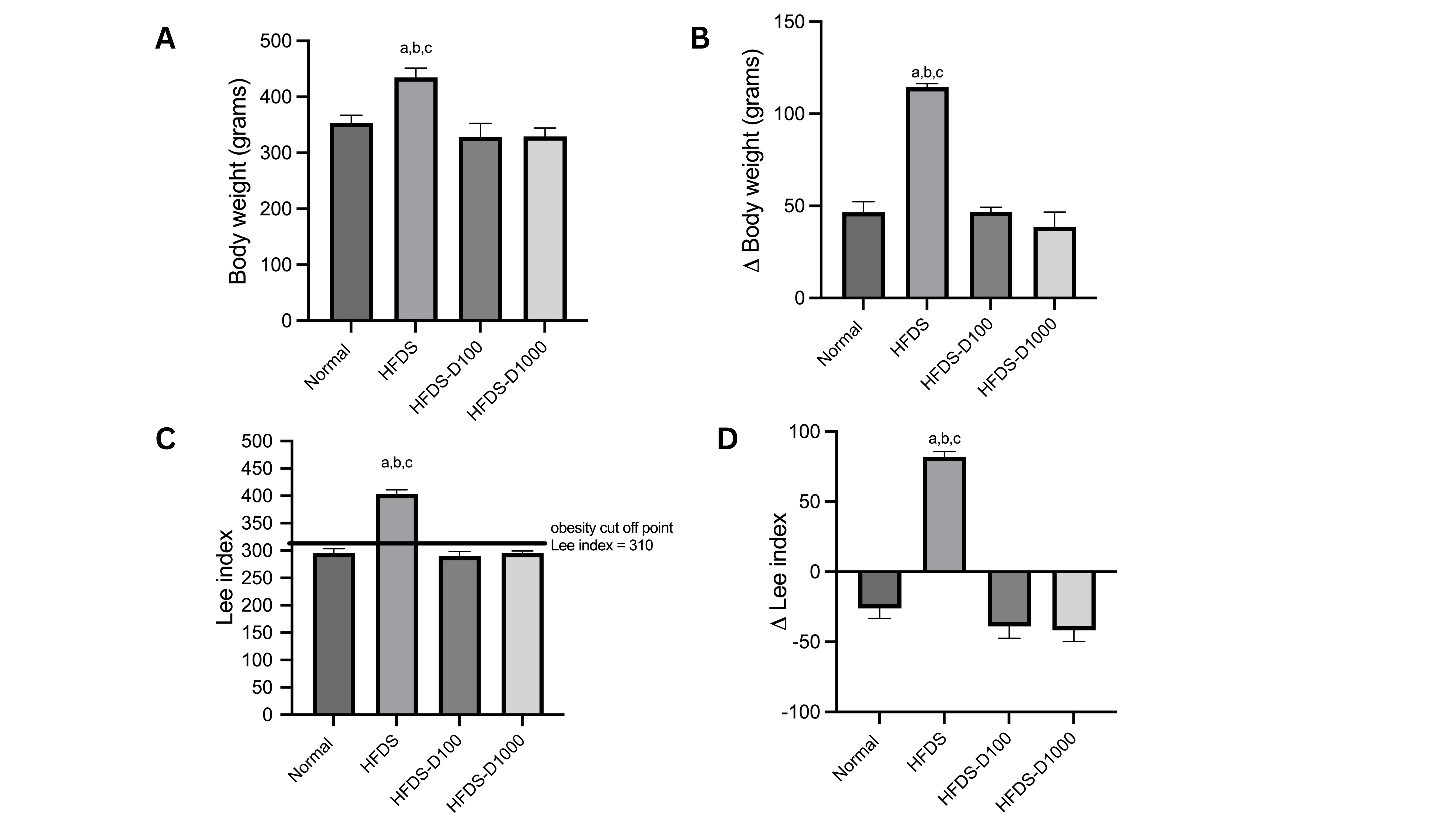
Figure 1 (A) Body weight after 12 weeks of treatment; (B) difference in body weight after and before treatment; (C) Lee index after 12 weeks of treatment; (D) difference in Lee index after and before treatment. HFDS: High-fat, high-glucose, and streptozotocin-induced rats; HFDS-D100: HFDS-induced rats + vitamin D 100 IU/kg BW; HFDS-D1000: HFDS-induced rats + vitamin D 1,000 IU/kg BW. a: P < 0.05 vs normal group; b: P < 0.05 vs. HFDS-D100; c: P < 0.05 vs. HFDS-D1000.
Our results confirmed previous research that high-fat-induced obesity affects vitamin D metabolism, leading to epigenetic modifications in liver vitamin D-metabolizing enzymes and resulting in vitamin D insufficiency. Vitamin D is fat-soluble and is typically absorbed with dietary fat through passive diffusion in the jejunum and ileum. Dietary vitamin D is packaged into chylomicrons with cholesterol, triglycerides, and other lipids within the intestinal wall, and this process depends on microsomal triglyceride transfer protein (MTP). Consequently, dietary vitamin D absorption in the small intestine may be influenced by fat consumption [18-20]. Therefore, supplemental vitamin D3 may be appropriate for obese individuals with excess abdominal fat on a high-fat diet to reduce obesity [19].
Vitamin D increases the adiponectin/leptin ratio by reducing leptin concentrations in the adipocytes
A high-fat, high-glucose diet markedly reduced adiponectin and the adiponectin/leptin ratio (p < 0.05). Supplementation with vitamin D at both doses reduced leptin concentration, thereby improving the adiponectin/leptin ratio. However, the reduction did not achieve the expected normal value (Figure 2).
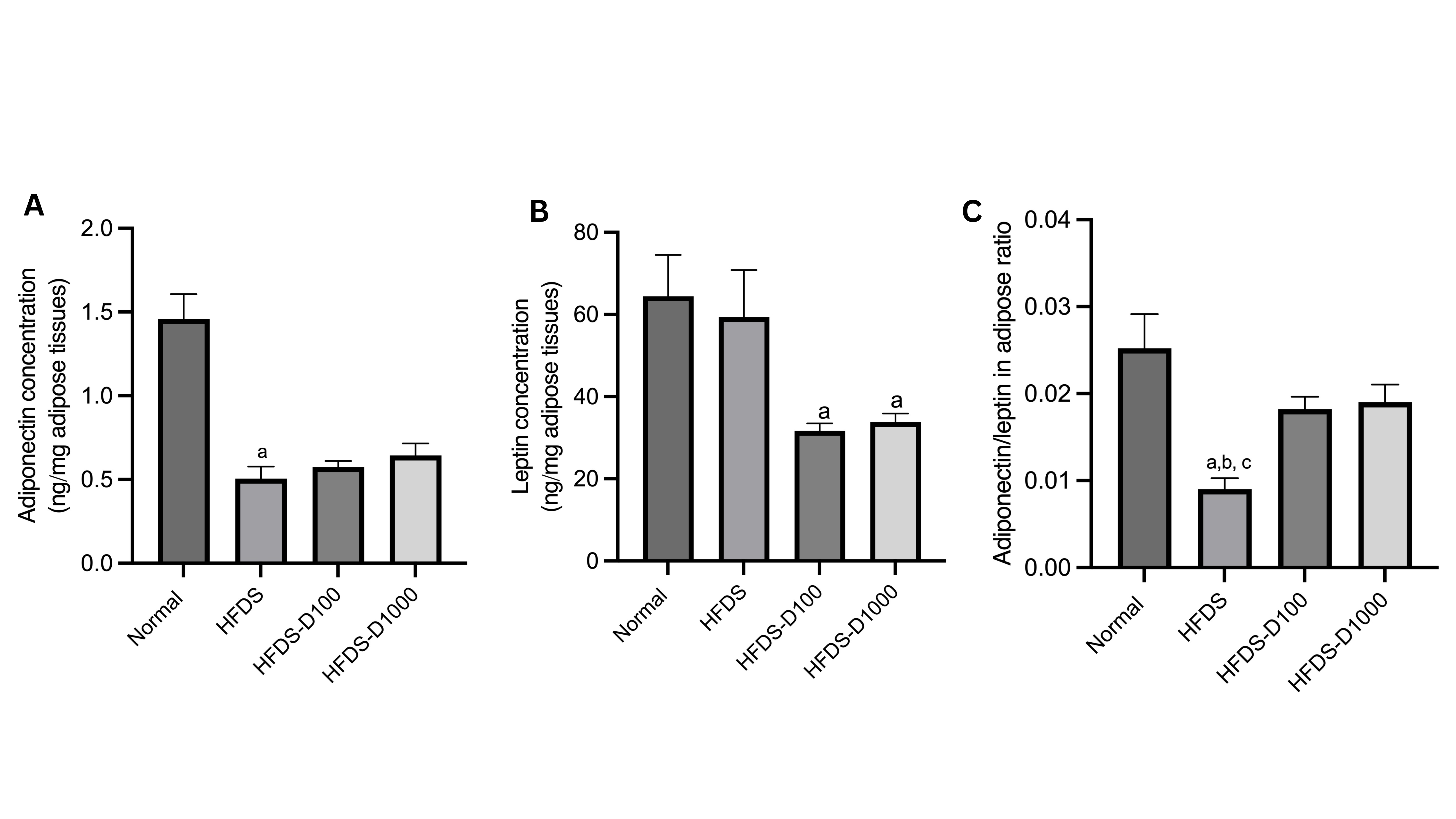
Figure 2 (A) adiponectin concentration; (B) leptin concentration; (C) adiponectin/leptin ratio, quantified 12 weeks after treatment. HFDS: High-fat, high-glucose, and streptozotocin-induced rats; HFDS-D100: HFDS-induced rats + vitamin D 100 IU/kg BW; HFDS-D1000: HFDS-induced rats + vitamin D 1,000 IU/kg BW. a: P < 0.05 vs. normal group; b: P < 0.05 vs. HFDS-D100; c: P < 0.05 vs. HFDS-D1000 HFDS: High-fat, high-glucose, and streptozotocin-induced rats; HFDS-D100: HFDS-induced rats.
Adipokines are considered the link between obesity and insulin resistance. They play active roles in energy balance as well as physiological functions such as immunity and inflammation. Adipokines include leptin, adiponectin, resistin and many others. Adiponectin is an insulin-sensitizing and anti-inflammatory adipokine, while leptin predominantly regulates satiety and energy equilibrium. In obesity, hyperleptinemia and leptin resistance frequently associated with pro-inflammatory signaling [21]. Thus, the adiponectin/leptin ratio has been suggested as an estimate of dysfunctional adipose tissue and cardiometabolic risk [22].
To regulate inflammation and insulin resistance, adipocytes release a range of pro-inflammatory and anti-inflammatory adipokines. The levels of pro-inflammatory adipokines are elevated in obese individuals and animal models, contributing to insulin resistance [23-25]. Vitamin D has been reported to inhibit inflammatory pathways and adipokine synthesis in human adipocytes through the VDR. Increasing vitamin D levels may help alleviate obesity-related metabolic issues by reducing adipose tissue inflammation [26,27].
Vitamin D restores the size of adipocytes in obese rats
Adipocytes in the obese groups were significantly larger than in the normal group. Obesity Vitamin D at 1,000 IU/kg BW resulted in the smallest adipocyte size compared to the healthy group or the treatment with Vitamin D at 100 IU/kg BW (Figure 3).
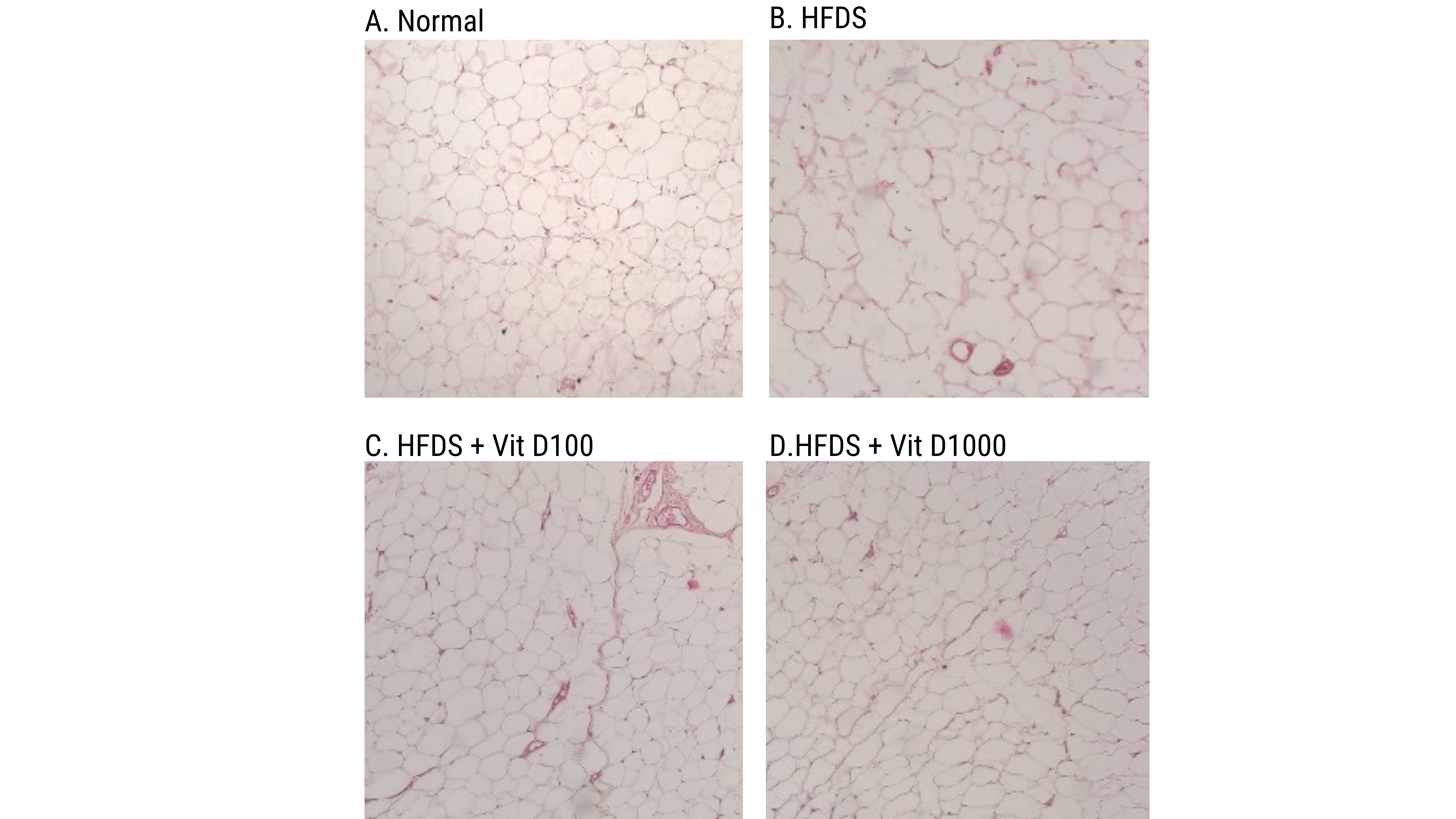
Figure
3
Histology of adipocytes taken from rats’ epididymis. Magnification
at 10× HFDS: High-fat, high-glucose, and streptozotocin-induced rats;
HFDS-D100: HFDS-induced rats + vitamin D 100 IU/kg BW; HFDS-D1000:
HFDS-induced rats + vitamin D 1,000 IU/kg BW.
HFDS: High-fat, high-glucose, and streptozotocin-induced rats;
HFDS-D100: HFDS-induced rats + vitamin D 100 IU/kg BW; HFDS-D1000:
HFDS-induced rats + vitamin D 1,000 IU/kg BW.
High-fat diets increase adipocyte size and number, which can contribute to adipose tissue dysfunction, limiting adipose tissue development, altering brown/beige ratios, and promoting irregular lipid deposits and insulin resistance [28]. In obesity, low circulating 25-hydroxyvitamin D has been associated with adipose tissue dysfunction, potentially reducing bioavailability due to the sequestration in adipose tissue [11]. Additionally, research in obese patients showed that impaired adipose tissue function was associated with low levels of vitamin D [29].
In our study, vitamin D3 supplementation was associated with reduced size of adipocytes, lower body Lee index, and a higher adiponectin/leptin ratio, leading to reduced body weight. Mechanistic links to systemic metabolic outcomes can be explained by the involvement of vitamin D in regulating energy metabolism in adipose tissue by influencing fatty acid oxidation, insulin resistance, and adipokine synthesis. Inflammation in adipose tissue can profoundly influence the metabolic problems commonly linked to obesity, while vitamin D may regulate the inflammatory response of immune cells and adipocytes in adipose tissue [30]. However, the links to the metabolic outcomes should be interpreted cautiously, in the absence of markers of adipocyte inflammatory remodeling, such as macrophage infiltration. Infiltration of adipose macrophages has been associated with insulin resistance and vascular endothelial dysfunction in obesity [31].
Vitamin D increases the diversity of the gut microbiome
HFDS has decreased the variety of the gut microbiome, evidenced by reduced richness and Chao1 indexes alongside an increase in the Shannon index. Vitamin D therapy at 100 IU/kg BW and 1,000 IU/kg BW increased sample richness and diversity, as demonstrated by improvements across all three alpha-diversity indicators (Figure 4).
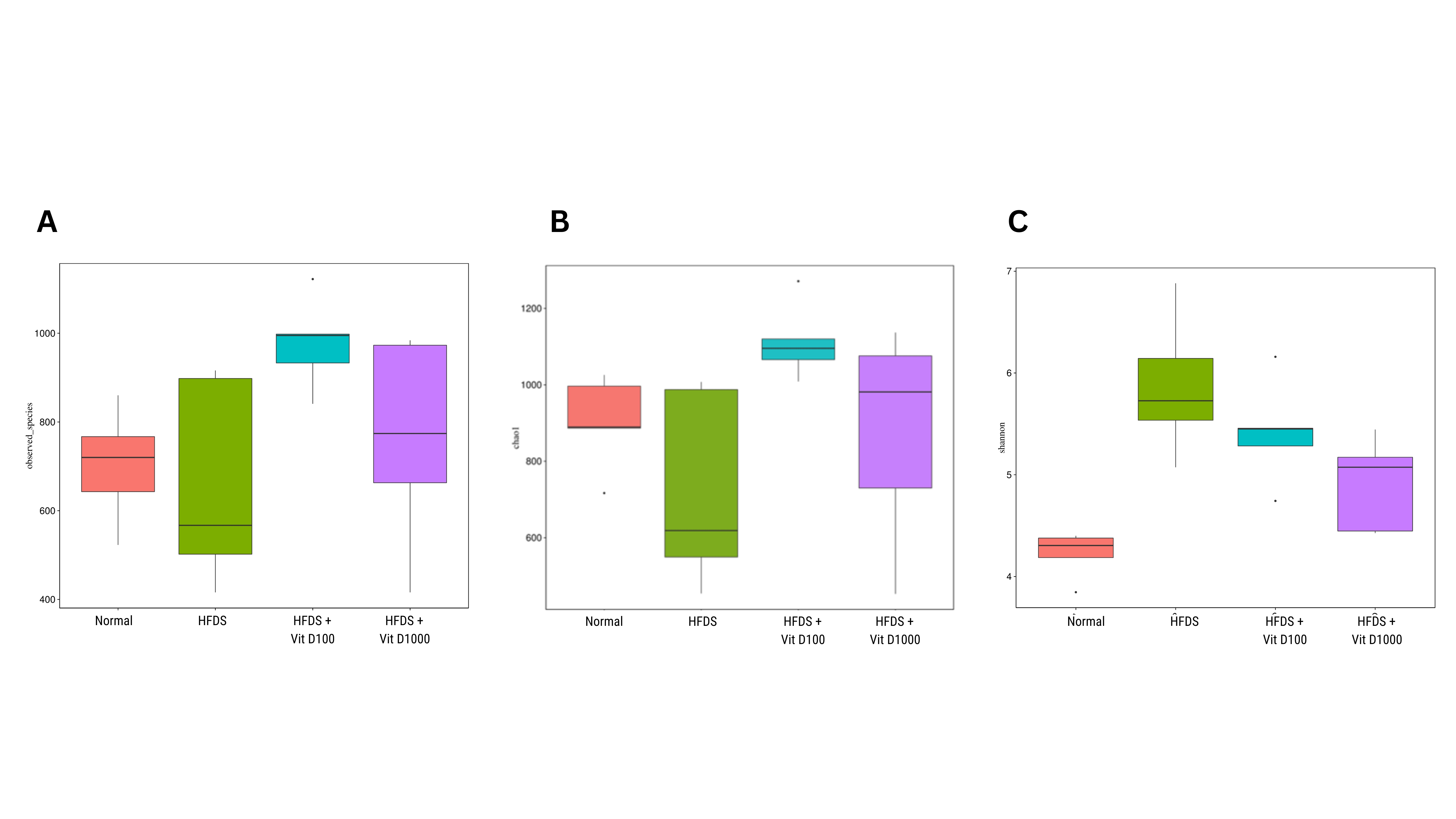
Figure 4 Alpha diversity of gut microbiomes as shown by (A) richness, (B) Chao1 index, and (C) Shannon index. HFDS: High-fat, high-glucose, and streptozotocin-induced rats; HFDS-D100: HFDS-induced rats + vitamin D 100 IU/kg BW; HFDS-D1000: HFDS-induced rats + vitamin D 1,000 IU/kg BW.
Beta diversity is a method used to analyze variations in the composition of microbial populations. These variations were evaluated using coordinate analysis (PcoA) with unweighted UniFrac and cluster analysis following principal component analysis (PCA). As shown in Figure 5, PCoA and PCA analyses indicate that administering vitamin D alongside a high-glucose, high-fat diet altered bacterial communities between treatment groups.
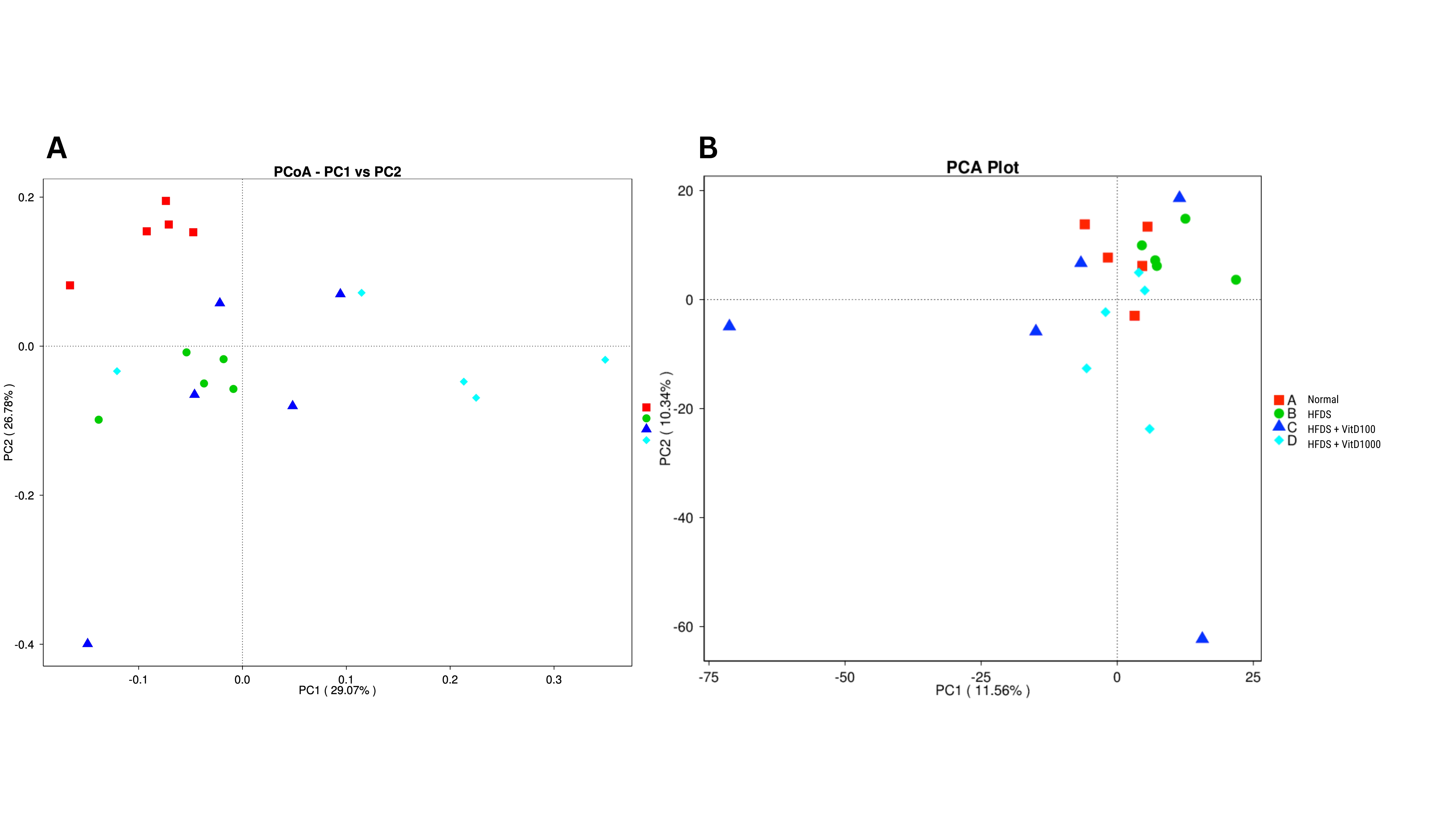
Figure 5 Beta diversity of gut microbiomes as shown by (A) Weighted Unifrac PCoA; (B) Weighted Unifrac PCA. Red points: Normal; green points (HFDS): High-fat, high-glucose, and streptozotocin-induced rats; blue points (HFDS-D100): HFDS-induced rats + vitamin D 100 IU/kg BW; light blue points (HFDS-D1000): HFDS-induced rats + vitamin D 1,000 IU/kg BW.
The microbiota in the human digestive system is essential for the body and comprises trillions of microorganisms. It is abundant in the gut and features thousands of distinct species. Changes in the composition and diversity of the microbiota have been associated with several disorders, including obesity and diabetes [32].
Alpha diversity reflects a sampleʼs richness and diversity, as shown by the richness, Chao1, and Shannon indices. In contrast, beta diversity is a method used to assess variation in microbial population composition [33]. In this study, high-fat, high-glucose delivery reduced the richness and Chao1 indexes compared to the control groups. In contrast, both levels of vitamin D treatment may enhance the microbial richness index. These findings support a prior study by Chen et al. [34] that found a decrease in the richness index in the prediabetic rat group [34]. Additionally, Ibrahim et al. [35] found a reduction in alpha diversity, as indicated by both the richness and Chao1 indices, in diabetic and obese rats compared to the control group [35].
Studies have shown that obesity-related gut microbiomes exhibit reduced diversity and altered species- or genus-level composition [36,37] Chen et al. [34] demonstrated that changes in alpha and beta diversity in diabetic rats induced by a high-fat diet may affect gut microbiota composition and the early stages of diabetes development [34] In the present study, the impact of high-fat, high-glucose administration also alters beta diversity, resulting in variations in the bacterial communities observed through PCoA analysis. These findings support recent work by Shan et al. [38] which identified that feeding prediabetic rats a high-fat diet led to distinct cluster regions in the Weighted UniFrac analysis [38].
Vitamin D modified the composition of the microbiome at the phylum and genus levels
A high-fat, high-glucose diet altered the intestinal microbiota composition at phylum and genus levels, particularly reducing Blautia, Ruminococcus, Fusicatenibacter, and Allobaculum. Compared to obese rats, vitamin D administration increased Lactobacillus, Romboutsia, and Duboisiella while decreasing Fusicatenibacter, UCG-005, and Ruminococcus (Figure 6).
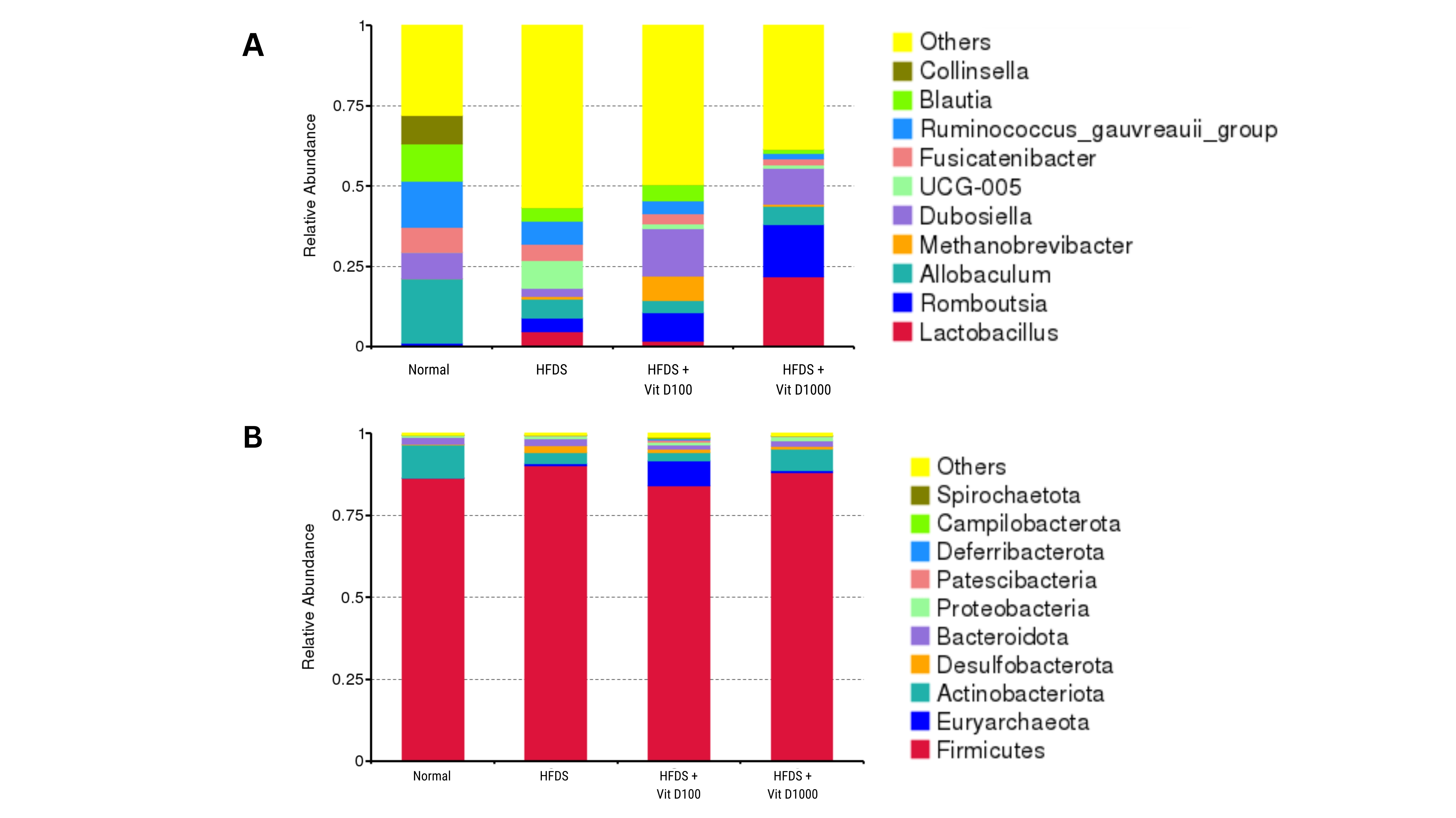
Figure 6 Proportion of gut microbiome in (A) phylum level; (B) genus level. HFDS: High-fat, high-glucose, and streptozotocin-induced rats; HFDS-D100: HFDS-induced rats + vitamin D 100 IU/kg BW; HFDS-D1000: HFDS-induced rats + vitamin D 1,000 IU/kg BW.
Compared with the healthy control group, HFDS treatment reduced the abundance of the Actinobacteria phylum. Previous studies have shown a decline in this phylum in people with obesity and type 2 diabetes. The Actinobacteria phylum has been linked to a lower incidence of obesity and diabetes [37,39]. Actinobacteria are gram-positive bacteria that produce inhibitor enzymes, such as amylase inhibitors, associated with the etiology of obesity in diabetes [37,40]. A Study by Mandal demonstrated that administering 1,000 IU of vitamin D may boost the Actinobacteria phylum [40].
In the present study, vitamin D supplementation at 1,000 IU/kg BW was associated with higher Lactobacillus abundance compared with the HFDS groups. Prior studies aligned have reported associations between vitamin D status/vitamin receptor signaling and Lactobacillus abundance, suggesting that vitamin D-related immune pathways may influence this genus. Vitamin D supplementation may lower TLR2 and TLR4 expression, promoting Lactobacillus, enhancing glycemic response, strengthening the integrity of the intestinal barrier, and limiting bacterial translocation to the systemic circulation. This translocation could increase pro-inflammatory cytokines that contribute to the development of obesity and diabetes [41].
Our results indicate that Allobaculum abundance was lower in the obese rats than in healthy controls, while vitamin D supplementation resulted in minimal change. However, the response of Allobaculum to metabolic disturbances and vitamin D supplementation appears to be highly model-dependent [35,42]. A study by Q Ma et al. [42] showed that the quantity of Allobaculum in diabetic rat models was lower than predicted [42]. This varies from another research by Ibrahim et al. [35] which found that diabetic rats show a rise in Allobaculum, whereas obese rats do not [35]. A recent study by Lee et al. [43] indicates that vitamin D administration may enhance the number of Allobaculum in cirrhotic rats [43]. These discrepancies suggest that Allobaculum may not serve as a universal biomarker of metabolic improvement but rather reflects context-specific host-microbiome interactions, influenced by disease state, dietary composition, and inflammatory milieu [35,38]. Allobaculum is a beneficial bacterium found in the intestines of mice that may help reduce weight gain and produce short-chain fatty acids with anti-inflammatory properties by stimulating the release of butyrate and mucus in the gastrointestinal system [35].
Our study further identified a reduction in Blautia in obese rats compared to healthy rats. Treating obese rats with vitamin D3 at 1,000 IU/kg BW reduces the number of Blautia. Our findings are consistent with previous research that observed a reduction in Blautia in individuals with high vitamin D levels [44].
Blautia are gram-positive bacteria belonging to the phylum Firmicutes. In humans, these bacteria are predominant in hyperglycemia and play an essential role in glucose metabolism by converting acetate, lactate, hydrogen, ethanol, and succinate in the gut [45]. Obesity reduces Blautia and elevates pro-inflammatory cytokines associated with TLR2 and TLR4 [46].
Although several studies have reported that Blautia spp. are associated with beneficial metabolic outcomes, including lower body weight, reduced fat accumulation, and improved lipid profiles, recent evidence indicates that Blautiaʼs metabolic role is highly context-dependent and influenced by host metabolic status, dietary composition, and glycemic status [46,47]. In metabolically impaired conditions such as obesity and insulin resistance, altered Blautia abundance has been associated with intestinal inflammation and dysregulated glucose metabolism [47,48]. Therefore, the reduction in Blautia observed following vitamin D3 supplementation at 1,000 IU/kg BW in the present study may reflect normalization of gut microbial composition rather than a detrimental effect. This microbial shift was accompanied by reduced body weight, smaller adipocyte size, and an improved adiponectin/leptin ratio, supporting a beneficial metabolic impact of vitamin D-mediated microbiome modulation [45].
In our study, vitamin D supplementation at both doses lowered the quantity of Ruminococcus. This is consistent with human research, showing that vitamin D administration reduces the amount of Ruminococcus [44]. Another study found that increased Ruminococcus in obese rats was associated with propionate metabolism and the development [47]. Furthermore, the level of UCG-005 in the vitamin D groups was lower than in the obese group. The results contrasted with prior research, which found a reduction in UCG-005 in diabetic patients. UCG-005 is claimed to be a beneficial gut bacterium that inhibits the development of diabetes and increases the synthesis of short-chain fatty acids with anti-inflammatory properties [48].
Although improvements in obesity parameters, adipokine profiles, and gut microbiome composition were observed concurrently following vitamin D supplementation, the present study does not allow causal inference regarding the directionality or mechanistic hierarchy of these associations. The observed microbiome shifts may represent downstream effects, parallel adaptations, or bidirectional interactions with host metabolic changes rather than primary drivers of obesity improvement [33,38].
Our study has several limitations. Microbiome analysis was restricted to colon samples and did not encompass the entire gastrointestinal tract, which may not fully capture overall gut microbiome variability. In addition, the experimental model lacked heterogeneity in sex and age, potentially underestimating the biological complexity of human physiological systems. Sex differences are well-described in metabolic regulation and gut microbiome composition [49], therefore, extrapolation from male-only data should be cautious and future work should include both sexes.
In addition, the comprehensive metabolic panels (fasting glucose, insulin, HOMA IR, lipids) as well as serum 25-hydroxyvitamin D3 concentrations, which has been presented in our previous results, were not correlated with the present results, thus limiting direct inference about systemic vitamin D and metabolic outcomes with adipokine/microbiome findings.
Furthermore, the cross-sectional, endpoint-based design precludes the establishment of causal relationships between vitamin D supplementation, adipokine modulation, gut microbiome alterations, and obesity-related outcomes, as the observed associations may reflect parallel or downstream effects rather than direct mechanistic links. Methodologically, although OTU-based clustering was employed in the present study, ASV-based pipelines such as DADA2 or Deblur offer higher taxonomic resolution and improved reproducibility. Both OTU-based and ASV-based pipelines are considered appropriate depending on study objectives and data characteristics. In the present study, the OTU-based approach was suitable for genus-level ecological analyses, and given the modest sample size, higher sequence resolution may increase sparsity without substantially altering biological interpretation [50,51]. Future studies incorporating longitudinal designs, larger and more diverse cohorts, whole-gastrointestinal-tract sampling, and mechanistic approaches such as microbiota transplantation or targeted microbial manipulation are warranted to delineate causal pathways and to better define microbial signatures relevant to vitamin D supplementation and human obesity.
Conclusions
Vitamin D3 supplementation was associated with improvements in obesity-related parameters, accompanied by changes in the adiponectin/leptin ratio and shifts in gut microbiome diversity and species composition. These findings support a potential interaction between vitamin D status, host metabolic signaling, and the gut microbiome, which warrants further mechanistic investigation. Another study is needed to comprehensively explore the interactions between host and gut microbial ecosystems and potential comorbidities in insulin-resistant conditions. Lastly, validation of these findings in obese individuals experiencing insulin resistance is a must before formal implementation in humans.
Acknowledgements
This study was supported by a Grant from the Directorate of Research and Community Engagement, Universitas Indonesia, Contract No. NKB-589/UN2.RST/HKP.05.00/2022.
Declaration of generative AI in scientific writing
No content in the present manuscript was created using generative AI. We used Grammarly to improve the readability and language of a manuscript. All authors remain fully responsible for the authenticity of the content.
CRediT author statement
Desak Gede Budi Krisnamurti: conceptualization, investigation, formal analysis; Melva Louisa: conceptualization, methodology, funding acquisition, writing – original draft; Felix Mesak: validation, visualization; Erni H Poerwaningsih: conceptualization, resources, project administration; Tri Juli Edi Tarigan: conceptualization, supervision, resources Vivian Soetikno: validation, writing - review and editing; Heri Wibowo: methodology, data curation; Fadilah Fadilah: software, data curation, validation; Linda Erlina: data curation, formal analysis; and Christian Marco Hadi Nugroho: investigation, visualization.
References
[1] RN Haththotuwa, CN Wijeyaratne and U Senarath. Worldwide epidemic of obesity. Second (Ed.). Elsevier, Germany, 2020, p. 3-8.
[2] NJ Temple. The origins of the obesity epidemic in the USA-lessons for today. Nutrients 2022; 14(20), 4253.
[3] S Moreno-Fernández, M Garcés-Rimón, G Vera, J Astier, JF Landrier and M Miguel. High fat/high glucose diet induces metabolic syndrome in an experimental Rat Model. Nutrients 2018; 10(10), 1502.
[4] B Arcidiacono, E Chiefari, A Foryst-Ludwig, G Currò, G Navarra, FS Brunetti, M Mirabelli, DM Corigliano, U Kintscher, D Britti, V Mollace, DP Foti, ID Goldfine and A Brunetti. Obesity-related hypoxia via miR-128 decreases insulin-receptor expression in human and mouse adipose tissue promoting systemic insulin resistance. eBioMedicine 2020; 59, 102912.
[5] N Hariri and L Thibault. High-fat diet-induced obesity in animal models. Nutrition Research Reviews 2010; 23(2), 270-299.
[6] YS Lee, P Li, JY Huh, IJ Hwang, M Lu, JI Kim, M Ham, S Talukdar A Chen, WJ Lu, GK Bandyopadhyay, R Schwendener, J Olefsky and JB Kim. Inflammation is necessary for long-term but not short-term high-fat diet-induced insulin resistance. Diabetes 2011; 60(10), 2474-2483.
[7] EA Greco, A Lenzi and S Migliaccio. Role of hypovitaminosis D in the pathogenesis of obesity-induced insulin resistance. Nutrients 2019; 11(7), 1506.
[8] CA Peterson, AK Tosh and AM Belenchia. Vitamin D insufficiency and insulin resistance in obese adolescents. Therapeutic Advances in Endocrinology and Metabolism 2014; 5(6), 166-189.
[9] CB Turer, H Lin and G Flores. Prevalence of vitamin D deficiency among overweight and obese US children. Pediatrics 2013; 131(1), 152-161.
[10] GE Walker, R Ricotti, M Roccio, S Moia, S Bellone, F Prodam and G Bona. Pediatric obesity and vitamin D deficiency: A proteomic approach identifies multimeric adiponectin as a key link between these conditions. PLoS One 2014; 9(1), 83685.
[11] RJA Argan, SG Alqatari, AJ Alwaheed, MA Hasan, SY AlQahtani, MDA Shubbar, AH Alnasser, SMA Abbas and NH AlYousef. Vitamin D deficiency in obesity: Epidemiological evidence, biological mechanisms, and clinical considerations. Obesity Medicine 2026; 59, 100680.
[12] JD Roizen, C Long, A Casella, L OʼLear, I Caplan, M Lai, I Sasson, R Singh, AJ Makowski, R Simmons and MA Levine. Obesity decreases hepatic 25-Hydroxylase activity causing low serum 25-Hydroxyvitamin D. Journal of Bone and Mineral Research 2019; 34(6), 1068-1073.
[13] A Sukik, J Alalwani and V Ganji. Vitamin D, gut microbiota, and cardiometabolic diseases—a possible three-way axis. International Journal of Molecular Sciences 2023; 24(2), 940.
[14] K Zuo, J Li, Q Xu, C Hu, Y Gao, M Chen, R Hu, Y Liu, H Chi, Q Yin, Y Cao, P Wang, Y Qin, X Liu, J Zhong, J Cai, K Li and X Yang. Dysbiotic gut microbes may contribute to hypertension by limiting vitamin D production. Clinical Cardiology 2019; 42(8), 710-719.
[15] N Kazemian, M Mahmoudi, F Halperin, JC Wu and S Pakpour. Gut microbiota and cardiovascular disease: Opportunities and challenges. Microbiome 2020; 8, 36.
[16] M Witkowski, TL Weeks and SL Hazen. Gut microbiota and cardiovascular disease. Circulation Research 2020; 127, 553-570.
[17] DGB Krisnamurti, M Louisa, EH Poerwaningsih, TJE Tarigan, V Soetikno, H Wibowo and CMH Nugroho. Vitamin D supplementation alleviates insulin resistance in prediabetic rats by modifying IRS-1 and PPARγ/NF-κB expressions. Front Endocrinol 2023; 14, 1089298.
[18] YS Jung, D Wu, D Smith, SN Meydani and SN Han. Dysregulated 1,25-dihydroxyvitamin D levels in high-fat diet-induced obesity can be restored by changing to a lower-fat diet in mice. Nutrition Research 2018; 53, 51-60.
[19] CY Park, Y Shin, JH Kim, S Zhu, YS Jung and SN Han. Effects of high fat diet-induced obesity on vitamin D metabolism and tissue distribution in vitamin D deficient or supplemented mice. Nutrition & Metabolism 2020; 17, 44.
[20] R Parsanathan and SK Jain. Glutathione deficiency induces epigenetic alterations of vitamin D metabolism genes in the livers of high-fat diet-fed obese mice. Scientific Reports 2019; 9, 14784.
[21] VJ Clemente-Suárez, L Redondo-Flórez, AI Beltrán-Velasco, A Martín-Rodríguez, I Martínez-Guardado, E Navarro-Jiménez, CC Laborde-Cárdenas and JF Tornero-Aguilera. The role of adipokines in health and disease. Biomedicines 2023; 11(5), 1290.
[22] G Frühbeck, V Catalán, A Rodríguez and J Gómez-Ambrosi. Adiponectin-leptin ratio: A promising index to estimate adipose tissue dysfunction. Relation with obesity-associated cardiometabolic risk. Adipocyte 2018; 7(1), 57-62.
[23] B Antuna-Puente, B Feve, S Fellahi and JP Bastard. Adipokines: The missing link between insulin resistance and obesity. Diabetes & Metabolism Journal 2008; 34(1), 2-11.
[24] YE Kang, JM Kim, KH Joung, JH Lee, BR You, MJ Choi, MJ Ryu, YB Ko, MA Lee, J Lee, BJ Ku, M Shong, KH Lee and HJ Kim. The roles of adipokines, proinflammatory cytokines, and adipose tissue macrophages in obesity-associated insulin resistance in modest obesity and early metabolic dysfunction. PLoS One 2016; 11(4), 0154003.
[25] K Zorena, O Jachimowicz-Duda, D Ślęzak, M Robakowska and M Mrugacz. Adipokines and Obesity. Adipokines and Obesity. Potential link to metabolic disorders and chronic complications. International Journal of Molecular Sciences 2020; 21(10), 3570.
[26] MJ Lee. Vitamin D regulation of adipogenesis and adipose tissue functions. Nutrients 2020; 17(3), 553.
[27] H Nimitphong, W Guo, MF Holick, SK Fried and MJ Lee. Vitamin D inhibits adipokine production and inflammatory signaling through the vitamin D receptor in human adipocytes. Obesity 2021; 29(3), 562-568.
[28] CY Park and SN Han. The role of vitamin D in adipose tissue biology: Adipocyte differentiation, energy metabolism, and inflammation. Journal of Lipid and Atherosclerosis 2021; 10(2), 130-144.
[29] AD Nisio, LD Toni, I Sabovic, MS Rocca, VD Filippis, G Opocher, B Azzena, R Vettor, M Plebani and C Foresta. Impaired release of vitamin D in dysfunctional adipose tissue: New cues on vitamin D supplementation in obesity. The Journal of Clinical Endocrinology & Metabolism 2017; 102(7), 2564-2574.
[30] FA Cimini, F Sentinelli, A Oldani, I Barchetta, and MG Cavallo. Adipose tissue dysfunction and metabolic diseases: The role of vitamin D/vitamin D receptor Axis. International Journal of Molecular Sciences 2025; 26(21), 10256.
[31] CM Apovian, S Bigornia, M Mott, MR Meyers, J Ulloor,M Gagua, M McDonnell, D Hess, L Joseph and N Gokce. Adipose macrophage infiltration is associated with insulin resistance and vascular endothelial dysfunction in obese subjects. Arteriosclerosis, Thrombosis, and Vascular Biology 2008; 28(9), 1654-1659.
[32] A Everard and PD Cani. Diabetes, obesity and gut microbiota. Best Practice & Research Clinical Gastroenterology 2013; 27(1), 73-83.
[33] J Zhao, X Zhang, H Liu, MA Brown and S Qiao. Dietary protein and gut microbiota composition and function. Current Protein & Peptide Science 2018; 20(2), 145-154.
[34] Z Chen, D Radjabzadeh, L Chen, A Kurilshikov, M Kavousi, F Ahmadizar, MA Ikram, AG Uitterlinden, A Zhernakova, J Fu, R Kraaij and T Voortman. Association of insulin resistance and Type 2 diabetes with gut microbial diversity: A microbiome-wide analysis from population studies. JAMA Network Open 2021; 4(7), 2118811.
[35] KS Ibrahim, N Bourwis, S Dolan, S Lang, J Spencer and JA Craft. Characterisation of gut microbiota of obesity and type 2 diabetes in a rodent model. Bioscience of Microbiota, Food and Health 2021; 40(1), 64-74.
[36] J Gong, Y Shen, H Zhang, M Cao, M Guo, J He, B Zhang and C Xiao. Gut microbiota characteristics of people with obesity by meta-analysis of existing datasets. Nutrients 2022; 14(14), 2993.
[37] M Pinart, A Dötsch, K Schlicht, M Laudes, J Bouwman, SK Forslund, T Pischon and K Nimptsch. Gut microbiome composition in obese and non-obese persons: A systematic review and meta-analysis. Nutrients 2021; 14(1), 12.
[38] K Shan, H Qu, K Zhou, L Wang, C Zhu, H Chen, Z Gu, J Cui, G Fu, J Li, H Chen, R Wang, Y Qi, W Chen and YQ Chen. Distinct gut microbiota induced by different Fat-to-Sugar-Ratio High-energy diets share similar Pro-obesity genetic and metabolite profiles in Prediabetic Mice. mSystems 2019; 4(5), 00219-19.
[39] J Long, Q Cai, M Steinwandel, MK Hargreaves, SR Bordenstein, WJ Blot, W Zheng and XO Shu. Association of oral microbiome with Type 2 diabetes risk. Journal of Periodontal Research 2017; 52(3), 636-643.
[40] S Mandal, KM Godfrey, D McDonald, WV Treuren, JV Bjørnholt, T Midtvedt, B Moen, K Rudi, R Knight, AL Brantsæter, SD Peddada and M Eggesbø. Fat and vitamin intakes during pregnancy have stronger relations with a pro-inflammatory maternal microbiota than does carbohydrate intake. Microbiome 2016; 4, 55.
[41] S Gheibi, K Kashfi and A Ghasemi. A practical guide for induction of Type-2 diabetes in rat: Incorporating a high-fat diet and streptozotocin. Biomedicine & Pharmacotherapy 2017; 95, 605-613.
[42] Q Ma, Y Li, J Wang, P Li, Y Duan, H Dai, Y An, L Cheng, T Wang, C Wang, T Wang and B Zhao. Investigation of gut microbiome changes in Type 1 diabetic mellitus rats based on high-throughput sequencing. Biomedicine & Pharmacotherapy 2020; 124, 109873.
[43] PC Lee, YC Hsieh, TI Huo, UC Yang, CH Lin, CP Li, YH Huang, MC Hou, HC Lin and KC Lee. Active vitamin D3 treatment attenuated bacterial translocation via improving intestinal barriers in Cirrhotic Rats. Molecular Nutrition & Food Research 2021; 65(3), 2000937.
[44] N Naderpoor, A Mousa, LFG Arango, HL Barrett, MD Nitert and B de Courten. Effect of vitamin D supplementation on faecal microbiota: A randomised clinical trial. Nutrients 2019; 11(12), 2888.
[45] L Egshatyan, D Kashtanova, A Popenko, O Tkacheva, A Tyakht, D Alexeev, N Karamnova, E Kostryukova, V Babenko, M Vakhitova and S Boytsov. Gut microbiota and diet in patients with different glucose tolerance. Endocrine Connections 2016; 5, 1-9.
[46] A Benítez-Páez, EMG del Pugar, I López-Almela, Á Moya-Pérez, P Codoñer-Franch and Y Sanz. Depletion of Blautia species in the microbiota of obese children relates to intestinal inflammation and metabolic phenotype worsening. mSystems 2020; 5(2), 00857-19.
[47] L Krych, DS Nielsen, AK Hansen and C Hansen. Gut microbial markers are associated with diabetes onset, regulatory imbalance, and IFN-γ level in NOD Mice. Gut Microbes 2015; 6(2), 101-109.
[48] GG Pircalabioru, J Liaw, O Gundogdu, N Corcionivoschi, I Ilie, L Oprea, M Musat and MC Chifiriuc. Effects of the lipid profile, Type 2 diabetes and medication on the metabolic syndrome—associated gut microbiome.
International Journal of Molecular Sciences 2022; 23(14), 7509.
[49] YS Kim, T Unno, BY Kim and MS Park. Sex differences in gut microbiota. World Journal of Menʼs Health 2020; 38(1), 48-60.
[50] JT Nearing, GM Douglas, AM Comeau and MGI Langille. Denoising the Denoisers: An independent evaluation of microbiome sequence error-correction approaches. PeerJ 2018; 6, 26566.
[51] BJ Callahan, PJ McMurdie and SP Holmes. Exact sequence variants should replace operational taxonomic units in marker-gene data analysis. The ISME Journal 2017; 11, 2639-2643.