
Trends
Sci.
2026;
23(9):
12987
Enhancement of Paclitaxel Efficacy by Tectona grandis Leaves Extract: Synergistic Antiproliferative and Antimigratory Actions in Triple-Negative Breast Cancer Cells
Sofy Permana1, Neyla Vista Maramy2, Maghfira Rahma Azizah3, Halida Adib Hanum3, Yoshiyuki Kawamoto4, Edwin Widodo5, Heni Endrawati6 and Agustina Tri Endharti6,7,8,*
1Department of Biology, Faculty of Mathematic and Natural Sciences, Universitas Brawijaya, East Java, Indonesia
2Post Doctoral Fellow, Universitas Brawijaya, East Java, Indonesia
3Medical Professional Study Program, Faculty of Medicine, Brawijaya University, East Java, Indonesia
4Department of Biomedical Sciences, Graduate School of Life and Health Sciences, Chubu University, Aichi, Japan
5Department of Physiology, Faculty of Medicine, Universitas Brawijaya, East Java, Indonesia
6Departement of Parasitology, Faculty of Medicine, Universitas Brawijaya, East Java, Indonesia
7Innovation Center of Integrative Jamu and Eco-pharmaca, Universitas Brawijaya, East Java, Indonesia
8Immunotherapy Research Group, Faculty of Medicine, Universitas Brawijaya, East Java, Indonesia
(*Corresponding author’s e-mail: [email protected])
Received:12 December 2025, Revised: 28 January 2026, Accepted: 10 February 2026, Published: 10 April 2026
Abstract
Triple negative breast cancer (TNBC) lacks ER, PR, and HER2 expression, leading to poor prognosis. Tectona grandis contains bioactive compounds with anticancer properties. This study investigated antiproliferative and antimigratory effect of T. grandis leaves extract (TGLE) and paclitaxel. This study involved computational study to determine the binding affinity of Mucin-1 and TGLE and in vitro study to examine the antiproliferative and antimigratory effect of TGLE and paclitaxel as well as the Mucin 1 (MUC1) and Matrix Metalloproteinase-9 (MMP9) expression in MDA-MB-231 cells. This study identified quercetin in TGLE as the strongest compound to bind with MUC1. In vitro study confirmed the inhibitory effect of TGLE and paclitaxel on MUC1 expression and cell proliferation. Furthermore, TGLE and paclitaxel exhibited a significant reduction in MMP-9 expression and cell migration. These findings suggest that TGLE and paclitaxel has promising antiproliferative and antimigratory properties against TNBC through its inhibition on MUC1 and MMP-9.
Keywords: Triple negative breast cancer, Tectona grandis, Antiproliferative, Antimigratory
Introduction
Triple negative breast cancer (TNBC) is a specific subtype of breast cancer, representing approximately 15% - 20% of all breast cancer cases [1]. It is characterized by the absence of estrogen receptor (ER), progesterone receptor (PR), and human epidermal growth factor receptor 2 (HER2) expression. The ER and PR are present in less than 0.01% of TNBC samples, while HER2 typically shows either no expression or a score of +1 based on immunohistochemistry (IHC) staining. Among the various breast cancer subtypes, TNBC is associated with the poorest prognosis [2]. Patients with TNBC have a mortality rate of approximately 40% within 5 years of diagnosis. The recurrence rate for TNBC is also relatively high at 25%. In comparison, patients without TNBC experience recurrence rates between 35 and 67 months, whereas those with TNBC face recurrence as early as 19 to 40 months after initial treatment. Furthermore, TNBC patients exhibit a significantly higher mortality rate, which can surpass 75% [1].
The TNBC subtype is resistant to endocrine therapy because it lacks the presence of ER, PR, and HER2 receptors. The standard treatment approaches for early TNBC involve administering neoadjuvant chemotherapy after surgery, which includes alkylating agents (cyclophosphamide), anthracyclines, antimetabolite fluorouracil, and anti-microtubule drugs (taxanes). Advanced TNBC is typically treated with chemotherapy drugs like gemcitabine and capecitabine (anti-metabolites), eribulin (non-taxane anti-microtubule), and platinum-based drugs (DNA cross-linkers) [3,4]. Nevertheless, chemotherapy also affects normal cells in the body, such as bone marrow, gastrointestinal (GI) mucosa, and hair follicles, leading to various side effects like nausea, stomatitis, bone marrow suppression, and hair loss [5]. It is crucial to develop therapies that target specific molecular markers or pathways to mitigate these undesirable effects.
Currently, there is ongoing research on targeting Mucin 1 (MUC1) as a potential therapeutic approach for TNBC. Approximately 90% of TNBC cells produce the heterodimer protein MUC1, which makes it an attractive target for treatment [6]. Several studies have provided evidence of MUC1’s involvement in the proliferation of TNBC cells. Activation of signaling pathways such as phosphatidylinositol-3-kinase (PI3K)/protein kinase B (AKT), extracellular signal regulated kinase (ERK), and receptor tyrosine kinases (RTKs) by MUC1 has been shown to promote TNBC cell proliferation. Additionally, the MUC1 protein enhances the uptake of glutamine and glucose, which are essential for TNBC cell proliferation, thus further promoting cellular proliferation [7].
The breakdown of the basement membrane during tumor formation is frequently required for tumors to metastasize. Matrix metalloproteinases (MMPs) are a type of enzyme that degrades the extracellular matrix (ECM) which then facilitates cell migration. Among these MMPs, MMP-9, which has been extensively investigated, plays critical functions in tumor metastasis [8]. Multiple extracellular matrix proteins are cut and degraded by protease hydrolysis in order to regulate the structure of the ECM by MMP-9. There is a considerable rise in MMP-9 protein levels at the tumor site, but this is not followed by a corresponding increase in the production of its inhibitor [9]. As a result, MMP-9 appears as a critical therapeutic target [10].
The leaf of Tectona grandis, commonly known as teak, is a potential herbal component that may exhibit anticancer properties. The anticancer activity of teak leaves is closely associated with its bioactive compounds, although their specific effects have not been fully understood [11]. The objective of this study is to investigate the anticancer potential of T. grandis leaves, focusing on their ability to inhibit cell proliferation and migration. Additionally, this study aims to analyze the interaction of teak leaf components with MUC1 proteins using computational methods.
Materials and methods
Computational analysis
The 3-dimensional crystal structure of MUC1 (CID 2ACM) was obtained from the RCSB PDB website (https://www.rcsb.org/) in PDB format [12]. To isolate the A chain of the protein, the structure was sterilized using PyMOL software, removing the entire molecules, contaminants, and water. Bioactive compounds derived from T. grandis leaves [11,13], were collected. These compounds were then screened for their anticancer activity using the PassOnline website (http://way2drug.com/PassOnline/index.php). The bioactive substances exhibiting anticancer activity, along with the standard ligand GO-203, were retrieved from the PubChem website (https://pubchem.ncbi.nlm.nih.gov/) in SDF format. Both the ligands and the protein were minimized using OpenBabel in PyRx software. Molecular docking was performed on the MUC1 protein using the AutoDock Vina program in PyRx, based on the results of the screening. Docking validation was carried out using standard methods to identify the ligand with the strongest protein binding, comparing the binding energy to the control. Additional factors such as LogP, hydrogen bond donor (HBD), hydrogen bond acceptor (HBA), volume, and rotational binding were considered in the analysis. The potency of the ligands in terms of binding energy and drug-likeness was evaluated using Lipinski’s rule of 5. Pharmacokinetic and drug-likeness analysis was conducted using the SwissADME website (http://www.swissadme.ch/). For better visualization of the binding site, the 3D structure of MUC1 and a specific ligand were displayed using PyMOL software. The docking analysis results and the 3-dimensional structure of the MUC1 protein were visualized. Additionally, the 2D visualization of the ligand’s location and binding type within the MUC1 domain was achieved using the ProteinPlus website (https://proteins.plus/).
Cell culture
MDA-MB-231 and T47D cells were obtained from the American Type Culture Collection (ATCC; Manassas, VA, USA). The cells were grown in Roswell Park Memorial Institute (RPMI; Invitrogen, Carlsbad, CA, USA) media supplemented with 10% fetal bovine serum (FBS) Gibco; ThermoFisher Scientific, Inc., Waltham, MA, USA), 100 IU/mL penicillin, and 100 g/mL streptomycin. The cells were kept alive in an incubator with 5% CO2 at 37 °C and a pH range of 7.2 - 7.4. The cells were then classified into 6 different groups, (1) negative control (NC); (2) standard control (SC) that given paclitaxel 10 μg/mL; (3) combination of paclitaxel 5 μg/mL (PAC) with T. grandis leaves extract (TGLE) 7.5 mg/mL (T1); (4) combination of PAC with TGLE 15 mg/mL (T2); (5) combination of PAC with TGLE 30 mg/mL (T3); (6) TGLE alone 60 mg/mL.
Mucin 1 and MMP9 protein expression
Treated and cultured cells were placed in a 24-well plate with a pre-mounted coverslip on the bottom. Once the 80% confluence had been reached, the media was aspirated and washed 3 times for 5 min each using a phosphate buffer saline (PBS) solution. The washing process was then continued with a 0.1% Triton-X 100 PBS solution for 5 min. The washed cells were subsequently incubated with a 1% bovine serum albumin (BSA) solution for 30 min at room temperature in a humid environment. After the incubation with 1% BSA, the cells were further incubated with the primary antibody in a blocking buffer solution overnight at 4 °C. The incubated cells were then washed 3 times for 5 min each with PBS. Subsequently, the washed cells were treated with the secondary antibody for 30 min at room temperature in a dark room. The cells were once again washed 3 times for 5 min each with PBS in the dark room. The cells that had been treated with both the primary and secondary antibodies were incubated with 4′,6-diamidino-2-phenylindole (DAPI) for 3 min at room temperature in the dark room. The DAPI-treated cells were washed 3 times for 5 min each with PBS in the dark room. Finally, the cells were covered with mounting media. The coverslip was removed, transferred onto a slide, and observed under a fluorescence microscope to visualize the location of the target protein expression.
Cell proliferation assay
The MTT solution was prepared by dissolving MTT powder in the PBS solution to achieve a concentration of 10 mg/mL. The cells were cultured in a 96-well plate at a density of 5,000 - 10,000 cells per well. The cells were then incubated in a cell incubator for 24 h. Subsequently, the cells were observed under a microscope to assess their distribution. Once 80% confluency was achieved, the cells were treated. The addition of paclitaxel was administered earlier, followed by the addition of the extract at the subsequent 4 h without removing the medium, according to the tested concentrations, with a total volume of 200 µL per well. Each experimental group was replicated 3 times. The cells were further incubated for 24, 48, and 72 h, and the proliferative activity of each experimental group was observed under an inverted microscope at 24, 48, and 72 h. The culture medium was then discarded and replaced with 200 µL of fresh culture medium and 10 or 20 µL of MTT solution. The cells were incubated again for 4 h while the formation of formazan was monitored. Subsequently, the cells were observed under an inverted microscope, and the medium containing MTT mixed with cells was discarded, leaving behind the formazan. The formazan crystals were dissolved in 100 µL of DMSO solution. The cells were incubated again for 30 min and then observed under an inverted microscope. The samples were read by a microplate reader at an absorbance of 450 nm. The readings obtained from the microplate reader provided absorbance data. Cell viability was then analyzed based on the absorbance results to determine the percentage of viable cells. The formula for calculating cell viability is explained below (OD = optical density).

The percentage of cell viability obtained is used to determine the IC50 value, which represents the concentration required to inhibit 50% of the proliferation activity of MDA-MB 231 cells. The percentage of cell viability in the groups treated with a combination of paclitaxel and extract is presented in the form of a graph, and a linear equation is derived from it.
Cell migration assay
The wound-healing assay, often known as the scratch test, was used to examine the migratory ability of MDA-MB-231 cells. The cells were cultured in 24-well plates until they formed a monolayer and were about 80% confluent. The cells were then given treatments. Following the treatments, the cells were then rinsed with PBS and a consistent width wound area was made in the middle of each cell monolayer by gently scratching it with a sterile 10L pipette tip. At 0 and 24 h, the cells within the scraped wound part were examined and documented under a microscope at a magnification of 200× for each experimental group.
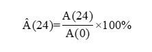
The migration quantification methods for the scratch assay were examined using the Area method, which is widely employed for quantifying cell migration. This method indirectly evaluates the migration rate by determining the percentage of the wound area through the utilization of the following formula. The percentage of the wound area (Â) is measured where A(24) is the wound area at 24 h and A(0) is the beginning area [14].
Results and discussion
Bioactive compounds in TGLE has the potential in targeting MUC1
Thirteen compounds contained in TGLE showed anticancer activity based on the PassOnline server. In this particular study, the analysis indicates that thirteen compounds found in T. grandis leaves exhibited 0 violation thus are considered as drug-like molecules. All the compounds were docked towards MUC1 using PyRx software. The results of the virtual screening revealed that quercetin, a compound found in T. grandis leaves, exhibited the lowest binding energy against MUC1. It is noteworthy that ligands with the most negative binding energy exhibit stronger interactions with the target protein and have a significant impact on its biological activity (Table 1). Therefore, quercetin and its interactions with MUC1 were then visualized using ProteinPlus website. Within the A-chain domain, quercetin compounds established hydrophobic bonds with Leu1089A and Leu1045A, along with hydrogen bonds involving Leu1045A, Ser1090A, and Gly1088A (Figure 1).
Table 1 The drug likeness and virtual screening results of TGLE targeting MUC1.
Compound |
Molecular mass (Da) |
Lipophilicity (LogP) |
HBD |
HBA |
Molecular reactivity |
Binding energy (kcal/mol) |
Quercetin |
302.24 |
1.23 |
5 |
7 |
78.03 |
–8.6 |
Ellagic acid |
302.19 |
1.00 |
4 |
8 |
75.31 |
–8.2 |
Ferulic acid |
194.18 |
1.36 |
2 |
4 |
51.63 |
–6.9 |
Cinnamic acid |
148.16 |
1.79 |
1 |
2 |
43.11 |
–6.8 |
P-coumarate |
163.15 |
1.35 |
1 |
3 |
43.24 |
–6.8 |
Sinapic acid |
224.21 |
1.31 |
1 |
3 |
43.24 |
–6.8 |
Vanillic acid |
168.15 |
1.08 |
2 |
4 |
41.92 |
–6.1 |
Gallic acid |
170.12 |
0.21 |
4 |
5 |
39.47 |
–5.6 |
Methyl decanoate |
186.29 |
3.36 |
0 |
2 |
56.28 |
–5.6 |
Glycerine diacetate |
176.17 |
0.17 |
1 |
5 |
39.49 |
–5.0 |
Butyl acetate |
116.16 |
1.53 |
0 |
2 |
32.24 |
–4.9 |
4-hydroxy-4-methyl-2-pentanone |
116.16 |
0.63 |
1 |
2 |
32.36 |
–4.4 |
Glycerine monoacetate |
134.13 |
-0.46 |
2 |
4 |
29.76 |
–4.1 |
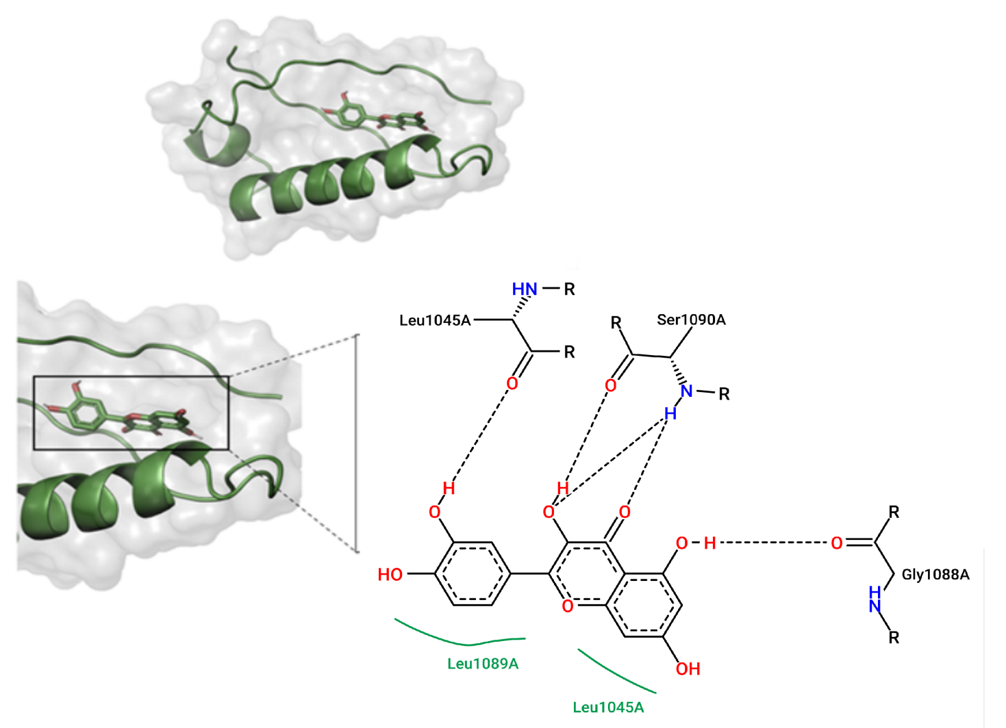
Figure 1 Structure of target protein (MUC1) and its chemical interactions with quercetin. Hydrophobic bonds are represented by green and hydrogen bonds represented by dashed lines. The hydrophobic bonds were produced at Leu1089A and Leu1045A, while hydrogen bonds involved with Leu 1045A, Ser1090A, and Gly1088A.
Mucin 1 protein is one of the most promising therapeutic targets for TNBC. This protein is expressed in nearly 90% of TNBC cells [6]. It plays a critical role in various kinds of cell proliferation pathways, such as Wnt/β-catenin, nuclear factor kappa B (NF-kB), STAT3, PI3K/AKT, and mitogen activated protein kinase (MAPK)/ ERK. In the Wnt/β-catenin pathway, the binding of MUC1 with Dvl and β-catenin leads to the activation of the Wnt/β-catenin pathway, contributing to cell proliferation and EMT. In the NF-kB pathway, the cytoplasmic domain of MUC1 (MUC1-C) forms a complex with NF-kB transcription factors, resulting in the activation of the NF-kB pathway involved in inflammation regulation, immune response, and cell proliferation. Additionally, MUC1 can interact with MAPK and STAT3, leading to excessive cell proliferation [15]. Moreover, MUC1 plays a role in increasing glutamine and glucose uptake, enhancing cell proliferation activity [7].
Computational analysis revealed quercetin as the compound within TGLE with the lowest binding energy towards MUC1, suggesting a potential interaction. It demonstrates the strongest affinity with MUC1 at the A-chain portion, forming hydrogen and hydrophobic bonds. Hydrophobic and hydrogen bonds are both involved in ligand-protein interaction. The hydrophobic interaction is the major driving force for the binding of the ligand to the protein [16], while hydrogen bonds play a significant role in the specificity of the interaction [17,18]. The presence of specific functional groups in the ligand molecule acts as a hydrogen bond donor or acceptor, and the formation of hydrogen bond interactions with specific amino acids in the protein is crucial for the binding of the ligand to the protein [17,18]. Furthermore, quercetin satisfies Lipinski’s rule of 5, making it a drug-like molecule. The potential of bioactive compounds derived from T. grandis leaves utilization as inhibitors of MUC1 has been emphasized. However, it should be noted that molecular docking provides a static prediction of binding propensity rather than direct evidence of biological inhibition. The observed differences in predicted binding energies were modest, and molecular dynamics simulations were not performed to assess interaction stability under physiological conditions. Although quercetin’s properties seem to be promising, these in silico findings should be interpreted as preliminary, supporting its potential for further mechanistic and experimental validation rather than confirming functional inhibition of MUC1.
The expression of MUC1 decreased upon the TGLE intervention
In this study, the indirect IF was utilized to examine the MUC1 expression in MDA-MB-231 cells. The concentration of TGLE employed was derived from Ghareeb et al. [24] study with modifications. The standard therapy group was administered with paclitaxel at 10 µg/mL (SC), whereas cells treated with TGLE at 7.5 (PAC + T1), 15 (PAC + T2), and 30 mg/mL (PAC + T3) were given paclitaxel at 5 µg/mL. Additionally, TGLE at 60 mg/mL (T4) was administered as a single agent. The results showed that there was a very strong negative correlation between the dosage of TGLE and MUC1 expression (p < 0.05; r = –0.880). Although Ghareeb et al. [24] reported antiproliferative effects of TGLE at µG/ml concentrations in HepG2 cells, the dose range applied in the present study was determined empirically based on IC50 analysis in MDA-MB-231 cells, around 15 mg/mL. Variation in cancer subtype, cellular sensitivity, and extract composition likely contributes to this inconsistency [19]. Accordingly, the selected concentrations were intended to consider the IC50 (0.5× - 4×) for exploratory in vitro evaluation rather than to reflect physiological exposure. Nevertheless, these dosing raise translational concerns, as crude plant extracts often exhibit limited bioavailability and may pose toxicity risks at the systemic level, in line with reported sub-chronic toxicity of TGLE in vivo at a dose of 40 mg/kg [20]. The 1-way ANOVA analysis revealed that the administration of paclitaxel and TGLE led to a significant reduction in MUC1 expression. Significant differences in MUC1 expression were observed between the PAC + T3 and PAC + T2 groups with the negative control (NC) group. Additionally, significant differences were found between the PAC + T3 group and the PAC + T1 as well as the SC groups (Figure 2).
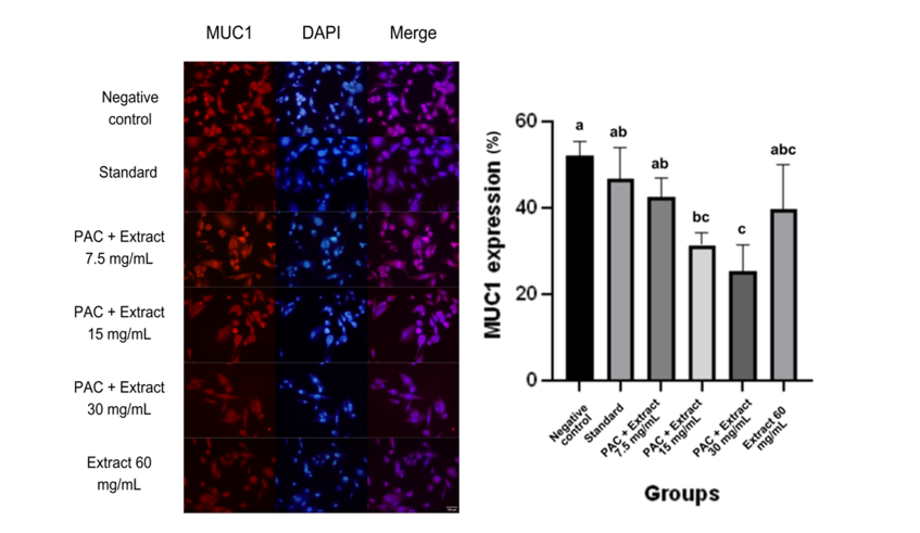
Figure 2 MUC1 expression upon extract and paclitaxel administration. The protein MUC1 is expressed in the cytoplasm. The expression of MUC1 protein decreases progressively with increasing doses of extract administered in combination with paclitaxel; PAC = paclitaxel 5 µg/mL.
In vitro study demonstrated that TGLE can reduce MUC1 expression in MDA-MB-231 cells, confirming the formulated hypothesis. These findings align with in silico analysis and the study by Wang et al. [21] as well, which showed that quercetin, one of the bioactive compounds present in TGLE, can inhibit MUC1 expression and cell proliferation in TNBC. Additionally, the study by Hiraki et al. [22] indicated that MUC1-C inhibition in TNBC cells induces apoptosis and decreases the expression of B-Cell Lymphoma 2-related Protein A1 (BCL2A1), which acts as an antiapoptotic factor. These findings support the potential of TGLE as an antiproliferative agent through the MUC1 pathway.
The TGLE intervention led to a reduction in MMP-9 expression
This investigation employed the indirect immunofluorescence method to assess MMP-9 expression in MDA-MB-231 cells. Various concentrations ranging from 0 to 60 mg/mL were utilized. The control group received paclitaxel at 10 µg/mL, while the treatment groups received paclitaxel at reduced doses (5 µg/mL) along with TGLE at concentrations of 7.5, 15, and 30 mg/mL. TGLE at 60 mg/mL was administered independently. The findings demonstrated a significant negative correlation between TGLE dosage and MMP-9 expression (p < 0.05; r = –0.886). One-way ANOVA analysis indicated a notable decrease in MMP-9 expression following the administration of paclitaxel and TGLE. Significant differences in MMP-9 expression were discovered between the PAC + 15 mg/mL and PAC + 30 mg/mL treatment groups, as well as the 60 mg/mL TGLE group, when compared to the negative control. Furthermore, there were statistically significant differences between the PAC + 30 mg/mL treatment group and the single-agent TGLE group (Figure 3).
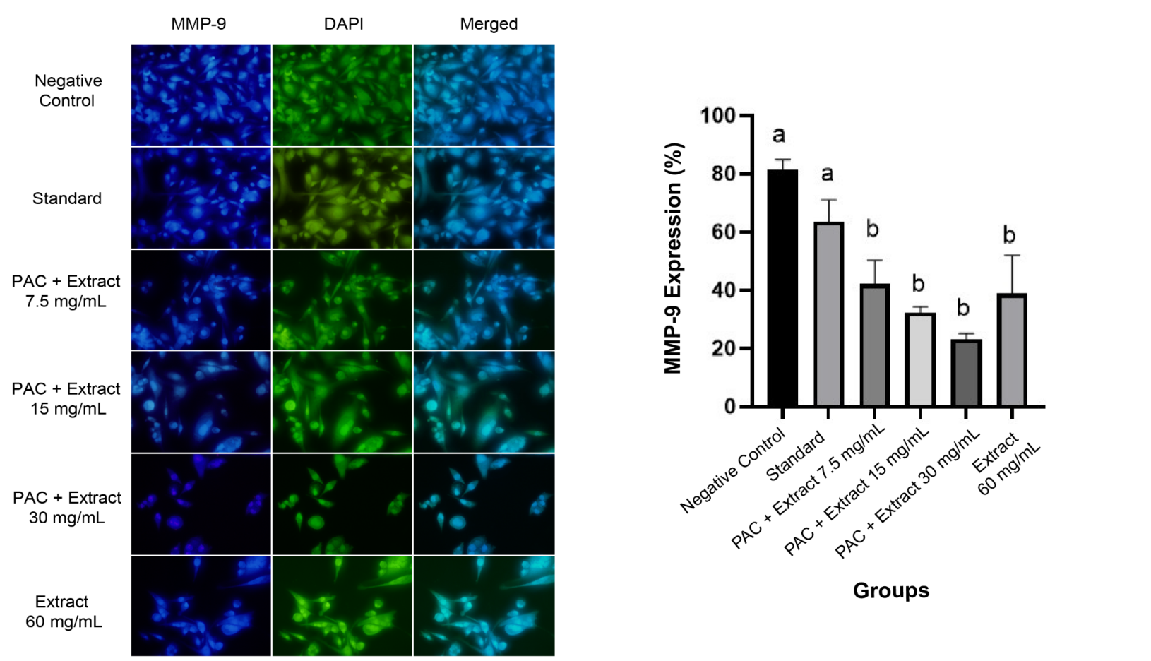
Figure 3 The impact of extract and paclitaxel co-administration on MMP-9 expression. The MMP-9 protein expression shows a gradual decrease as the doses of the extract administered alongside paclitaxel increase; PAC = paclitaxel 5 µg/mL.
The investigation indicated that TGLE effectively decreases MMP-9 expression in MDA-MB-231 cells, validating the initial premise. These results are consistent with research conducted by Brala et al. [23], which demonstrated that quercetin can hinder MMP-9 expression involved in cell migration. Furthermore, this study revealed that the combination of quercetin and 5-fluorouracil led to a 0.77-fold decrease in MMP-9 expression. The gelatinase enzyme, which includes MMP-9, is an important element in the metastatic progression of breast cancer cells, necessitating cell migration as one of its processes. Therefore, TGLE has the potential to impede cell migration through the effect of reducing MMP-9.
Tectona grandis leaves extract decreased cell proliferation and cell migration of MDA-MB-231 cell
In this study, the 48 h MTT assay was conducted on MDA-MB-231 cells to determine the IC50 of TGLE in combination with paclitaxel at 5 µg/mL. Based on the cytotoxicity test results, the IC50 of the extract was found to be 14.519 mg/mL, indicating that the IC50 fell within the range of doses to be tested. Based on the results, the cell proliferation was dose-dependently decreased upon the administration of TGLE (p < 0.05; r = –0.864). It was observed that cell viability decreased significantly compared to the NC (p < 0.0002 or p < 0.0001). Significant differences in cell viability were found between PAC + T3 VS SC group (p < 0.0332) as well as PAC + T3 VS PAC + T1 group (p < 0.0332). In the T4 group, a decrease in cell viability was observed, though it remained higher than that in PAC + T3 group. These findings suggest that the administration of paclitaxel and TGLE can reduce the proliferation activity of MDA-MB-231 cells (Figure 4).
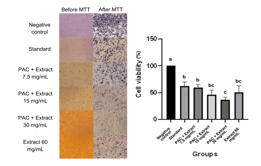
Figure 4 Cell Cytotoxicity Assay at 48 h MTT. There was significant reduction of cell proliferation compared to negative control. Significant reduction also found in PAC + TGLE 30 mg/mL VS standard and PAC + TGLE 7,5 mg/mL; PAC = paclitaxel 5 µg/mL.
The 24, 48, and 72 h MTT proliferation assay was conducted, followed by a comparative analysis to compare the cell viability between NC, SC, and PAC + T2 groups at these time points. Based on the comparative analysis, significant differences were found between the treatment groups at 24, 48, and 72 h (p < 0.05). Further post hoc tests were conducted for the 24, 48, and 72-hour data. In the SC group, a significant reduction of cell proliferation was observed after 48 h of MTT (p < 0.0021). On the other hand, in PAC + T2 group, significant decrease of cell viability was already evident at 24 h of MTT (p < 0.0332) (Figure 5).
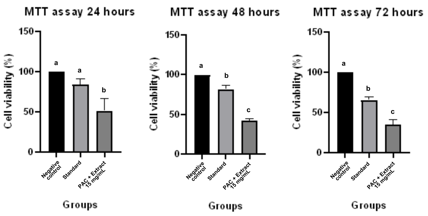
Figure 5 Cell Proliferation Assay at 24, 48, and 72 h MTT. There were significant differences between the treatment groups at 24, 48, and 72 h. In the SC group, a significant reduction of cell proliferation was observed after 48 h of MTT, while in PAC + TGLE 15 mg/mL group, significant decrease of cell viability was already evident at 24 h of MTT; PAC = paclitaxel 5 µg/mL
In this study, the MTT assay was performed to investigate the effect of TGLE on the proliferation activity of MDA-MB-231 cells. The results demonstrated that the TGLE exhibited antiproliferative effects both as a single agent and as a complementary therapy with paclitaxel, in line with the formulated hypothesis. At a dosage of 15 mg/mL, the combination of paclitaxel and TGLE showed significantly faster antiproliferative activity (observed at MTT 24 h) compared to single-agent paclitaxel therapy (observed at MTT 48 h). These findings are consistent with Ghareeb et al. [24], which showed that methanol, butanol, and ethanol extracts of T. grandis leaves possessed cytotoxic activity against HepG2 hepatocarcinoma cells.
Collectively, the observed antiproliferative and migration-associated effects of TGLE were correlated with reduced expression of MUC1 and MMP-9, respectively. Previous studies showed the potential of quercetin, a bioactive compound found in TGLE, to have an antiproliferative effect in TNBC [21,25]. In this study, quercetin possessed the lowest binding affinity among others, indicating the most stable interaction. Quercetin has been shown to interact with α-fetoprotein (AFP), leading to the competitive inhibition of MUC1 binding with AFP, ultimately reducing β-catenin protein levels and cell proliferation [25]. Furthermore, quercetin can also inhibit cell proliferation by downregulating MUC1 [21]. However, the present study does not establish that these pathways are the primary mediators of TGLE activity. Instead, the data support a correlation, rather than a causative relationship, between the expression of MUC1 and MMP-9 with the proliferation and migratory activity of the cells.
In addition to MUC1, the TGLE can potentially inhibit cell proliferation through other molecular pathways. Several bioactive compounds that have been demonstrated to play a significant role in cell proliferation are flavonoids, phenolic acids, and tannins [26]. Flavonoids act as antioxidants and inhibit mutant P53 protein and heat shock protein (HSP) production [27]. Phenolic acids in T. grandis leaves have been shown to inhibit several molecular pathways involved in cell proliferation and apoptosis, such as PI3K/AKT/epidermal growth factor receptor (EGFR), MAPK, and tumor necrosis factor receptor 1 (TNFR1) [28-30]. Additionally, tannins in T. grandis leaves act as antioxidants [31].
To evaluate the cell migration ability of MDA-MB-231 cells treated with TGLE, a scratch test was conducted. The results of the scratch test indicated that TGLE exhibited an inhibitory effect on cell migration, which was compatible with the hypothesis. The scratch test showed that increasing the extract dose in combination groups resulted in an increase in cell distance, indicating a decrease in MDA-MB-231 cell migration ability (p < 0.05; r = 0.956). However, the statistical difference was found to be non-significant, thus no post-hoc test was conducted (Figure 6). These findings align with the research conducted by Brala et al. [23], which investigated the combination of quercetin with 5-fluorouracil. This study demonstrated a reduction in the migration rate of breast cancer cells by 62%.
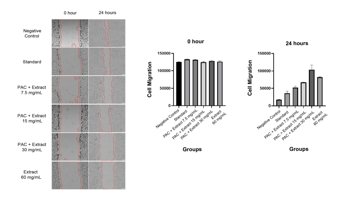
Figure 6 Assessing Cell Migration using Scratch Test. The observation results of triple negative breast cancer cells MDA-MB-231 were conducted using a scratch test to assess the changes in cell distance at 0 and 24 h. An increase in cell distance was observed with the increase in extract dosage in the PAC + extract group, indicating a decrease in cell migration ability; PAC = paclitaxel 5 µg/mL.
Apart from its role in proliferation inhibition, quercetin has been observed to downregulate MMP-9 expression, consequently impeding cell migration. Quercetin’s angiogenic properties contribute to the reduction in MMP-9 expression. Multiple studies suggest that quercetin plays a significant role in inhibiting various types of cancer cells, including breast cancer cells [32].
Flavonoids exhibit high diversity in chemical structure and function and can play a role in the tumor microenvironment. The diversity in chemical structure results in various mechanisms of action, including anticancer effects [32]. The study by Rudin et al. [27] indicated that flavonoids found in TGLE, such as quercetin, are bioactive compounds with antioxidant properties. During the initiation stage, flavonoids can stabilize free radicals formed by carcinogens such as oxygen, peroxides, and superoxide, preventing them from oxidizing deoxyribonucleic acid (DNA) and thereby preventing DNA mutations. Additionally, flavonoids play a role in suppressing mutant P53 protein expression and inhibiting HSP production. These 3 mechanisms contribute to the antiproliferative activity.
Two types of phenolic acids in T. grandis leaves that have been studied for their anticancer effects on TNBC are gallic acid and cinnamic acid. Gallic acid has the potential to be a TNBC therapy agent by arresting the G1 phase, reducing cyclin-dependent kinase (CDK)-4 and CDK2 mediators, exerting antioxidant effects, and regulating the PI3K/AKT/EGFR and MAPK signaling pathways. Gallic acid reduces cell viability in a dose-dependent manner and has been shown to be selective for non-cancerous cells such as MCF-10F [28,29]. On the other hand, cinnamic acid induces apoptosis through the TNFR1 extrinsic pathway. Cinnamic acid exhibits anticancer effects on TNBC, glioblastoma, melanoma, prostate cancer, and lung carcinoma [30].
The tannin compounds present in T. grandis leaves are classified as hydrolyzable tannins, including gallic acid, and ellagic acid. Gallic acid has been proven to have anticancer effects on TNBC cells [28,29]. Furthermore, ellagic acid, a heterocyclic organic compound derived from the formal dimerization of gallic acid, exerts anticancer effects through antioxidant and antiproliferative activities. One type of cancer that has shown a response to ellagic acid therapy is follicular lymphoma [33].
This research has several limitations. The study was only conducted in vitro cell proliferation and cell migration assays, which means that other factors such as absorption, distribution, metabolism, and excretion cannot be evaluated. The toxicological profile of TGLE and the specific bioactive components of TGLE responsible for reducing cell proliferation and cell migration could not be assessed through in vitro studies. This study did not use non-TNBC cells nor normal mammary epithelial cells to assess selectivity and physiological safety, leading to a limitation of specificity interpretation. Therefore, further research is needed to determine the efficacy and toxicological profile of TGLE as a complementary therapy for TNBC compared to non-TNBC cells and healthy cells through in vivo studies, followed by clinical trials. Future studies should also normalize dosing to calculated bioactive constituents, especially quercetin equivalents, to improve translational relevance and enable pharmacokinetic evaluation. Furthermore, further investigation is necessary to assess the bioactive compounds present in the TGLE that play a role as antiproliferative and antimigratory agents. Functional studies, such as MUC1 knockdown and MMP-9 inhibition, along with rescue experiments, are also required to confirm its role in antiproliferative and antimigratory activity, respectively.
Conclusions
Based on the research findings, it can be concluded that TGLE contains several bioactive compounds possessing anticancer activity. Quercetin has the strongest binding affinity towards MUC1, making it a promising anti-MUC1 candidate. Tectona grandis leaves extracts could reduce MUC1 expression and significantly decrease TNBC cell proliferation in vitro, whether used as a standalone therapy or as a complementary treatment with paclitaxel. There exists a very strong inverse correlation between the dosage of TGLE and both MUC1 expression and cell proliferation. Moreover, TGLE has exhibited in vitro efficacy in reducing MMP-9 expression and cell migration when utilized in combination with paclitaxel or as a monotherapy, with a robust correlation observed between TGLE dosage and the reduction in MMP-9 expression and cell migration capacity. Additional investigations, such as molecular dynamic simulation and pathway prediction utilizing the Stitch database are recommended. Analysis regarding the atoms involved in protein-ligand bonding is needed to further validate this study. Furthermore, the potential of TGLE should be verified through in vivo study and clinical trials to establish the efficacy and correct formulation.
Acknowledgements
The authors would like to express their gratitude to the Funding of Posdocoral Fellow of Universitas Brawijaya, Indonesia, for the support provided.
CRediT author statement
Sofy Permana: Conceptualization; Methodology; Writing - Original Draft; Writing - Review & Editing. Neyla Vista Maramy: Conceptualization; Methodology; Investigation; Formal analysis; Writing - Original Draft. Maghfira Rahma Azizah: Conceptualization; Methodology; Investigation; Formal analysis; Writing - Original Draft. Halida Adib Hanum: Conceptualization; Methodology; Investigation; Formal analysis; Writing - Original Draft. Yoshiyuki Kawamoto: Conceptualization; Methodology; Writing - Review & Editing. Edwin Widodo: Investigation; Methodology; Original Draft; Writing - Review & Editing. Heni Endrawati: Investigation; Methodology; Original Draft; Writing - Review & Editing. Agustina Tri Endharti: Conceptualization; Methodology; Investigation; Writing - Original Draft; Writing - Review & Editing; Supervision; Project administration.
References
[1] L Yin, JJ Duan, XW Bian and SC Yu. Triple-negative breast cancer molecular subtyping and treatment progress. Breast Cancer Research 2020; 22(1), 61.
[2] AC Wolff, MEH Hammond, KH Allison, BE Harvey, PB Mangu, JMS Bartlett, M Bilous, IO Ellis, P Fitzgibbons, W Hanna, RB Jenkins, MF Press, PA Spears, GH Vance, G Viale, LM McShane and M Dowsett. Human epidermal growth factor receptor 2 testing in breast cancer: American Society of Clinical Oncology/College of American Pathologists clinical practice guideline focused update. Archives of Pathology & Laboratory Medicine 2018; 142(11), 1364-1382.
[3] JW Chang, Y Ding, M Tahir ul Qamar, Y Shen, J Gao and LL Chen. A deep learning model based on sparse auto-encoder for prioritizing cancer-related genes and drug target combinations. Carcinogenesis 2019; 40(5), 624-632.
[4] KA Won and C Spruck. Triple-negative breast cancer therapy: Current and future perspectives. International Journal of Oncology 2020; 57(6), 1245-1261.
[5] MA Clark, R Finkel, JA Rey and K Whalen. Lippincott’s illustrated reviews: Pharmacology. 5th ed. Wolters Kluwer, Philadelphia, United States, 2012.
[6] T Maeda, M Hiraki, C Jin, H Rajabi, A Tagde, M Alam, A Bouillez, X Hu, Y Suzuki, M Miyo, T Hata, K Hinohara and D Kufe. MUC1-C induces PD-L1 and immune evasion in triple-negative breast cancer. Cancer Research 2018; 78(1), 205-215.
[7] G Goode, V Gunda, NV Chaika, V Purohit, F Yu and PK Singh. MUC1 facilitates metabolomic reprogramming in triple-negative breast cancer. PLoS One 2017; 12(5), e0176820.
[8] H Huang. Matrix metalloproteinase-9 (MMP-9) as a cancer biomarker and MMP-9 biosensors: Recent advances. Sensors 2018; 18(10), 3249.
[9] G Barillari. The impact of matrix metalloproteinase-9 on the sequential steps of the metastatic process. International Journal of Molecular Sciences 2020; 21(12), 4526.
[10] Y Li, J He, F Wang, X Wang, F Yang, C Zhao, C Feng and T Li. Role of MMP-9 in epithelial-mesenchymal transition of thyroid cancer. World Journal of Surgical Oncology 2020; 18(1), 181.
[11] SMB Asdaq, N Nayeem, MT Alam, SI Alaqel, M Imran, EWE Hassan and SI Rabbani. Tectona grandis Lf: A comprehensive review on its patents, chemical constituents, and biological activities. Saudi Journal of Biological Sciences 2022; 29(3), 1456-1464.
[12] RB Kilaru, KR Valasani, NK Yellapu, HP Osuru, CS Kuruva, B Matcha and NR Chamarthi. Design, synthesis, in silico and in vitro studies of novel 4-methylthiazole-5-carboxylic acid derivatives as potent anti-cancer agents. Bioorganic & Medicinal Chemistry Letters 2014; 24(18), 4580-4585.
[13] V Suryanti, T Kusumaningsih, SD Marliyana, HA Setyono and EW Trisnawati. Identification of active compounds and antioxidant activity of teak (Tectona grandis) leaves. Biodiversitas 2020; 21(3), 946-952.
[14] AV Ponce Bobadilla, J Arévalo, E Sarró, HM Byrne, PK Maini, T Carraro, S Balocco, A Meseguer and T Alarcón. In vitro cell migration quantification method for scratch assays. Journal of the Royal Society Interface 2019; 16(151), 20180709.
[15] A Hosseinzadeh, P Merikhian, N Naseri, MR Eisavand and L Farahmand. MUC1 is a potential target to overcome trastuzumab resistance in breast cancer therapy. Cancer Cell International 2022; 22(1), 110.
[16] R Patil, S Das, A Stanley, L Yadav, A Sudhakar and AK Varma. Optimized hydrophobic interactions and hydrogen bonding at the target-ligand interface leads the pathways of drug-designing. PLoS One 2010; 5(8), e12029.
[17] D Chen, N Oezguen, P Urvil, C Ferguson, SM Dann and TC Savidge. Regulation of protein-ligand binding affinity by hydrogen bond pairing. Science Advances 2016; 2(3), e1501240.
[18] RF De Freitas and M Schapira. A systematic analysis of atomic protein–ligand interactions in the PDB. MedChemComm 2017; 8(10), 1970-1981.
[19] M Alamoodi. Factors affecting pathological complete response in locally advanced breast cancer cases receiving neoadjuvant therapy: A comprehensive literature review. European Journal of Breast Health 2024; 20(1), 8.
[20] CD Hamdin, D Galanova, DC Saputro, IM Munir, A Nurrochmad and I Puspitasari. Safety concerns of Tectona grandis Lf leaf extract as a natural food colorant: evidence of irreversible organ pathology in subchronic toxicity study. The Indonesian Biomedical Journal 2025; 17(1), 78-89.
[21] R Wang, L Yang, S Li, D Ye, L Yang, Q Liu, Z Zhao, Q Cai, J Tan and X Li. Quercetin inhibits breast cancer stem cells via downregulation of aldehyde dehydrogenase 1A1 (ALDH1A1), chemokine receptor type 4 (CXCR4), mucin 1 (MUC1), and epithelial cell adhesion molecule (EpCAM). Medical Science Monitor 2018; 24, 412-420.
[22] M Hiraki, T Maeda, N Mehrotra, C Jin, M Alam, A Bouillez, T Hata, A Tagde, A Keating, S Kharbanda, H Singh and D Kufe. Targeting MUC1-C suppresses BCL2A1 in triple-negative breast cancer. Signal Transduction and Targeted Therapy 2018; 3(1), 13.
[23] C Jakobušić Brala, A Karković Marković, A Kugić, J Torić and M Barbarić. Combination chemotherapy with selected polyphenols in preclinical and clinical studies - An update overview. Molecules 2023; 28(9), 3746.
[24] MA Ghareeb, AH Hussein, MFM Hassan, AR Laila, AM Mona and MS Amal. Antioxidant and cytotoxic activities of Tectona grandis Linn leaves. International Journal of Phytopharmacology 2014; 5(2), 143-157.
[25] B Lin, Q Wang, K Liu, X Dong, M Zhu and M Li. Alpha-fetoprotein binding mucin and scavenger receptors: an available bio-target for treating cancer. Frontiers in Oncology 2021; 11, 625936.
[26] PK Kolli, S Obbalareddy, RP Yejella, LD Athili and S Ponnada. A review on Tectona grandis. International Journal of Research in Pharmaceutical and Biomedical Sciences 2022; 12, 359-366.
[27] NA Rudin and R Rahmawati. The potential utilization of young teak leaves (Tectona grandis Linn. f.) as basic ingredients on producing anti-cancer herbal tea. Jurnal Pendidikan Biologi Undiksha 2022; 9(1), 10-23.
[28] HL Lee, CS Lin, SH Kao and MC Chou. Gallic acid induces G1 phase arrest and apoptosis of triple-negative breast cancer cell MDA-MB-231 via p38 mitogen-activated protein kinase/p21/p27 axis. Anti-Cancer Drugs 2017; 28(10), 1150-1156.
[29] S Lin, HZ Qin, ZY Li, H Zhu, L Long and LB Xu. Gallic acid suppresses the progression of triple-negative breast cancer HCC1806 cells via modulating PI3K/AKT/EGFR and MAPK signaling pathways. Frontiers in Pharmacology 2022; 13, 1049117.
[30] A Pal, P Tapadar and R Pal. Exploring the molecular mechanism of cinnamic acid-mediated cytotoxicity in triple negative MDA-MB-231 breast cancer cells. Anti-Cancer Agents in Medicinal Chemistry 2021; 21(9), 1141-1150.
[31] E Sieniawska and T Baj. Tannins. In: S Badal and R Delgoda (Eds.). Pharmacognosy. Academic Press, Massachusetts, United States, 2017.
[32] WH Talib, A Abuawad, S Thiab, A Alshweiat and AI Mahmod. Flavonoid-based nanomedicines to target tumor microenvironment. OpenNano 2022; 8, 100081.
[33] National Center for Biotechnology Information. Ellagic acid, Available at: https://pubchem.ncbi.nlm.nih.gov/compound/Ellagic-Acid, accessed November 2025.