
Trends
Sci.
2026;
23(9):
12876
Unveiling the Anti-Inflammatory Potential of Urena lobata: Insights into Nitric Oxide Suppression and Protein Targets
Yudi
Purnomo1,
Andri Tilaqza1,
Doti Wahyuningsih2
and
Dinia
Rizqi Dwijayanti3,4,*,

1Department of Pharmacy, Faculty of Medicine, University of Islam Malang, Malang 65144, Indonesia
2Department of Biochemistry, Faculty of Medicine, University of Islam Malang, Malang 65144, Indonesia
3Department of Biology, Faculty of Mathematics and Natural Science, Brawijaya University, Malang 65145, Indonesia
4Innovation Center of Integrative Jamu and Eco-pharmaca, Brawijaya University, Malang 65145, Indonesia
(*Corresponding author’s e-mail: [email protected])
Received: 3 December 2025, Revised: 14 January 2026, Accepted: 24 January 2026, Published: 25 March 2026
Abstract
Urena lobata is recognized for its traditional medicinal use, particularly in addressing inflammation-related ailments. This study examined the anti-inflammatory properties of U. lobata extract and its fractions, focusing on their ability to suppress nitric oxide (NO) synthesis, a key mediator of inflammation. In vitro experiments using LPS-stimulated RAW 264.7 macrophages showed that the crude extract of U. lobata had an IC50 of 58.05 ± 5.87 µg/mL, while the ethyl acetate fraction showed greater suppression of NO formation with an IC50 of 31.40 ± 10.56 µg/mL. On the other hand, the n-butanol fraction and the water-soluble fraction did not have a significant effect on inhibition. Toxicity tests also showed that the n-hexane fraction was good at reducing NO formation (IC50 of 49.73 ± 9.49 µg/mL), but was toxic to cells at higher concentrations (> 200 µg/mL). The ethyl acetate fraction also inhibited the formation of important pro-inflammatory mediators, such as inducible nitric oxide synthase (iNOS), interleukin (IL)-12, and tumor necrosis factor-alpha (TNF-α). Protein target analysis revealed 40 proteins associated with the inflammatory cascade as potential targets for active chemicals in U. lobata. Molecular docking studies identified quercetin, apigenin, and luteolin as key bioactive compounds with strong binding affinity for inflammation-related proteins such as AKR1B1, NOX4, and CDK5, which subsequently influence NO suppression during inflammation, suggesting a multi-target mechanism of action. These results emphasize the therapeutic potential of U. lobata and its bioactive chemicals as candidates for natural anti-inflammatory drug formulations, while also underscoring the need to evaluate potential cytotoxicity in future studies.
Keywords: Urena lobata, Inflammation, Nitric oxide, AKR1B1, NOX4, CDK5, Natural therapeutics
Introduction
Inflammation is the fundamental basis for the pathogenesis of various diseases, encompassing both degenerative and non-degenerative conditions [1]. Chronic inflammation is a key factor in the development of many diseases, such as cardiovascular disorders [2], diabetes mellitus [3], neuropathic pain [4], and cancer [5]. Unregulated inflammation, frequently instigated by microbial infections or intrinsic stressors, results in the excessive synthesis of pro-inflammatory mediators, including nitric oxide (NO), tumor necrosis factor-alpha (TNF-α), and interleukin-6 (IL-6), which
exacerbate tissue damage and facilitate disease advancement [6].
The rising incidence of chronic inflammatory diseases, along with the limitations of existing therapies, has generated considerable interest in developing safer and more effective anti-inflammatory medications. Nonsteroidal anti-inflammatory drugs (NSAIDs) and corticosteroids are two common types of anti-inflammatory drugs. Despite their widespread use, NSAIDs are associated with clinically relevant adverse effects, such as gastrointestinal toxicities, cardiovascular risks, renal injuries, hepatotoxicity, hypertension, and other minor disorders, especially when used for a long time [7-9]. Therefore, there is an increasing demand for alternative medicines that provide similar efficacy with less toxicity.
Natural products, especially those from medicinal plants, are becoming a viable source of bioactive chemicals that can help with inflammation. These plants frequently possess a varied array of phytochemicals, such as flavonoids, terpenoids, alkaloids, and polyphenols, capable of modulating several molecular pathways associated with inflammation [10,11]. Additionally, plant-derived chemicals frequently exhibit reduced toxicity profiles, rendering them appealing options for prolonged utilization in the management of chronic inflammatory disorders [12].
Urena lobata, a well-known herb in traditional medicine, has been getting more and more scientific interest for its possible use in reducing inflammation. U. lobata, or Caesar weed, is a plant that has been used in folk medicine for a long time in many places because it can reduce inflammation, fight free radicals, and help wounds heal. A preliminary study [13,14] found that this plant had anti-bacterial and anti-inflammatory properties. Other research has shown that U. lobata stops the growth of free radicals such superoxide radicals, hydroxyl radicals, and lipid peroxidation because it is high in flavonoids [13,15]. Gossypetin (GPE) is a flavonoid identified in this herb, with prior research demonstrating several pharmacological actions, including anti-inflammatory properties [15-18]. Nevertheless, the inhibitory efficacy of U. lobata and its phytoconstituents on pro-inflammatory mediators has been investigated solely in crude extracts [14].
While prior research indicates that U. lobata possesses bioactive substances with anti-inflammatory properties, the precise molecular pathways and targets that mediate its actions have yet to be thoroughly investigated. inflammation-related proteins through in silico methodologies. This study enhances comprehension of the application of plant extracts in the formulation of innovative anti-inflammatory drugs, presenting new solutions for the management of inflammation-related disorders.
Materials and methods
Extraction and fractionation of U. lobata leaf
Simplicia of U. lobata leaves were obtained from Laboratory UPT Materia Medika Batu, Malang, Indonesia with certificate number 074/027/101.8/2015. The simplicia powder (1,000 g) was extracted with 5,000 mL methanol (Smart Lab) for 6 h using a water-bath and a shaker (Memmert) and evaporated to produce a paste form. Therefore, methanol extract (80 g) was performed liquid-liquid partition fractionation using n-hexane solvent (Smart Lab), ethyl acetate (Smart Lab), n-butanol (E. Merck) and water (Bratachem), respectively.
RAW 264.7 cell culture preparation
The murine macrophage RAW 264.7 cell line was obtained from the Laboratory of Animal Physiology, Structure and Development, Faculty of Mathematics and Natural Sciences, Brawijaya University. Cells were maintained in Dulbeccoʼs Modified Eagle Medium (DMEM, D6430, Sigma Aldrich, Merck, Germany) supplemented with 10% fetal bovine serum (FBS, F2442, Sigma Aldrich, Merck, Germany) and 1% penicillin-streptomycin solution (100 U/mL and 100 µg/mL, respectively, A5955, Sigma Aldrich, Merck, Germany). Cells were then seeded in 24-well plates at a density of 1×10⁵ cells per well and incubated at 37 °C in a 5% CO₂ atmosphere for 24 h for adhesion and stabilization.
Determination of nitric oxide (NO) production
Nitric oxide production was assessed using a modified Griess assay to measure nitrite levels in culture media according to Dwijayanti et al. [19], RAW 264.7 cells were treated with lipopolysaccharide (LPS) and extracts or fractions for 24 h at 37 °C. After treatment, 75 µL of culture supernatant or sodium nitrite (NaNO₂) was mixed with 75 µL of Griess reagent (G4410, Sigma Aldrich) in a 96-well plate. The mixture was incubated for 5 min at 25 °C, and absorbance was measured using a microplate reader at 570 nm. The experiment was performed in triplicate, with cells from different passages. The IC50 value of NO inhibition was calculated based on the concentration of extract or fraction required to reduce nitrite levels by 50%. The concentrations used in subsequent experiments were selected based on the dose-response curves obtained from the NO inhibition and cytotoxicity assays.
Cell viability assay
To assess cell viability, the WST-1 assay was performed following the NO production assay. The medium from the treated RAW 264.7 cell culture was aspirated and replaced with 120 µL of fresh medium containing 5% WST-1 reagent (11644807001, Roche). The cells were then incubated at 37 °C in a 5% CO₂ for 20 min. After incubation, 100 µL of the supernatant was transferred to a 96-well plate, and the absorbance was measured at 450 nm. The assay was performed in triplicate with a minimum of 3 replicates [19].
Flow cytometry analysis of iNOS, IL-12, and TNF-α
The macrophage RAW 264.7 cells were seeded at a density of 1×10⁵ cells/well in 24-well plates and incubated at 37 °C in a 5% CO2 incubator for 24 h. After incubation, the cells were divided into three groups: (1) Control group, which was treated only with DMEM medium; (2) LPS group, which was treated with 4 µg/mL lipopolysaccharide (LPS); and (3) LPS + ethyl acetate fraction (EAF) of U. lobata group, which was simultaneously treated with 4 µg/mL LPS and IC₅₀ concentration of U. lobata EAF. The IC₅₀ value was chosen for flow cytometry analysis as it represents the minimal effective concentration that produces a clear biological response while minimizing potential cytotoxic effects. After 24 h, cells were harvested, washed with PBS, and stained with fluorochrome-conjugated antibodies specific to inducible nitric oxide synthase (iNOS; Cat. bs-20601R; Bioss; ThermoFisher Scientific, Waltham, MA, USA), interleukin-12 (IL-12; Cat. bs-0767R; Bioss; ThermoFisher Scientific, Waltham, MA, USA), and tumor necrosis factor-alpha (TNF-α; Cat. bs-2081R; Bioss; ThermoFisher Scientific, Waltham, MA, USA). After 20 min, the cells were added by 300 μL PBS and analyzed by flow cytometry using a BD FACS Calibur™ (BD Biosciences, San Jose, CA, USA) [19]. All flow cytometry experiments were performed in at least three independent biological replicates. Flow cytometric data were processed using BD CellQuest™ Pro Software. Cells were first gated based on forward scatter (FSC) and side scatter (SSC) to exclude debris, followed by singlet discrimination. The final gates were applied to quantify the percentage of positive cells based on the corresponding fluorescence channels.
Statistical analysis
Data from the nitric oxide (NO) assay were collected from 5 independent experiments, each utilizing different passages of the RAW 264.7 cell line to ensure variability and reproducibility. The results are presented as means ± standard deviation (SD). Statistical analyses were conducted using Studentʼs t-test to compare the means between groups. Significance levels were determined at p < 0.05 and p < 0.01, indicating the threshold for statistical significance in the findings.
Data mining of bioactive compounds
The bioactive compounds present in U. lobata were retrieved from the KNApSAcK database (https://www.knapsackfamily.com/KNApSAcK/) and relevant scientific literature. The canonical SMILES (Simplified Molecular Input Line Entry System) for each compound were obtained from the PubChem database (https://pubchem.ncbi.nlm.nih.gov/), which facilitated their further analysis [20].
Toxicity prediction
Toxicity prediction for the identified compounds was performed using the database of Protox II (https://tox-new.charite.de/protox_II/index.php?site= compound_input). Protox II was used to evaluate toxicity endpoints, including Lethal Dose 50 (LD50), hepatotoxicity, carcinogenicity, immunotoxicity, mutagenicity, and cytotoxicity, of the compounds.
Screening of bioactive compounds based on drug-likeness and probable activity
Drug-likeness screening was conducted using the SWISS ADME web server (https://www.swissadme.ch/) to assess the medicinal potential of the compounds according to Lipinski rules. To evaluate the probable activity of the compounds, especially their potential involvement in anti-inflammatory mechanisms, the PASS Online web server (https://www.way2drug.com/passonline/) was used. Compounds showing high potential for anti-inflammatory activity were selected for further studies [20].
Protein target prediction
Compounds that met the criteria for anti-inflammatory bioactivity were subjected to protein target prediction. The SWISS Target Prediction web server (https://www.swisstargetprediction.ch/) was used to identify direct protein targets based on structural similarities with known bioactive molecules. For indirect protein interactions, the STRING 11.0 database (https://string-db.org/) was employed with a confidence threshold of 0.4 and a maximum of five interactions per target. The protein-protein interaction network was visualized using Cytoscape 3.8.2 (https://cytoscape.org/) [20].
Molecular docking simulation
Molecular docking simulations were carried out to evaluate the binding affinity between the bioactive compounds and their target proteins. The three-dimensional (3D) structures of the compounds were retrieved from the PubChem database (https://pubchem.ncbi.nlm.nih.gov/) and prepared using OpenBabel within the PyRx software. Protein structures were obtained from the RCSB Protein Data Bank (https://www.rcsb.org/) and prepared by removing water molecules and co-crystallized ligands using Biovia Discovery Studio 2019 software (Dassault Systèmes Biovia, San Diego, CA, USA). Docking simulations were performed using AutoDock Vina integrated into PyRx 0.8, where the binding site for the compounds was set to match the drug control. The docking results were visualized and analyzed using Biovia Discovery Studio 2019 software [20].
Results
Inhibition of NO production and cytotoxicity of U. lobata extract on LPS-stimulated cells
Based on Figure 1(A), the U. lobata extract shows a concentration-dependent inhibition of nitric oxide (NO) production in LPS-stimulated RAW 264.7 cells. At concentrations of 25, 50, and 100 µg/mL, the extract significantly reduces NO levels compared to the untreated control, with the highest inhibition observed at 100 µg/mL. The calculated IC50 value of 58.05 ± 5.87 µg/mL indicates the concentration required to inhibit 50% of NO production. Furthermore, the U. lobata extract does not appear to exhibit significant toxicity at concentrations up to 100 µg/mL (Figure 1B). However, at the higher concentrations of 200 and 400 µg/mL, the cell viability was below 80%. Taken together, these results indicate that the U. lobata extract exerts a notable inhibitory effect on NO production at non-cytotoxic concentrations, supporting its potential anti-inflammatory activity.
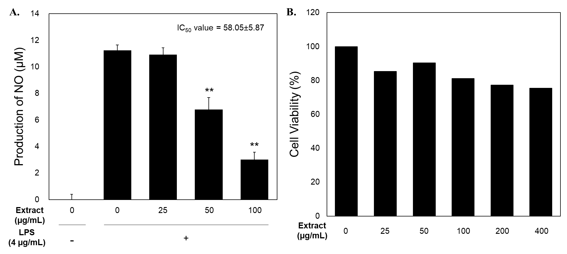
Figure 1 The effect of U. lobata extract on NO Production (A) and Cell Viability (B) of LPS-Treated RAW 264.7 Cell Line.
Anti-inflammatory effects of U. lobata fractions and cytotoxicity evaluation
The potential of U. lobata extract to inhibit NO production under inflammatory conditions was further investigated by testing its various fractions. The ethyl acetate and n-hexane-soluble fractions displayed dose-dependent inhibition of NO production in LPS-stimulated RAW 264.7 cells. The IC50 values were determined to be 31.40 ± 10.56 µg/mL for the ethyl acetate fraction and 49.73 ± 9.49 µg/mL for the n-hexane fraction, indicating the concentration needed to reduce NO production by 50%. In contrast, the n-butanol and water-soluble fractions showed no inhibition of NO production (Figure 2(A)). Despite the promising of NO inhibitory activity of the ethyl acetate and n-hexane fractions, cytotoxicity analysis revealed that the n-hexane fraction caused toxicity at 200 µg/mL (red highlighted in Figure 2(B)), along with the water fraction, which also exhibited cytotoxic effects at both 200 and 400 µg/mL. Notably, the ethyl acetate fraction (EAF) of U. lobata at a dose 31.40 ± 10.56 µg/mL also inhibited the production of key proinflammatory mediators, including inducible nitric oxide synthase (iNOS), interleukin-12 (IL-12), and tumor necrosis factor-alpha (TNF-α), in LPS-treated RAW 264.7 cells (Figure 2(C)).
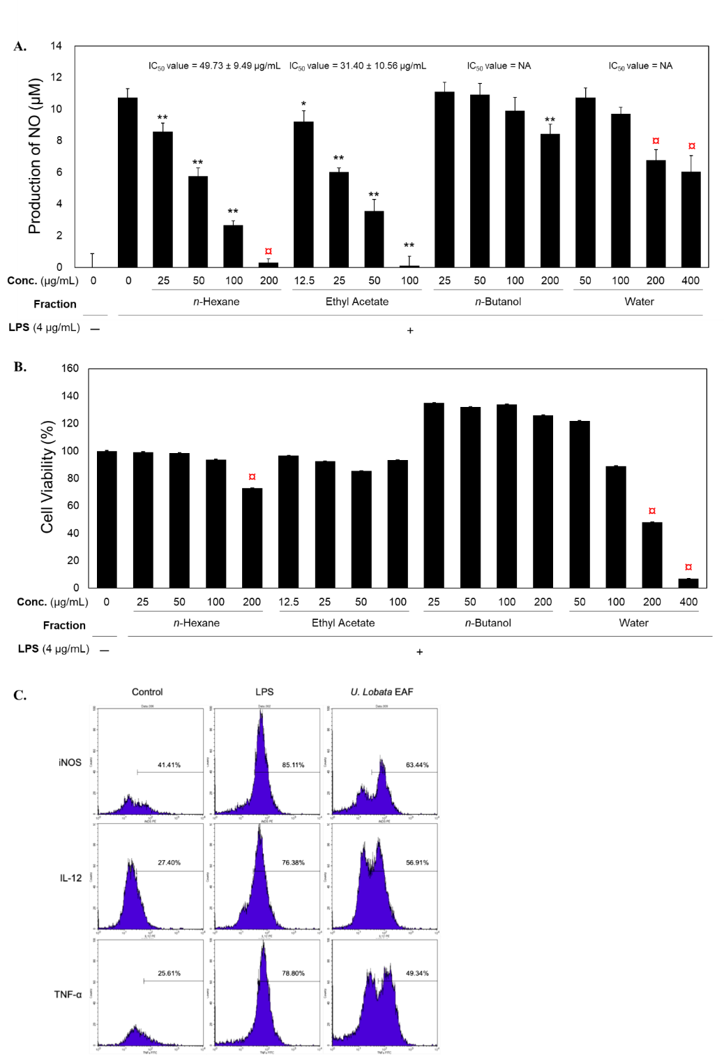
Figure 2 The effect of U. lobata fractions on NO Production (A), Cell Viability (B), and Pro-Inflammatory Production (C) of LPS-Treated RAW 264.7 Cell Line. Red circle indicate toxic dose.
Phytochemical composition of U. lobata extract
Based on the KNApSAcK database and the literature study, U. lobata contained 29 compounds presented in Table 1. The chemical composition of U. lobata reveals a diverse array of bioactive compounds, classified into several groups. Flavonoids represent the largest category, comprising eight compounds (29.63%). Following this, sterols, which include four compounds (14.81%). Fatty acids account for six compounds (22.22%). Additionally, eight compounds (29.63%) fall under the other organic compounds. Finally, a single nucleoside derivative (3.70%) is also present. This rich composition suggests that U. lobata may have significant therapeutic potential, particularly in the context of inflammation-related diseases.
Table 1 Bioactive compounds contained in U. lobata.
Compounds |
CID |
Molecular Formula |
Molecular Weight |
Gossypin |
5280661 |
C21H20O13 |
480.4 |
Mangiferin |
5281647 |
C19H18O11 |
422.3 |
Β-Sitosterol |
222284 |
C29H50O |
414.7 |
Stigmasterol |
5280794 |
C29H48O |
412.7 |
Stigmasta-3,5-diene |
6857463 |
C29H48 |
396.7 |
Stigmastan-3,5,22-trien |
6857464 |
C29H46 |
394.7 |
Undec-10-ynoic acid, pentadecyl ester |
5364555 |
C10H8N2O3 |
392.7 |
9-Octadecenoic acid (Z)-, 2,3-dihydroxypropyl ester |
445639 |
C21H40O4 |
356.5 |
Gossypetin |
5280660 |
C15H10O8 |
318.23 |
Quercetine |
5280343 |
C15H10O7 |
302.23 |
Hypolaetin |
5280662 |
C15H10O7 |
302.23 |
Chrysoeriol |
5280663 |
C16H12O6 |
300.26 |
Luteolin |
5280445 |
C15H10O6 |
286.24 |
Kaempferol |
5280863 |
C15H10O6 |
286.24 |
Oleic acid |
445639 |
C18H34O2 |
282.5 |
9,12-Octadecadienoic acid |
5280450 |
C10H8N2O3 |
280.4 |
Hexadecanoic acid, methyl ester |
8181 |
C10H8N2O3 |
270.5 |
Apigenin |
5280443 |
C15H10O5 |
270.24 |
n-Hexadecanoic acid |
985 |
C16H32O2 |
256.42 |
2H-Pyran-2-one, tetrahydro-6-undecyl- |
5318601 |
C16H30O2 |
254.41 |
Floxuridine |
3385 |
C9H11FN2O5 |
246.19 |
1H-Indole, 5-methyl-2-phenyl- |
14497 |
C15H13N |
207.27 |
Carbonic acid, monoamide, N-(2-ethylphenyl)-, propyl ester |
3082216 |
C12H17NO2 |
207.27 |
Pyrazol-5-ol, 3-(3,4-methylenedioxyphenyl)- |
10164 |
C10H8N2O3 |
204.18 |
Piperidine, 3-(bromomethyl)- |
10456 |
C6H12BrN |
178.07 |
phenol, 2,6-dimethoxy- |
3467 |
C8H10O3 |
154.16 |
2-methoxy-4-vinylphenol |
637678 |
C9H10O2 |
150.17 |
4h-pyran-4-one, 2,3-dihydro-3,5-dihydroxy-6-methyl- |
5280644 |
C6H8O4 |
144.12 |
2-Isopropoxyethylamine |
2734163 |
C5H13NO |
103.16 |
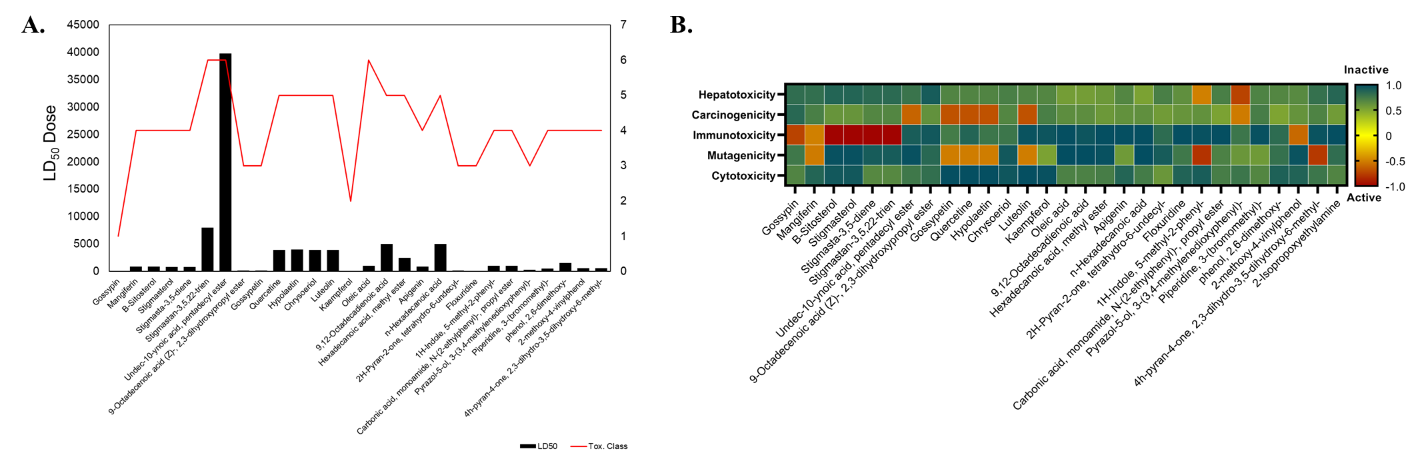
Figure 3 (A) The prediction of LD50 dose. (B) The toxicity profile prediction of compounds contained in U. lobata.
Toxicity classification of U. lobata compounds based on predicted LD50 values and toxicity profiles
Furthermore, this study analysed the predicted toxicity of various compounds from U. lobata, based on their LD50 values and toxicity profile. The LD50 values were classified into 6 categories according to the Globally Harmonized System of Classification and Labelling of Chemicals (GHS). As presented in Figure 3(A), Mangiferin, with an LD50 of 2 mg/kg, is classified as Class 1 (extremely toxic), while Oleic acid (48 mg/kg) falls under Class 2 (highly toxic). Compounds like Quercetine and Gossypetin, with an LD50 of 159 mg/kg, belong to Class 3 (toxic). Piperidine, 3-(bromomethyl) also falls into Class 3 with an LD50 of 295 mg/kg. Less toxic compounds include β-Sitosterol, Stigmasterol, and similar compounds, with LD50 values around 800 - 900 mg/kg, placing them in Class 4 (harmful). Gossypin and Apigenin, with LD50 values of 5,000 mg/kg and 2,500 mg/kg, respectively and some compounds such as luteolin and kaempferol, fall under Class 5, which indicates they may be harmful at high doses but generally have low acute toxicity. Finally, compounds like Undec-10-ynoic acid and 9-Octadecenoic acid are considered Class 6 (non-toxic or relatively harmless) with LD50 values exceeding 5,000 mg/kg.
Based on toxicity profile prediction, several com-pounds contained in U. lobata were considered toxic (Figure 3(B)). β-Sitosterol, Stigmasterol, Stigmas-ta-3,5-diene, Stigmastan-3,5,22-trien, and Oleic acid display low toxicity across various parameters, indicating they may be relatively safe for further research or application. However, Mangiferin, Gossypetin, Quercetine, Hypolaetin, and Luteolin are active in immunotoxicity and cytotoxicity, signaling potential risks in immune response and cellular toxicity that warrant careful consideration in therapeutic uses. Notably, 1H-Indole, 5-methyl-2-phenyl-, and Pyrazol-5-ol, 3-(3,4-methylenedioxyphenyl)- have significant carcinogenic profiles, raising red flags for cancer risk, while 4h-pyran-4-one exhibits both mutagenic and carcinogenic activity, suggesting it could pose serious health risks. Compounds like Apigenin and 2-Isopropoxyethylamine are inactive across all toxicity categories, implying minimal toxicity concerns. Overall, this analysis highlights promising compounds from U. lobata extract with low toxicity and others with toxicity profiles, especially regarding immunotoxicity, cytotoxicity, mutagenicity, and carcinogenicity.
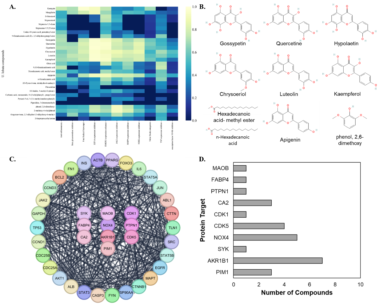
Figure 4 (A) Probable activity. (B) Compounds that meet anti-inflammatory criteria. (C-D) Prediction of Protein Targets for U. lobata Compounds.
Pharmacological properties of U. lobata compounds
Based on the analysis of the Table 1 for the bioactivities of compounds in U. lobata, it becomes clear that these compounds exhibit significant pharmacological properties, particularly in their anti-inflammatory, antioxidant, and signaling pathway modulation effects shown in Figure 4(A). Key compounds such as gossypetin, quercetin, hypolaetin, chrysoeriol, luteolin, kaempferol, and apigenin demonstrated strong inhibition of pro-inflammatory markers like HIF1A, JAK2, MMP9, NOS2, and TNF, alongside enhancement of the antioxidant enzyme HMOX1. These compounds also exhibited nitric oxide antagonism and NF-κB inhibition, supporting their role in mitigating oxidative stress and inflammatory responses. Other constituents such as hexadecanoic acid derivatives and phenol, 2,6-dimethoxy- showed moderate but relevant bioactivities. Overall, Gossypetin, Quercetine, Hypolaetin, Chrysoeriol, Luteolin, Kaempferol, Hexadecanoic acid- methyl ester, Apigenin, n-Hexadecanoic acid, and phenol, 2,6-dimethoxy, (Figure 4(B)) stand out as key bioactive compounds in U. lobata, with high probable activities across multiple inflammation-related pathways. These results indicate their potential as therapeutic agents for controlling inflammatory responses and oxidative stress.
Target protein analysis reveals key anti-inflammatory mechanisms of U. lobata active compounds
Based on the protein target analysis, 40 proteins involved in the inflammatory pathway were identified as targets of U. lobataʼs active compounds (Figure 4(C)). Among these, 10 proteins—AKR1B1, CDK1, CDK5, NOX4, MAOB, PTPN1, FABP4, SYK, CA2, and PIM1—were directly targeted (central node), while the remaining 30 proteins were indirectly affected (outer node). Notably, gossypetin, quercetin, and hypolaetin were predicted to target key proteins such as AKR1B1, PIM1, and NOX4, which are critical in the inflammatory response. Luteolin and apigenin showed similar target profiles, particularly targeting NOX4 and AKR1B1, while chrysoeriol, kaempferol, and apigenin were predicted to interact with CDK5. Additionally, hexadecanoic acid, methyl ester, targets CA2 and PTPN1, and n-hexadecanoic acid targets FABP4. Based on these findings, AKR1B1 emerged as the most targeted protein, being affected by 7 of the 10 active compounds in U. lobata. Five active compounds targeted NOX4, and CDK5 by 4 compounds (Figure 4(D)), highlighting the prominent role of U. lobata as a potential anti-inflammatory agent.
Table 2 Binding affinities (kcal/mol) of U. lobata compounds with key inflammatory pathway proteins.
No
|
Compounds |
Binding Affinity (kcal/mol) with Targeted Proteins |
|||||||||
AKR1B1 |
NOX4 |
CDK5 |
PIM1 |
CA2 |
MAOB |
FABP4 |
PTPN1 |
CDK1 |
SYK |
||
1. |
Native Ligand |
‒10 |
‒9.3 |
‒7.5 |
−7.4 |
−6.1 |
‒6.5 |
‒10.3 |
‒6.7 |
‒6.9 |
‒8 |
2. |
Gossypetin |
‒9.5 |
- |
- |
−8.3 |
- |
- |
- |
- |
- |
‒.84 |
3. |
Quercetine |
‒10.1 |
‒8.6 |
- |
−8.4 |
- |
- |
- |
- |
- |
- |
4. |
Hypolaetin |
‒9.7 |
‒8.8 |
- |
−8.4 |
- |
- |
- |
- |
- |
- |
5. |
Chrysoeriol |
‒9 |
- |
‒8.5 |
- |
- |
- |
- |
- |
‒7.8 |
- |
6. |
Luteolin |
‒10.2 |
‒8.7 |
‒8.5 |
- |
- |
- |
- |
- |
- |
- |
7. |
Kaempferol |
‒8.3 |
‒8.3 |
- |
- |
‒7 |
- |
- |
- |
- |
- |
8. |
Hexadecanoic acid, methyl ester |
- |
- |
- |
- |
‒5.2 |
- |
- |
‒5.2 |
- |
- |
9. |
Apigenin |
‒10.4 |
‒8.3 |
‒8.4 |
- |
- |
- |
- |
- |
- |
- |
10. |
n-Hexadecanoic acid |
- |
- |
- |
- |
- |
- |
‒5.7 |
- |
- |
- |
11. |
phenol, 2,6-dimethoxy- |
- |
- |
‒4.9 |
- |
‒5.5 |
‒5.6 |
- |
- |
- |
- |
Comparative binding affinities of U. lobata compounds with native ligand for inflammatory pathway proteins
The binding affinities of various compounds isolated from U. lobata were assessed through molecular docking to evaluate their potential as inhibitors of key proteins involved in inflammatory pathways. Table 2 provides an overview of the binding affinities (kcal/mol) of these compounds with multiple targeted proteins, including AKR1B1, NOX4, CDK5, PIM1, CA2, MAOB, FABP4, PTPN1, CDK1, and SYK. The results were compared to the native ligand, which served as a reference. Quercetin exhibited the strongest binding affinity for AKR1B1 (‒10.1 kcal/mol) and NOX4 (‒8.6 kcal/mol), surpassing the native ligandʼs affinity for AKR1B1 (‒10.0 kcal/mol) and closely approaching its affinity for NOX4 (‒9.3 kcal/mol). This suggests quercetin is a potent inhibitor of these proteins, particularly in modulating oxidative stress and inflammatory pathways. Apigenin also displayed significant binding to AKR1B1 (‒10.4 kcal/mol) and NOX4 (‒8.3 kcal/mol), with its affinity for AKR1B1 being higher than both quercetin and the native ligand. Additionally, apigenin strongly binds to CDK5 (‒8.4 kcal/mol), indicating its potential in modulating cell cycle-related inflammation.
Moreover, Luteolin and hypolaetin showed strong binding to AKR1B1, with binding affinities of ‒10.2 kcal/mol and ‒9.7 kcal/mol, respectively. Both also exhibited considerable interaction with NOX4, indicating their capacity to target multiple inflammatory proteins. Luteolin also demonstrated competitive binding to CDK5 (‒8.5 kcal/mol), which is relevant to its role in inflammation control. Chrysoeriol showed moderate binding to CDK1 (‒7.8 kcal/mol) and PIM1 (‒8.5 kcal/mol), highlighting its potential to interact with kinases involved in inflammation. Gossypetin exhibited moderate binding affinity to AKR1B1 (‒9.5 kcal/mol) and PIM1 (‒8.3 kcal/mol), though weaker than quercetin or apigenin. In contrast, compounds such as hexadecanoic acid, methyl ester, and phenol, 2,6-dimethoxy-, demonstrated much lower binding affinities to the majority of proteins, particularly in comparison to the native ligand, suggesting a limited role in modulating the targeted inflammatory proteins. Molecular docking simulation can be seen in Figure 5.
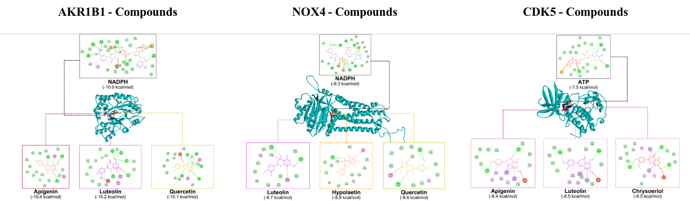
Figure 5 Molecular docking simulation results.
Discussion
This study reveals the anti-inflammatory activity of U. lobata extract. The ethyl acetate-soluble fraction of U. lobata emerged as the most potent in suppressing NO production in LPS-treated RAW 264.7 cells, with an IC50 value of 31.40 ± 10.56 µg/mL. This significant inhibitory effect contrasts with the toxicity observed in other fractions, particularly the n-hexane fraction, which exhibited cytotoxic effects at higher concentrations. Interestingly, in addition to NO suppression, the ethyl acetate fraction also reduced the production of other key proinflammatory mediators, iNOS, IL-12, and TNF-α, indicating its broader role in modulating inflammatory responses. The selective efficacy of the ethyl acetate fraction suggests that it contains bioactive compounds capable of targeting multiple inflammatory pathways without eliciting significant toxicity, making it a promising candidate for further therapeutic exploration. NO, produced by nitric oxide synthases (NOSs) from Arginine, acts as a signaling free radical that contributes to chronic inflammation when excessively accumulated. These cellular effects suggest that the ethyl acetate fraction of U. lobata interferes with upstream regulatory nodes controlling oxidative stress and inflammatory gene transcription. Since the expression of iNOS and pro-inflammatory cytokines is largely governed by redox-sensitive signaling pathways and kinase-mediated inflammatory cascades, we hypothesized that the active constituents of U. lobata may target key molecular regulators involved in these processes. This hypothesis is further supported by our in silico analysis, which predicts that major constituents of U. lobata target key regulatory proteins involved in these pathways.
To further elucidate the mechanisms underlying the anti-inflammatory effects of Urena lobata, an in silico analysis identified 10 active compounds—Gossypetin, Quercetin, Hypolaetin, Chrysoeriol, Luteolin, Kaempferol, Hexadecanoic acid methyl ester, Apigenin, n-Hexadecanoic acid, and phenol, 2,6-dimethoxy—that demonstrated strong potential as anti-inflammatory agents based on their Pa values. These compounds were predicted to target key inflammation-related proteins, such as aldose reductase 1B1 (AKR1B1), NADPH oxidase 4 (NOX4), and cyclin-dependent kinase 5 (CDK5), which are central to the regulation of oxidative stress and inflammatory signaling pathways [21-23]. Consistent with in vitro findings, the ethyl acetate-soluble fraction of U. lobata was identified as the most potent, aligning with prior studies that reported various bioactivities of ethyl acetate fractions from medicinal plants, including hepatoprotective, antioxidant, antimicrobial, antidiabetic, and anticancer effects [24-27]. Although previous studies have reported the antioxidant and anti-inflammatory potential of U. lobata and several flavonoids, such as quercetin and luteolin, most of these works have been limited to phenotypic observations without providing a clear molecular explanation. In contrast, the present study extends current knowledge by integrating in vitro functional assays with in silico target prediction and molecular docking, thereby proposing a multi-target mechanistic model involving AKR1B1, NOX4, and CDK5. To the best of our knowledge, this is the first study to explicitly link the anti-inflammatory activity of U. lobata to the simultaneous modulation of these key regulators of oxidative stress and inflammatory signaling pathways.
Molecular docking analysis further supports the anti-inflammatory potential of Urena lobata compounds by demonstrating their strong binding affinities to key inflammatory proteins, particularly AKR1B1, NOX4, and CDK5. Quercetin exhibited dual inhibitory potential with binding affinities of −10.1 kcal/mol for AKR1B1 and −8.6 kcal/mol for NOX4, suggesting its capability to modulate both oxidative stress and inflammation [30]. Apigenin also showed promising interactions, binding strongly to AKR1B1 (−10.4 kcal/mol) and CDK5 (−8.4 kcal/mol), supporting its role in dampening oxidative and inflammatory signaling. Luteolin emerged as a potent multitarget compound with high affinities for AKR1B1 (−10.2 kcal/mol), NOX4 (−8.7 kcal/mol), and CDK5 (−8.5 kcal/mol), indicating its broad-spectrum activity in mitigating inflammatory responses. Gossypetin, a key fingerprint compound of U. lobata, demonstrated slightly lower binding than the aforementioned flavonoids but still showed significant interactions with AKR1B1 (−9.5 kcal/mol) and PIM1 (−8.3 kcal/mol), implicating its role in reducing oxidative stress and potentially modulating cell survival pathways [21]. In contrast, hexadecanoic acid methyl ester and phenol, 2,6-dimethoxy-, exhibited weak binding affinities, suggesting a limited role in the direct inhibition of inflammation-related proteins.
From a macrophage biology perspective, the identified protein targets (AKR1B1, NOX4, and CDK5) play critical roles in regulating inflammatory activation and oxidative stress responses. In LPS-stimulated macrophages, excessive ROS production and kinase-driven signaling cascades are central drivers of NF-κB activation, leading to the transcription of iNOS and pro-inflammatory cytokines. AKR1B1 contributes to inflammatory amplification by promoting oxidative stress and redox imbalance, while NOX4 serves as a major enzymatic source of ROS that sustains inflammatory signaling and iNOS induction. Meanwhile, CDK5 functions as a regulatory kinase that modulates inflammatory transcriptional programs, including NF-κB signaling, thereby influencing the production of TNF-α, IL-6, and NO [28,29]. Therefore, the predicted inhibition of AKR1B1, NOX4, and CDK5 by quercetin, apigenin, and luteolin provides a biologically plausible explanation for the observed suppression of NO, iNOS, TNF-α, and IL-12 in LPS-stimulated macrophages. The strong binding of these compounds to the identified targets supports a multi-target modulation model, in which simultaneous attenuation of oxidative stress and inflammatory signaling pathways leads to reduced activation of pro-inflammatory transcriptional programs and, ultimately, decreased production of NO and inflammatory cytokines.
Furthermore, AKR1B1, NOX4, and CDK5 have important interactions with peripheral proteins (indirect protein target of U. lobata compounds) especially INS, JAK2, TP53, IL6, FN1, BCL2, ALB, and EGFR, particularly in the context of inflammation, oxidative stress, and metabolic regulation. AKR1B1 is integral to inflammatory pathways and oxidative stress, modulated indirectly by INS and JAK2. INS regulates key metabolic processes, including glucose and lipid metabolism, influencing AKR1B1ʼs role in inflammation-related glucose toxicity [30]. JAK2, a critical mediator in cytokine signaling, enhances pro-inflammatory cytokine production, such as IL6, which activates AKR1B1 and promotes NO and inflammatory cytokine synthesis, linking metabolic dysregulation to oxidative stress [21,31]. AKR1B1 also plays a significant role in inflammatory signaling by modulating pro-inflammatory pathways, which influence the expression of pro-inflammatory cytokines and iNOS. Since iNOS produces large amounts of NO during inflammation, dysregulation of AKR1B1 can lead to alter NO levels [21]. Additionally, AKR1B1 contributes to oxidative stress by increasing the production of ROS. Elevated ROS further activates inflammatory pathways that upregulate iNOS, resulting in excessive NO production and potentially worsening cellular damage in inflammatory conditions [21]. Thus, AKR1B1 dysregulation can indirectly influence NO levels through both inflammatory signaling and oxidative stress pathways.
NOX4 is essential for ROS production and is activated by IL6, TP53, and JAK2. IL6 enhances ROS generation, where NOX4 contributes directly to oxidative stress [32,33]. TP53, while primarily regulating the cell cycle and apoptosis, can also induce conditions that activate NOX4, thereby linking apoptotic pathways to inflammatory ROS production. Additionally, JAK2 signaling promotes the release of pro-inflammatory mediators, further amplifying oxidative stress via NOX4. Inhibition of NOX4 reduces oxidative stress by decreasing the production of ROS, preventing the interaction of ROS with NO, which would otherwise form harmful reactive nitrogen species. Additionally, NOX4 inhibition downregulates inflammatory pathways, including the expression of iNOS, leading to a decrease in excessive NO production during inflammation [33]. Thus, inhibiting NOX4 can help balance NO levels, reducing both pathological overproduction and oxidative depletion of NO.
CDK5 interacts with TP53, BCL2, and EGFR, regulating pro-inflammatory pathways. TP53 influences CDK5 indirectly by modulating stress responses that trigger inflammation, while BCL2, which governs cell survival and apoptosis, can modulate CDK5 activity in chronic inflammatory contexts. EGFR further influences CDK5 by promoting cell proliferation and survival under inflammatory conditions, linking it to chronic inflammatory diseases [34]. CDK5 is also known to regulate NF-κB pathway, which plays a central role in controlling pro-inflammatory gene expression. Activation of CDK5 leads to the phosphorylation of proteins that influence NF-κB activity, thereby affecting the production of pro-inflammatory cytokines like TNF-α, IL-1β, and IL-6 [23]. Inhibiting CDK5 can reduce NO production, particularly in inflammation-related contexts. CDK5 is also involved in regulating inflammatory signaling, and studies have shown that its activation promotes the expression of iNOS, an enzyme responsible for producing large amounts of NO during inflammation [23]. By inhibiting CDK5, the signaling pathway that upregulates iNOS is disrupted, leading to a decrease in NO production. This reduction in NO helps to control inflammation and minimize cell damage caused by excessive NO levels.
The current study primarily focused on evaluating the individual effects of the U. lobata extract and its major compounds. Exploring combinatorial effects would provide valuable insights into the overall therapeutic potential. In future studies, combination testing using well-established methods, such as isobolographic or checkerboard assays, will be performed to evaluate interactions between compounds. Additionally, network pharmacology approaches will be employed to analyze potential synergistic or antagonistic effects among the active constituents. Mechanistically, this multi-target inhibition model provides a coherent explanation linking the molecular docking predictions with the cellular anti-inflammatory responses observed in vitro, particularly the suppression of iNOS-dependent NO production and pro-inflammatory cytokine release. In summary, this study demonstrated that quercetin, apigenin, and luteolin which are contained in an ethyl acetate-soluble fraction of U. lobata, emerge as the most promising compounds from U. lobata, with strong binding affinities to AKR1B1, NOX4, and CDK5 which further have a significant role in suppressing NO levels during inflammation condition. These findings suggest that the ethyl acetate-soluble fraction of U. lobata, enriched in these flavonoids, holds significant potential as a natural anti-inflammatory agent, targeting multiple proteins contributing to inflammation and oxidative stress. The multi-targeted approach of these compounds provides a solid foundation for further research into the therapeutic potential of U. lobata in managing inflammation-related diseases.
However, despite their promising anti-inflammatory effects, some of these compounds exhibit predicted toxicity, particularly in immunotoxicity and cytotoxicity. For example, Quercetine, and Luteolin have moderate to high immunotoxicity and cytotoxicity scores, raising concerns about potential adverse effects. While these properties suggest caution in therapeutic use, they also present a dual role: the cytotoxicity observed in these compounds may contribute to their effectiveness in targeting aberrant or overactive immune cells, which are often involved in chronic inflammatory diseases or cancer. This selective action could, therefore, enhance their therapeutic value when applied in controlled, disease-specific contexts. Moreover, toxicity is often dose-dependent, and it is likely that these compounds could provide substantial therapeutic benefits at lower, carefully regulated doses [35]. For instance, these compounds may demonstrate a favorable safety profile in doses designed to modulate inflammation rather than exert broad cytotoxic effects. This is a critical consideration, as many conventional anti-inflammatory drugs, such as NSAIDs and corticosteroids, are themselves associated with significant side effects, yet their benefits often justify their use in clinical practice [9]. Similarly, the flavonoids and fatty acids studied here may offer valuable alternatives or adjuncts to current therapies, provided their dosing is optimized to minimize toxicity.
The relationship between the bioactive compounds in the ethyl acetate-soluble fraction and their targeted protein interactions suggests a nuanced approach to therapeutic development. The ethyl acetate fractionʼs ability to inhibit NO production without significant toxicity may be attributed to the selective action of certain compounds that effectively modulate inflammatory pathways. Conversely, the presence of compounds with higher toxicity profiles highlights the need for further characterization of the fractions to isolate the most beneficial components. Given this context, further investigation is warranted to explore the therapeutic window of these compounds. In vivo and clinical studies will be essential to evaluate their efficacy in suppressing inflammation while minimizing their toxicity risks. Understanding the appropriate dosing regimens and their pharmacodynamic properties could unlock the full potential of these compounds as safe and effective anti-inflammatory agents. This balance between efficacy and safety highlights the importance of ongoing research into the pharmacological properties of natural compounds, such as those studied here.
Although U. lobata showed anti-inflammatory activity at non-cytotoxic concentrations, reduced cell viability at higher doses indicates the need for careful dose optimization and further fractionation. Since the present work is limited to in vitro experiments, in vivo studies are required to confirm efficacy and safety. Furthermore, the in-silico analyses are predictive in nature and therefore require further experimental validation. Despite these limitations, this study integrates cellular and computational evidence to provide a coherent mechanistic framework that supports the anti-inflammatory potential of U. lobata and guides future in-depth investigations.
Conclusions
The findings from both in vitro and in silico analyses underscore the potential of the ethyl acetate fraction of U. lobata as a viable candidate for the development of anti-inflammatory therapies. The ability of its compounds to target critical proteins like AKR1B1, NOX4, and CDK5, alongside the suppression of NO production, presents a compelling case for further research. Future studies should aim to isolate and characterize the specific compounds within the ethyl acetate fraction to fully understand their mechanisms of action and assess their safety profiles. This comprehensive approach will be crucial for developing effective and safe therapeutic strategies for managing inflammation and related disorders.
Acknowledgements
This work was financially supported by DRTPM (Directorate General of Higher Education, Ministry of Education and Culture, Republic of Indonesia) under the Penelitian Dasar Unggulan Perguruan Tinggi (PDUPT) scheme.
Declaration of generative AI in scientific writing
Generative AI tools were used solely to improve the readability, grammar, and clarity of the manuscript. The authors bear full responsibility for the integrity and originality of the manuscript.
CRediT author statement
Yudi Purnomo: Conceptualization; Methodology; Project administration; Funding acquisition; Writing - Review & Editing. Andri Tilaqza: Data curation; Writing - Review & Editing. Doti Wahyuningsih: Validation; Writing - Review & Editing. Dinia Rizqi Dwijayanti: Methodology; Investigation; Formal analysis; Visualization; Writing - Original Draft.
References
[1] YP Dhokane and U Akhtar. Pharmacological evaluation of lactuca canadensis. linn extract on anti-inflammatory activity in Experimental Animals. Research Journal of Pharmacology and Pharmacodynamics 2024; 16(1), 15-18.
[2] P Bansal, D Verma, SS Tamber and R Sharma. An update on pathogenesis of inflammatory disorders with its management. RPS Pharmacy and Pharmacology Reports 2023; 2(2), 011.
[3] DR Dwijayanti, T Shimada, T Ishii, T Okuyama, Y Ikeya, E Mukai and M Nishizawa. Bitter melon fruit extract has a hypoglycemic effect and reduces hepatic lipid accumulation in Ob/Ob mice. Phytotherapy Research 2020; 34(6), 1338-1346.
[4] ED Ahire, KR Surana, KR Jadhav, SJ Kshirsagar and D Sonawane. Current therapeutic targets for neuropathic pain. Asian Journal of Pharmaceutical Research 2022; 12(1), 96-104.
[5] A Nishida and A Andoh. The role of inflammation in cancer: Mechanisms of tumor initiation, progression, and metastasis. Cells 2025; 14(7), 488.
[6] CLR Soares, P Wilairatana, LR Silva, PS Moreira, NMMV Barbosa, PR da Silva, HDM Coutinho, IRA de Menezes and CFB Felipe. Biochemical aspects of the inflammatory process: A narrative review. Biomedicine & Pharmacotherapy 2023; 168, 115764.
[7] DB Johnson, R Sivasakthi, M Ashwini and R Venkatanarayanan. Anti-inflammatory activity screening of methanolic extract of Bryophyllum pinnatum root. Research Journal of Pharmacology and Pharmacodynamics 2022; 14(1), 1-3.
[8] MG Shinde, KD Sayyad, GV Swami, PB Savant, PR Yalmar and ST Mane. A review on inflammation and its pharmacotherapy. Asian Journal of Pharmacy and Technology 2023; 13(3), 201-206.
[9] I Ghlichloo and V Gerriets. Nonsteroidal Anti-Inflammatory Drugs (NSAIDs). StatPearls, USA, 2025.
[10] J Srikanth and P Muralidharan. Analgesic and anti-inflammatory activity pericarps of Sapindus emarginatus vahl. Research Journal of Pharmacology and Pharmacodynamics 2009; 1(1), 21-24.
[11] PR Kumar, SS Priyadarsini and M Thirumal. Nitric oxide radical scavenging activity of the ethanolic and chloroform extracts of Ocimum basilicum Linn. Leaves – A comparative study. Research Journal of Pharmacognosy and Phytochemistry 2017; 9(4), 215-218.
[12] OO Oguntibeju. Medicinal plants with anti-inflammatory activities from selected countries and regions of Africa. Journal of Inflammation Research 2018; 11, 307-317.
[13] A Adewale, A David, O Abiodun and A Craig. Studies on anti-microbial, antioxidant and phytochemical analysis of Urena lobata leaf extract. Journal of Physics and Natural Sciences 2007; 1(2), 1-9.
[14] Y Purnomo and A Tilaqza. Inhibitory potential of pulutan (Urena lobata) leaf extract on inducible nitric oxide synthase as anti-inflammatory agent: in vitro and in silico approaches. Tropical Journal of Natural Product Research 2024; 8(8), 8195-8201.
[15] Y Purnomo, A Tilaqza, DM Mirza and AZ Mustopa. In vitro and in silico assessment of the antidiabetic and antioxidant potential of Urena lobata leaf fraction. Tropical Journal of Natural Product Research 2025; 9(10), 5016-5024.
[16] Y Purnomo and M Nishizawa. Dechlorophyllized Urena lobata leaf fraction inhibits nitric oxide production in IL-1β-induced rat hepatocytes. Tropical Journal of Natural Product Research 2025; 9(4), 1492-1495.
[17] Y Purnomo, DW Soeatmadji, SB Sumitro and MA Widodo. Anti-hiperglycemic effect of Urena lobata leaf extract by inhibition of dipeptidyl peptidase-4 (DPP-4) on diabetic rat. International Journal of Pharmacognosy and Phytochemical Research 2015; 7(5), 1073-1079.
[18] AB Kunnumakkara, AS Nair, KS Ahn, MK Pandey, Z Yi, M Liu and BB Aggarwal. Gossypin, a pentahydroxy glucosyl flavone, inhibits the transforming growth factor beta-activated kinase-1-mediated NF-KappaB activation pathway, leading to potentiation of apoptosis, suppression of invasion, and abrogation of osteoclastogenesis. Blood 2007; 109(12), 5112-5121.
[19] DR Dwijayanti, MH Widyananda, FE Hermanto, A Soewondo, M Afiyanti and N Widodo. Revealing the anti-inflammatory activity of Euphorbia hirta extract: Transcriptomic and nitric oxide production analysis in LPS-induced RAW 264.7 cells. Food and Agricultural Immunology 2024; 35(1), 2351360.
[20] DR Dwijayanti, A Mamamia, RYN Maulana, N Rosyadah and N Widodo. Beyond a spice: Harnessing the anti-inflammatory potential of Indonesian ginger ̶ insights into gingerenones and isogingerenone. Cogent Food & Agriculture 2025; 11(1), 2568198.
[21] SK Srivastava, UCS Yadav, ABM Reddy, A Saxena, R Tammali, M Shoeb, NH Ansari, A Bhatnagar, MJ Petrash, S Srivastava and KV Ramana. Aldose reductase inhibition suppresses oxidative stress-induced inflammatory disorders. Chemico-Biological Interactions 2011; 191(1-3), 330-338.
[22] A Lozhkin, AE Vendrov, H Pan, SA Wickline, NR Madamanchi and MS Runge. NADPH oxidase 4 regulates vascular inflammation in aging and atherosclerosis. Journal of Molecular and Cellular Cardiology 2017; 102, 10-21.
[23] P Pfänder, AK Eiers, U Burret and S Vettorazzi. Deletion of Cdk5 in macrophages ameliorates anti-inflammatory response during endotoxemia through induction of C-Maf and Il-10. International Journal of Molecular Sciences 2021; 22(17), 9648.
[24] T Venkatachalam, VK Kumar, PS Kumar, P Kalaiselvi, M Chitra and NS Kumar. In-vitro anti oxidant and antimicrobial activities of ethyl acetate extract of Evodia lunu-Ankenda (Gaertn) Merr. Bark. Research Journal of Pharmacognosy and Phytochemistry 2009; 1(3), 201-203.
[25] DJ Sawarkar, C Vijaya, AO Turaskar, VS Shende, VK Chatap, VA Sawant and SN Borkar. Hepatoprotective activity of ethanolic and ethyl acetate extract of Ziziphus mauriatiana on liver damaged caused by Paracetamol in Rats. Research Journal of Pharmacognosy and Phytochemistry 2009; 1(3), 194-197.
[26] BE Jose and P Panneerselvam. In vivo anti diabetic activity of ethyl acetate leaf extract of Azima tetracantha lam in streptozotocin-induced Diabetic Mice. Research Journal of Pharmacy and Technology 2019; 12(2), 660-664.
[27] JD Milton and MA Jose. Anticancer activity of ethyl acetate root extract of Xanthium strumarium Linn. in N-nitroso N-methyl urea induced mammary carcinoma in female sprague-dawley Rats. Research Journal of Pharmacy and Technology 2016; 9(8), 1115-1118.
[28] Y Zhao, M Zhang, H Li, Y Yang, L Xiaofy, Y Junjing and P Lei. Aldo-keto reductase 1B: Much learned, much more to do. hLife 2024; 2(4), 154-178.
[29] Y Li, H Li, X Chen, X Li, J Su, S Yang, W Xiao and Z Deng. CDK5 regulates PPARγ/NF-κB signaling to exacerbate obesity-related osteoarthritis via modulating macrophage polarization and chondrocyte apoptosis. Cellular & Molecular Biology Letters 2025; 30, 135.
[30] FJ Sánchez-Gómez, B Díez-Dacal, E García-Martín, JAG Agúndez, MA Pajares and D Pérez-Sala. Detoxifying enzymes at the cross-roads of inflammation, oxidative stress, and drug hypersensitivity: Role of glutathione transferase P1-1 and aldose reductase. Frontiers in Pharmacology 2016; 7, 00237.
[31] JJ O'Shea and PJ Murray. Cytokine signaling modules in inflammatory responses. Immunity 2008; 28(4), 477-487.
[32] U Weyemi and C Dupuy. The emerging role of ros-generating NADPH oxidase NOX4 In DNA-damage responses. Mutation Research/Reviews in Mutation Research 2012; 751(2), 77-81.
[33] S Piera-Velazquez and SA Jimenez. Oxidative stress induced by Reactive Oxygen Species (ROS) and NADPH oxidase 4 (NOX4) in the pathogenesis of the fibrotic process in systemic sclerosis: A promising therapeutic target. Journal of Clinical Medicine 2021; 10(20), 4791.
[34] PA Do and CH Lee. The role of CDK5 in Tumours and Tumour Microenvironments. Cancers 2021; 13(1), 101.
[35] CB Rajashekar. Dual role of plant phenolic compounds as antioxidants and prooxidants. American Journal of Plant Sciences 2023; 14(1), 15-28.